Injections of acrylic cement into a diseased femoral head and femoral canal have usually been involved in a procedure of total hip replacement (THR). Severe com- plications resulting from this procedure are infrequent.
However, this procedure can cause localized problems such as infection or cement leakage into the femoral vein to inferior vena cava. We report a case of sympto- matic foreign body formation in the right cardiac cham- bers caused by acrylic cement migration following a THR.
Case Report
A 60-year-old woman was hospitalized for hip pain which made standing on her own difficult. The patient had a history of rheumatoid arthritis in both hips for 15 years. Hip joint problems gradually worsened until the patient was not able to stand by herself. Radiologic find-
ings from both hip joints confirmed advanced rheuma- toid arthritis.
Initial chest radiography showed no remarkable find- ings. Initial blood gas analysis showed mild elevation of pO2(111.6 mmHg), and normal range of pCO2(35.4 mm Hg). Other blood chemistry results were not remark- able. Preoperative echocardiography showed no re- markable findings.
Left hip replacement was performed through the pos- terolateral approach using a bone cement injection gun under general anesthesia. The acrylic cement had been prepared at room temperature (22℃) and mixed with 1g of tungsten powder. There were no systemic complica- tions after left hip replacement. One month later, right hip replacement was performed using the same method.
A few days after the right hip replacement, the patient was bothered by a cough and complained of mild dysp- nea. Chest radiography showed no remarkable finding and blood gas analysis revealed both pO2(84.7 mmHg) and pCO2 (38.0 mmHg) to be within normal ranges.
Doppler sonography of both lower extremities showed no evidence of deep vein thrombosis. Echocardiography revealed a linear echogenic structure adjacent to the tri- cuspid valve with mild tricuspid regurgitation, mild pul-
J Korean Radiol Soc 2005;53:175-178
─ 175 ─
Intracardiac Foreign Body Formation from Bone Cement Material Following Total Hip Replacement: A Case Report1
Jin Hee Moon, M.D., In Jae Lee, M.D., Hyun Beom Kim, M.D., Eun Young Ko, M.D., Sung Hye Koh, M.D., Keon Ha Kim, M.D.
1Department of Radiology, Hallym University College of Medicine Received March 30, 2005 ; Accepted July 5, 2005
Address reprint requests to : In Jae Lee, M.D., Department of Radiology, Hallym University Sacred Heart Hospital,
896 Pyungchon-dong, Anyang, Kyungki-do 431-070, Korea.
Tel. 82-31-380-3885 Fax. 82-31-380-3878 E-mail: [email protected]
A linear intracardiac foreign body was identified following a total hip replacement (THR) on chest CT and transesophageal echocardiography in a 60-year-old woman with rheumatoid arthritis. Leakage and migration of bone cement during arthroplasty is a possible explanation for this rare complication. Therefore, adequate preparation and handling of cement using biplane fluoroscopy are recommended during arthro- plasty.
Index words :Chest, CT
Heart, echocardiography Heart, foreign bodies
monary hypertension and a moderate amount of peri- cardial effusion (Fig. 1A).
Exact delineation of the linear structure adjacent to the tricuspid valve was difficult to obtain with transtho- racic echocardiograpy. Consequently, a transesophageal echocardiography was performed. It revealed an abnor- mal linear structure that seemed to originate in the right atrial appendage extending into the right ventricle (Fig.
1B). Because the structure was less mobile than cardiac movement, it was considered to be a foreign body with a hard structure. The cardiac ejection fraction decreased following THR from 62% to 39%.
Non-enhanced chest CT scan showed a focal high at- tenuation structure within the right cardiac chamber (Fig. 1C). Chest CT with three-dimensional reconstruc- tion of images revealed a linear high attenuation lesion
6.4 cm in length with an irregular contour in the right atrium and ventricle (Fig. 1D). The linear structure was regarded as bone cement material related to the THR procedure. The patient was treated with theophylline, aspirin, and prednisolon. There was mild improvement of cardiopulmonary symptoms following medical treat- ment. Because of the deterioration of cardiac contractili- ty, surgical removal of bone cement was recommended for the patient. However, the patient refused the opera- tion and was discharged.
In this case, the intracardiac foreign body was not sur- gically confirmed to be bone cement material. However, there does not appear to be any other factors that may have caused this condition. Therefore, we regarded the intracardiac foreign body as bone cement material relat- ed to the THR procedure.
Jin Hee Moon, et al: Intracardiac Foreign Body Formation from Bone Cement Material Following Total Hip Replacement
─ 176 ─
A B
C D
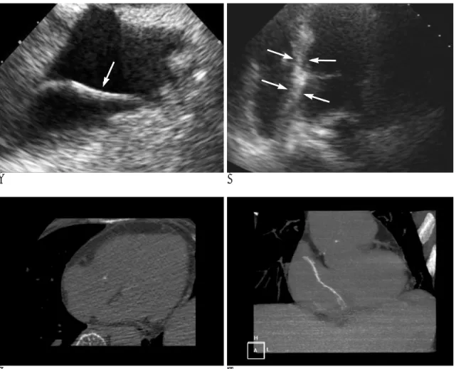
Fig. 1. A 60-year-old woman with an intracardiac foreign body from bone cement material migrated into the right cardiac chamber following THR.
A. Echocardiogram shows a linear echogenic structure in the right cardiac chamber.
B. Transesophageal echocardiogram also shows a linear echogenic structure in the right atrium and ventricle crossing the tricuspid valve.
C. Unenhanced chest CT scan shows a focal high attenuation structure within the right cardiac chamber.
D. Three-dimensional CT reformation image shows a linear high attenuation structure in the right atrium and ventricle.
Discussion
Although THR using a cement injection gun is consid- ered to be a less invasive procedure compared with open surgery, complications can occur during the proce- dure. Many authors have described an association be- tween the use of bone cement (polymethylmethacry- late) in human hip arthroplasty and cardiovascular com- plications such as hypotension, bradycardia, asystole, and bronchospasm. The etiology of these effects is not entirely clear, but mechanisms such as fat embolism as- sociated with increased intramedullary pressure, air em- bolism, a neurogenic reflex, the release of vasoactive mediators such as histamine, direct depressive effects on the myocardium, peripheral vasodilatation, and acti- vation of the coagulation cascade within the lungs have been suggested (1, 2). Other complications reported in literature include cardiac arrhythmias, myocardial in- farction and cardiac arrest (3). Although many reports have documented symptomatic conditions caused by acrylic cement, there have been no radiologic reports which deal with symptomatic intracardiac foreign body formation.
The patient had cardiopulmonary symptoms includ- ing cough, mild dyspnea and lowered ejection fraction.
An abnormal linear structure in the right cardiac cham- ber on echocardiography suggested bone cement had toxic effect on cardiopulmonary system.
The acrylic cement leakage and foreign body forma- tion can be explained by the fluid consistency of the acrylic cement at the moment of injection. More exten- sive leaks may occur if the tip of the injection gun is po- sitioned incorrectly or the procedure is performed under high pressure with a large volume of acrylic cement (1, 2).
In this case, leakage may have been caused by insuffi- cient polymerization of the acrylic cement at the time of injection, which allowed leakage of the cement into the inferior vena cava and right cardiac chamber. For this reason, the acrylic cement must be mixed to the proper consistency (i.e., a stage of advanced polymerization) be- fore injection, so that the several difficulties to evaluate factors that affect the polymerization rate (e.g., room temperature) may be better controlled.
Good quality lateral fluoroscopy is essential during in- jection of the bone cement material because it can detect
even minimal cement leakage into the femoral vein im- mediately. Jensen et al (4) recommend a barium/tung- sten combination for adequate visualization of needle positioning and venous flow during fluoroscopy.
Usually the cement is mixed only with tungsten powder to achieve opacification during fluoroscopy. In addition, biplane fluoroscopy or intermittent anteroposterior fluo- roscopy may also help to overcome this problem.
Venous leaks are more frequent with hypervascular lesions (4). Although rheumatoid arthritis is not a hyper- vascular lesion, in many reported cases cardiac prob- lems following THR were associated with rheumatoid arthritis (4). Acrylic cement leakage causing cardiac problems or pulmonary embolisms have been reported after vertebroplasty more often than after THR, al- though much more cement is used during arthroplasty than is required for vertebroplasty (5).
With regard to prophylaxis, in addition to correct han- dling of the cement and its insertion at the proper time under low pressure with manual packing, careful moni- toring and pre- and postoperative systemic evaluation including evaluation of the heart are necessary (6, 7).
In conclusion, intracardiac foreign body formation can occur by bone cement material after THR. To avoid such a complication, careful handling of the cement ma- terial and monitoring of its leakage are essential.
References
1. Bernard P, Olivier K, Philippe B, Paula P. Pulmonary embolism caused by acrylic cement: a rare complication of percutaneous ver- tebroplasty. AJNR Am J Neuroradiol 1999;20:375-377
2. Timothy JK, Mary EJ, Gabriele F, Lena LG, William FM, Daved FK. Cardiovascular effects of polymethylmethacrylate use in per- cutaneous vertebroplasty. AJNR Am J Neuroradiol 2002;23:601-604 3. Kourani R, Azzi A. Acrylic cement in hip arthroplasty. Middle East
J Anesthesiol 1985;8:425-435
4. Jensen ME, Avery JE, Masthis JM, Kallmes DF, Cloft JF, Dio JE.
Percutaneous polymethylmethacrylate vertebroplasty in the treat- ment of osteoporotic vertebral body compression fracture: techni- cal aspects. AJNR Am J Neuroradiol 1997;18:1897-1904
5. Burwell RG, Dennis CN, Ross AF, Barnes JM, Barnes R, Braden M, et al. Acrylic cement and the cardiovascular system. Lancet 1974;26:1002-1004
6. Prospst JW, Siegel LC, Schnittger I, Foppiano L, Goodman SB, Brock-Utne JG. Segmental wall motion abnormalities in patients undergoing total hip replacement: correlation with intraoperative events. Anesth Analg 1993;77:743-749
7. Charnley J, Follacci FM, Hammond BT. The long-term reaction of bone to self-curing acrylic cement. J Bone Joint Surg Br 1968;50:
822-829 J Korean Radiol Soc 2005;53:175-178
─ 177 ─
Jin Hee Moon, et al: Intracardiac Foreign Body Formation from Bone Cement Material Following Total Hip Replacement
─ 178 ─
대한영상의학회지 2005;53:175-178
전고관절치환술 후 골 시멘트에 의한 심장내 이물질 형성: 증례 보고1
1한림대학교 의과대학 방사선과학교실
문진희・이인재・김현범・고은영・고성혜・김건하
전고관절치환술에서 대퇴골두나 대퇴골관으로 아크릴 시멘트를 주입하게 된다. 이 시술의 심각한 합병증은 드물 지만 주위조직의 감염이나 대퇴정맥을 통한 하대정맥으로의 시멘트의 유출이 발생할 수 있다. 저자들은 전고관절 치환술 후 호흡곤란 증세를 보인 환자에서 심초음파와 3차원 재구성 흉부 CT를 시행한 결과 골 시멘트에 의해 오 른쪽 심장내 이물질이 형성된 1예를 경험하였기에 문헌고찰과 함께 보고한다.