한림대학교 의과대학 1내과학교실 및 폐연구소, 2흉부외과학교실
김영묵1, 이주용1, 이명구1, 이창률1, 김고운1, 손경민1, 양하나1, 김대용1, 최현희1, 김형수2
A Case of Severe Acute Respiratory Distress Syndrome Treated with Extracorporeal Life Support
Young Mook Kim, M.D.
1, Jue Yong Lee, M.D.
1, Myung-Goo Lee, M.D.
1, Chang Youl Lee, M.D.
1, Go Woon Kim, M.D.
1, Kyoung Min Sohn, M.D.
1, Ha Na Yang, M.D.
1, Dae Yong Kim, M.D.
1, Hyun Hee Choi, M.D.
1, Hyoung Soo Kim, M.D.
2Departments of
1Internal Medicine,
2Thoracic and Cardiovascular Surgery, Hallym University School of Medicine, Chuncheon, Korea
The incidence of acute respiratory distress syndrome (ARDS) has been estimated worldwide to range from 1.7 to 75 cases per 100,000. There are many treatments for ARDS, but only the low tidal volume strategy is based on strong clinical evidence from randomized clinical trials. The efficacy of extracorporeal life support (ECLS) in adults remains controversial. Ongoing clinical trials and research have shown a benefit for its use to salvage severe ARDS patients that are in failure with conventional treatment. We encountered a 41-year-old woman who developed ARDS induced by pneumococcal pneumonia. Despite conventional mechanical ventilation in the emergency room, severe hypoxia remained. We treated the patient immediately with ECLS. The patient has almost fully recovered, and was discharged from a 177-day stay at our hospital. (Tuberc Respir Dis 2007;63:526-530)
Key Words: Acute respiratory distress syndrome, Extracorporeal life support
Address for correspondence: Myung-Goo Lee, M.D.
Division of Pulmonary, Allergy and Critical Care Medicine, Department of Internal Medicine, Hallym University School of Medicine, 153, Gyo-dong, Chuncheon 200-704, Korea Phone: 82-33-240-5812, Fax: 82-33-255-4291
E-mail: [email protected] Received: Sep. 28, 2007 Accepted: Nov. 1, 2007
서 론
급성호흡곤란증후군은 전세계적으로 100,000명당 1.5 에서 75명의 발생률을 보이고 있으며 상당한 이환율, 사 망률 및 사회적인 비용을 유발하고 있다1.
현재까지 급성호흡곤란증후군의 치료에 관한 다양한 접근이 있었으나 사망률을 의미 있게 낮추는 것은 낮은 일회환기량을 통해 폐를 보호하는 방법뿐이었고 다른 치 료법에 대해서는 엇갈리는 결과 또는 의미 있는 결과를 도출하지 못했다1,2.
특히 기존의 치료에 반응하지 않을 경우 시도하게 되는 체외생명보조(extracorporeal life support)는 무작위 전향 적 임상연구에서 그 효용성을 입증하지 못하였다3,4. 그러
나 최근 들어 체외생명보조의 중증급성호흡곤란증후군에 대한 좋은 치료성적을 보고하는 연구가 증가하고 있으며 영국에서 2006년 완료된 128명을 대상으로 한 무작위대 조연구인 CESAR 임상시험의 결과가 2007년 발표될 예정 이다5,6.
저자들은 중증급성호흡곤란증후군이 발생한 41세 여자 환자를 체외생명보조로 치료하여 완치한 1예를 경험하여 보고하고자 한다.
증 례
환 자: 다나○○이꼬, 41세 여자, 국적 일본 주 소: 호흡곤란
현병력: 한달 전부터 기침, 가래로 종합병원 외래에서 치료 받던 중 내원 1주일 전부터 위의 증상이 심해져 입원 하여 치료 받았으나 호전이 없고 내원 당일 심한 호흡곤란 이 발생하여 응급실로 이송되었다.
가족력: 특이 소견 없음.
과거력: 4남 1녀의 출산력
신체검사 소견: 내원 시 활력징후 혈압 160/80 mmHg,
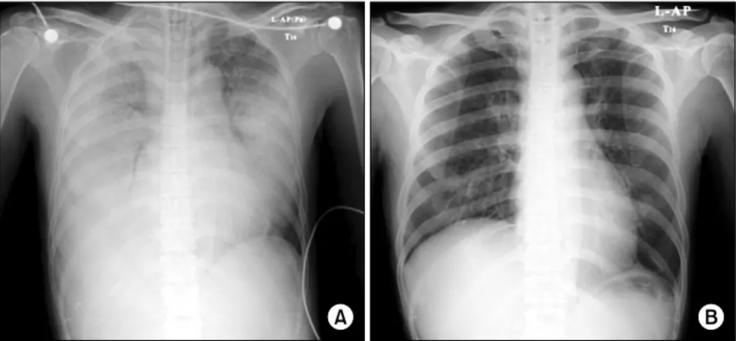
Figure 1. Chest PA at (A) admission and (B) discharge.
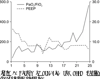
Figure 2. Veno-venous extracorporeal membrane oxygenation.
맥박 142/분, 호흡수 60/분, 체온 38.3oC, 맥박산소측정법 으로 측정한 산소포화도는 32%였다. 양쪽 폐에서 수포음 이 청진되었고 매우 아픈 모습으로 과호흡을 하고 있었다.
검사실 소견: 동맥혈가스검사는 pH 7.31, PaCO2 36 mmHg, PaO2 19 mmHg, bicarbonate 18.1 mmol/L, 산소 포화도 24%이었다. 말초혈액검사는 백혈구 6,670/mm3 (호중구 89.4%), 생화학검사에서 AST 72 IU/L, LDH 940 IU/L로 상승되어 있었고 총단백 5.7 g/dl, 알부민 2.9 g/dl 로 약간 감소되어 있었다. 혈액응고 검사에서 INR 1.30으 로 약간 상승된 소견이었으며 소변검사에서는 단백이 2+
이었다.
방사선 소견: 흉부X-선 촬영 및 흉부전산화단층촬영에 서 양쪽 폐에서 광범위한 경화가 관찰되었다(Figure 1A).
치료 및 경과: 최초 동맥혈가스검사에서 산소포화도가 24%의 저산소증을 보여 PEEP 16 cmH2O, FiO2 1.0으로 기계환기를 하였음에도 pH 7.29, PaCO2 42 mmHg, PaO2
36 mmHg, 산소포화도는 61%에 머물렀으며 A-aDO2는 628 mmHg, PaO2/FiO2 36 mmHg으로 중증급성호흡곤란 증후군으로 판단되었다.
기존의 고전적인 치료법으로는 소생이 불가능할 것으 로 판단되었고 연령, 급성호흡곤란증후군의 발병시점 및 중증도를 고려하여 체외생명보조를 시행하였다. 정맥 대 정맥의 방법을 사용하였으며 응급실 도착 3시간 후에 시 술되었다(Figure 2). 시술 후 FiO2 1.0에서 동맥혈가스검 사는 pH 7.26, PaCO2 23 mmHg, PaO2 115 mmHg, 산소 포화도는 97%로 호전되었다. 이후 13일간 체외생명보조
Figure 3. Change of PaO2/FiO2 and PEEP (positive end-expiratory pressure).
를 시행하였고 이탈 시의 동맥혈검사는 FiO2 0.6, PEEP 8 cmH2O에서 pH 7.43, PaCO2 41 mmHg, PaO2 87 mmHg, 산소포화도가 97%로 고전적 치료로 유지되고 있 었다. 입원 20일, 25일 후에 각각 PaO2/FiO2가 200 mmHg, 300 mmHg 이상으로 회복 되었다(Figure 3).
혈액배양검사에서
Streptococcus pneumoniae
가 동정 되었으며 중환자실로 입원한 직후부터 폐동맥 카테터로 측정된 전신혈관저항계수(SVRI)는 1,756 dynesㆍsecㆍ m2/cm5 (1,970∼2,390)으로 감소된 소견의 폐혈쇼크가 있어 도파민을 입원 후 3일 동안 사용하였다. 입원 2일 후부터 무뇨와 급성신부전이 발생하여 지속적 정정맥 혈 액여과기를 107일간 사용하였으며 사용중지 후 퇴원 시까 지 정상 신기능을 유지하였다. 입원 7일 후까지 급성호흡 곤란 증후군의 중증도가 지속되어 아직 효과에 대한 논란 이 있지만 스테로이드 치료를 시작하였으며 킬로그램 당 2 mg을 부하용량으로 투여 후 2주간 하루에 킬로그램 당 2 mg을 4회에 나누어 정맥투여 하였고 이후 1주간 하루에 킬로그램 당 1 mg을 2회에 나누어 정맥투여 후 점차적으 로 용량을 줄여가면서 종료하였다.177일간의 입원 기간 중 임상경과가 악화되어 3차례의 기계환기를 다시 하게 되는 상황까지 있었으나 결국 호전 되어 2차 병원으로 전원하였다. 퇴원 시 흉부X-선은 다음 과 같다(Figure 1B).
고 찰
급성폐손상은 1) 수일 이내의 급성으로 발병하고 2) PaO2/FiO2 300 mmHg 이하이며 3) 흉부X-선에서 양측 폐 침윤이 보이고 4) 폐모세혈관쐐기압이 18 mmHg 이하 이
거나 좌심방압이 증가된 증거가 없는, 위의 4가지 조건을 모두 만족하는 경우에 진단할 수 있다. 위의 급성폐손상 의 조건을 만족하고 PaO2/FiO2가 200 mmHg 이하인 경우 급성호흡곤란증후군으로 진단할 수 있다7.
급성호흡곤란증후군은 사망률이 매우 높은 질환으로 1997년 대한 결핵 및 호흡기학회 학술위원회의 조사에 의 하면 국내 사망률이 71.9%에 이른다는 결과가 있었다8. 급성호흡곤란증후군의 치료로는 원인 질환에 대한 치 료, 영양 공급, 수액 공급, 기계 환기 등의 보존적 치료가 주가 되고 있으며 이러한 치료들은 최근의 사망률의 저하 에 큰 역할을 해왔다1. 약물치료로는 부신피질호르몬, 계 면활성제, 프로스타글란딘(prostaglandin), ketoconazole, nitric oxide, lisofylline 등이 시도되었으나 효과가 입증되 지 못하였다. 그러나 몇몇 연구에서 부신피질호르몬이 7 일 이상 지속된 급성호흡곤란증후군 환자에서 사망률을 낮추는 결과를 보였지만1, 이 후 후속연구에서는 사망률에 차이가 없고 오히려 2주가 경과한 후의 사용은 사망률을 증가시킬 가능성이 있음을 시사하였다9.
기계 환기에 있어 여러 가지 시도가 있었는데 폐포동원 술, 제트 환기법, 복와위 환기법, 액체환기법 등이 있으나 효능을 입증하는데 실패하였지만1,2 호기말 양압을 걸어주 고 일회호흡량을 작게 하면서 고이산화탄소증은 허용하 자는 이른바 개방폐(open lung) 전략에 의한 환기법이 급 성호흡곤란증후군의 치료에서 제한적이나마 효과를 보였 으며10 이후 다기관 무작위대조검사로 진행된 ARDS net- work 임상시험에서 낮은 일회환기량으로 환기시키는 것 이 사망률을 감소시킨다는 것을 입증하였다11.
이러한 기존의 치료에도 반응하지 않는 경우 체외생명 보조를 사용해 볼 수 있는데, 신생아에서 발생된 중증 호 흡곤란에서는 그 효과가 입증되었으나12 성인의 경우 1979년, 1994년 발표된 2번의 임상시험에서 효용성을 입 증하지 못하여3,4 성인에서의 급성호흡곤란증후군의 일차 치료로 채택되지 못했으며 최후의 치료방법으로 남겨져 있는 상황이다. 1979년의 무작위 전향적 임상시험에서는 90명의 환자가 등록되어 치료군과 대조군에서 각각 90%
의 사망률을 보였으며 1994년의 임상시험에서는 40명의 환자가 등록되었는데 두 군(치료군 vs. 대조군)에서 각각 67%, 58%의 사망률을 보여(p=0.8) 체외생명보조의 효용 성이 입증되지 못했다.
그러나 1994년 임상시험 이후 지속적으로 체외생명보 조를 이용하여 급성호흡곤란증후군에 대한 치료의 성과 가 보고 되고 있으며 사망률이 24%에서 48%로 지난 두
Table 1. The indications for extracorporeal life support Michigan medical center
PaO2/FiO2<100 mmHg or A-aDO2>600 mmHg or transpulmonary shunt fraction>30%
Age<70 years
Time on mechanical ventilation<10 days CESAR trial inclusion criteria
Age (18∼65 years)
Murray score≥3.0 or uncompensated hypercapnea with a pH≤7.20
Time on mechanical ventilation≤7 days
연구보다 낮은 수준을 보이고 있다13.
실제로 체외생명보조는 대부분의 병원에서 기존의 급 성호흡곤란증후군의 치료에 반응하지 않는 중증의 경우 에 사용하고 있으며 2006년에 연구가 종료된 CESAR 임상 시험 및 Michigan 병원에서의 적응증을 살펴보면 Table 1과 같다.
체외생명보조를 적용 후 이탈까지의 치료는 폐의 휴식 이 가능하도록 인공호흡기를 설정하는 것이고 기존의 보 존적 치료가 주가 된다. CESAR 임상시험의 경우 인공 환기 의 경우 최고기도압 20 cmH2O, 호기말기도압 10 cmH2O, 호흡수 10회/분, FiO2 0.3을 목표로 하고 헤모글로빈은 14 g/dl, 혈소판은 >100,000/ml를 유지하도록 하였다. 또한 건조 중량(dry weight)까지 이뇨시키도록 하였다6. 본 환 자의 경우 지속적 정정맥 혈액여과기를 이용하여 체내의 수분을 충분히 제거하였으나 헤모글로빈은 9∼10 g/dl로 빈혈은 충분히 교정되지 못하였다. 향후 체외생명보조의 치료에 있어서는 폐가 충분히 휴식되도록 기계 환기 및 보존적 치료를 진행해야 할 것이다.
체외생명보조로부터 이탈은 기존의 급성호흡곤란증후 군의 치료에 안정적인 경우 시도하게 된다. CESAR 임상 시험에서는 흉부X-선과 폐탄성이 호전되고 최고압이 30 cmH2O 미만, FiO2가 0.6 미만인 경우에 이탈하도록 하였 으며6 Hemmila 등5은 혈역학적으로 안정되고 패혈증이 모두 해결되었으며 0.5 미만의 FiO2에서도 적절한 산소화 가 가능할 때 이탈 하도록 하였다. 본 환자의 경우도 체외 순환기를 일시 정지한 후에도 고전적 기계환기에도 충분 한 산소포화농도가 유지되고 패혈증이 호전되어 입원 13 일에 이탈할 수 있었다.
요 약
중증급성호흡곤란증후군으로 전원 된 41세 여자환자를 기존의 기계환기 방법으로 치료했으나 저산소증이 호전 되지 않아 내원 후 3시간 내에 신속한 체외생명보조를 시 행하여 성공적으로 치료한 예를 경험하여 문헌고찰과 함 께 보고하는 바이다.
참 고 문 헌
1. Wheeler AP, Bernard GR. Acute lung injury and the acute respiratory distress syndrome: a clinical review.
Lancet 2007;369:1553-64.
2. Hemmila MR, Napolitano LM. Severe respiratory failure:
advanced treatment options. Crit Care Med 2006;34:
S278-90.
3. Zapol WM, Snider MT, Hill JD, Fallat RJ, Bartlett RH, Edmunds LH, et al. Extracorporeal membrane oxygen- ation in severe acute respiratory failure: a randomized prospective study. JAMA 1979;242:2193-6.
4. Morris AH, Wallace CJ, Menlove RL, Clemmer TP, Orme JF Jr, Weaver LK, et al. Randomized clinical trial of pressure-controlled inverse ratio ventilation and ex- tracorporeal CO2 removal for acute respiratory distress syndrome. Am J Respir Crit Care Med 1994;149:295- 305.
5. Hemmila MR, Rowe SA, Boules TN, Miskulin J, McGillicuddy JW, Schuerer DJ, et al. Extracorporeal life support for severe acute respiratory distress syndrome in adults. Ann Surg 2004;240:595-607.
6. Peek GJ, Clemens F, Elbourne D, Firmin R, Hardy P, Hibbert C, et al. CESAR: conventional ventilatory sup- port vs extracorporeal membrane oxygenation for se- vere adult respiratory failure. BMC Health Serv Res 2006;6:163.
7. Bernard GR, Artigas A, Brigham KL, Carlet J, Falke K, Hudson L, et al. The American-European Consensus Conference on ARDS. Definitions, mechanisms, rele- vant outcomes, and clinical trial coordination. Am J Respir Crit Care Med 1994;149:818-24.
8. Scientific Subcommittee for National Survey of Acute Respiratory Distress Syndrome in Korean Academy of Tuberculosis and Respiratory Disease. The national survey of acute respiratory distress syndrome in Korea.
Tuberc Respir Dis 1997;44:25-43.
9. The National Heart, Lung, and Blood Institute Acute Respiratory Distress Syndrome (ARDS) Clinical Trials
Network. Efficacy and safety of corticosteroids for per- sistent acute respiratory distress syndrome. N Engl J Med 2006;354:1671-84.
10. Amato MB, Barbas CS, Medeiros DM, magaldi RB, Schettino GP, Lorenzi-Filho G, et al. Effect of a pro- tective-ventilation strategy on mortality in the acute res- piratory distress syndrome. N Engl J Med 1998;338:
347-54.
11. ARDS Clinical Trials Network. Ventilation with lower ti- dal volumes as compared with traditional tidal volumes
for acute lung injury and acute respiratory distress syndrome. N Engl J Med 2000;342:1301-8.
12. Bartlett RH, Roloff DW, Cornell RG, Andrews AF, Dillon PW, Zwischenberger JB. Extracorporeal circu- lation in neonatal respiratory failure: a prospective randomized study. Pediatrics 1985;76:479-87.
13. Kopp R, Dembinski R, Kuhlen R. Role of extracor- poreal lung assist in the treatment of acute respiratory failure. Minerva Anestesiol 2006;72:587-95.