• 검색 결과가 없습니다.

Multiple Hypercoagulability Disorders at Presentation of Non-Small-Cell Lung Cancer

N/A
N/A
Protected

Academic year: 2021

Share "Multiple Hypercoagulability Disorders at Presentation of Non-Small-Cell Lung Cancer"

Copied!
4
0
0

로드 중.... (전체 텍스트 보기)

전체 글

(1)

Multiple Hypercoagulability Disorders at Presentation of Non-Small-Cell Lung Cancer

Jeong Min Lee, M.D., Jun Hyeok Lim, M.D., Jung-Soo Kim, M.D., Ji Sun Park, M.D., Azra Memon, B.S., Seul-Ki Lee, B.S., Hae-Seong Nam, M.D., Jae-Hwa Cho, M.D., Seung-Min Kwak, M.D., Hong Lyeol Lee, M.D., Hyun-Jung Kim, R.N., Geun-Jeong Hong, R.N. and Jeong-Seon Ryu, M.D.

Department of Internal Medicine, Inha University College of Medicine, Incheon, Korea

Hypercoagulability disorders are commonly encountered in clinical situations in patients with a variety of cancers.

However, several hypercoagulability disorders presenting as first symptoms or signs in cancer patients have rarely been reported. We herein described a case of a woman with adenocarcinoma of the lung presenting with deep vein thrombosis, nonbacterial thrombotic endocarditis, recurrent cerebral embolic infarction, and heart failure.

Keywords: Venous Thrombosis; Heart Failure; Thrombophilia; Endocarditis, Non-Infective; Carcinoma, Non-Small-Cell Lung

as deep-vein thrombosis (DVT) and pulmonary embolism (PE), is more commonly observed than arterial thrombosis.

However, hypercoagulability disorders are a rare presenting symptom or sign of cancer patients, therefore, coexistence of DVT at the time of diagnosis of lung cancer was reported to be 0.1−0.4% in data from California Cancer Registry

2

.

NBTE, which was introduced in 1936 by Gross and Fried- berg

5

, is characterized by deposition of thrombi on aortic or mitral valves in the absence of bacterial infection

6

. No defini- tive clinical features suggest NBTE, in general. However, when a patient shows negative blood culture result and multiple disseminated embolisms, NBTE is more strongly suspected rather than infective endocarditis

7,8

. Incidence of NBTE is largely not well evaluated; it was 1.25% of cancer patients in an autopsy study

7

. Systemic embolization to the central nervous system and coronary arteries are often observed in NBTE patients

8,9

. However, acute heart failure caused by NBTE with cardiac valve lesions is a rare clinical event

10

.

Association with hypercoagulability disorders is a clinical concern because these cause delay in treatment and worse survival of cancer patients and have a significant adverse ef- fect on quality of life

2,11

. We herein report on a lung cancer patient who presented with DVT, NBTE, recurrent cerebral embolic infarction, and heart failure.

Copyright © 2014

The Korean Academy of Tuberculosis and Respiratory Diseases.

All rights reserved.

Introduction

As a paraneoplastic syndrome, hypercoagulability disorders are known to be commonly associated with a variety of cancer types including lung cancer

1-3

. Approximately 15% of cancer patients suffer from hypercoagulability disorders during their clinical course; 50% of cancer patients show evidence of hy- percoagulability disorders on autopsy

3,4

. Cancer-associated hypercoagulability status may also present as Trousseau’s syndrome (a migratory superficial thrombophlebitis), arte- rial thrombosis, disseminated intravascular coagulation, or nonbacterial thrombotic endocarditis (NBTE, formerly called marantic endocarditis). Venous thromboembolism, such

CASE REPORT

http://dx.doi.org/10.4046/trd.2014.77.1.34

ISSN: 1738-3536(Print)/2005-6184(Online) • Tuberc Respir Dis 2014;77:34-37

34

Address for correspondence: Jeong-Seon Ryu, M.D., Ph.D.

Department of Internal Medicine, Center for Lung Cancer, Inha University Hospital, 366 Seohae-daero, Jung-gu, Incheon 400-103, Korea Phone: 82-32-890-3738, Fax: 82-32-882-6578

E-mail: [email protected] Received: Dec. 18, 2013 Revised: Mar. 18, 2014 Accepted: Apr. 18, 2014

cc
It is identical to the Creative Commons Attribution Non-Commercial

License (http://creativecommons.org/licenses/by-nc/3.0/).

(2)

Severe form of hypercoagulability disorders in lung cancer

http://dx.doi.org/10.4046/trd.2014.77.1.34 35

www.e-trd.org

Case Report

A 61-year-old Korean woman was admitted to the Depart- ment of Vascular Surgery, presenting with a tingling sensation on both lower legs and vague chest discomfort for one month.

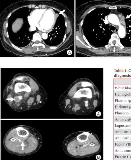
She was a never smoker and had dyspnea (3 on Medical Research Council scale) for two weeks but no fever. She had undergone surgical excision of a benign mass of the left breast and hysterectomy due to uterine prolapse, 15 and five years ago, respectively; denied any postoperative complications and history of renal or hepatic disease. Computed tomogram (CT) angiography of the chest did not show evidence of pulmonary thromboembolism. However, 2×1.2-cm-sized nodular lesion in left lingular division abutting to mediastinal pleura and lymphadenopathies on the left paraaortic and supraclavicular area and both prevascular and subaortic area were noted (Fig-

ure 1). Clots in the popliteal and calf vein of the right leg were noted on a CT angiogram for the pelvis and legs (Figure 2).

The patient was immediately started on treatment with con- tinuous administration of intravenous heparin. An extensive laboratory work-up was performed in search of underlying hypercoagulability disorders (Table 1). On the fourth day, she was transferred to Department of Center for Lung Cancer for further evaluation of lung cancer. No endobronchial lesion was observed on bronchoscopic examination. Adenocarci- noma was diagnosed pathologically on a biopsy specimen of supraclavicular lymph node. Thyroid transcription factor-1 was positive on immunohistochemistry and activating muta- tion of epidermal growth factor receptor gene was not demon- strated. On the fifth day, palsy of the lateral gaze of both eyes and double vision was developed suddenly. She underwent brain magnetic resonance angiogram, which demonstrated subacute stage of embolic infarctions on territories of both the middle and posterior cerebral arteries and the right cerebel- Figure 1. A 2×1.2-cm-sized nodular lesion in the left lingular division abutting to mediastinal pleura (A, arrow) and lymph- adenopathies on the left paraaortic area, on chest CT scan at the time of diagnosis (B, arrow).

Table 1. Coagulation-related laboratory data at the time of diagnosis

Value Reference

White blood cell, /mL 7,360 4,000–10,000

Hemoglobin 12.3 -

Platelet, /mL 184,000 150,000–400,000

D-dimer, mg/mL 29.0 0.0–0.4

Phospholipid 188 150–230

Anti-β2 glycoprotein Negative -

Lupus anticoagulant Negative -

Anti-cardiolipin IgM Negative -

Anti-cardiolipin IgG Negative -

Factor VIII, % 136 60–150

Antithrombin III, % 70 80–120

Protein C activity, % 77 70–140

Protein S activity, % 74 71–103

Figure 2. Computed tomogram angiogram for the legs showing

venous thrombosis at the popliteal vein (A, arrow) and calf vein (B,

arrow) of the right leg.

(3)

JM Lee et al.

36 Tuberc Respir Dis 2014;77:34-37 www.e-trd.org

lar hemisphere (Figure 3). Neither stenotic flow nor visible plaque was demonstrated in Doppler ultrasound examination for both carotid arteries. No evidence of distant metastasis was observed on brain magnetic resonance image (MRI), bone scan, and positron emission tomography. Finally, her disease was staged as IIIB; however, she did not want to receive any treatment for her lung cancer. On Transthoracic echocardio- gram, vegetation measuring 9 mm in size was observed on the posterior mitral leaflet with moderate mitral regurgitation and the basal inferior wall was akinetic with 50% of left ventricular ejection fraction (Figure 4A). Blood culture of the patient re- vealed no evidence of bacteremia. Diffuse irregular stenosis (30% of diameter) of the distal portion of the left anterior de- scending artery and diffuse irregular stenosis (80% of diam- eter) of the posterolateral branch of the right coronary artery were observed on coronary angiogram (Figure 4B). Intracoro- nary injection was administered with ReoPro (Abciximab,

Eli-Lilly, Indianapolis, IN, USA) and thrombus was aspirated from the right coronary artery. On the eighth day, heparin was switched to warfarin. On the 15th day, follow-up transthoracic echocardiogram showed a decrease in size of the vegetation (4 mm), improvement of mitral regurgitation (mild degree), and normalization of left ventricular ejection fraction. She was discharged on the 17th day. Three weeks later, she visited to the emergency room with sudden development of dysarthria, aphasia, and right hemiplegia. A newly demonstrated hy- peracute embolic infarction in the operculum area of the left frontal- parietal junction was observed on her brain MRI. She is receiving rehabilitation treatment for her hemiplegia.

Discussion

Hypercoagulable state (also called thrombophilia or pro- thrombotic state) is an abnormal condition that increases the risk of thrombosis. A hypercoagulability of malignancy occurs due to activation of coagulation system by cancer cells.

Prothrombotic mechanisms in malignancy associated with procoagulant/fibrinolytic substances secreted by cancer cells, tumor cell-host cell interactions and other nonspecific fac- tors such as necrosis or hemodynamic compromise. Venous thromboembolism is more common in mucin-producing adenocarcinoma of pancreas and gastrointestinal tract, lung cancer, ovarian cancer

12

.

After presence of a hypercoagulable state in stomach can- cer patients was found by Trousseau in 1865, venous throm- bosis has been a common clinical feature in patients with a variety of cancer types, including lung cancer

13

. Association of venous thrombosis, such as DVT and PE, in cancer patients has been well established and can have an adverse effect on survival

11,14

. However, in general, NBTE and its emboliza- tion to coronary and/or cerebral arteries have not been well evaluated; most were reported after the diagnosis of cancer

7,10

. With our limited knowledge, this case shows a unique clinical presentation of DVT, NBTE, recurrent cerebral embolic infarc-

Figure 4. Vegetation measuring 9 mm in size on the posterior mitral leaflet shown on transthoracic echocardiogram (A, arrow), and diffuse stenosis of the pos- terolateral branch of the right coronary artery shown on coronary angiogram (B, arrow).

Figure 3. Brain magnetic resonance image showing a subacute stage

infarction involving a territory of the right middle cerebral artery.

(4)

Severe form of hypercoagulability disorders in lung cancer

http://dx.doi.org/10.4046/trd.2014.77.1.34 37

www.e-trd.org

tion, and heart failure at the time of diagnosis of lung cancer.

In addition, involvement of major organs, including brain and heart at presentation may cause a delay in diagnosis of cancer because patients could initially be treated by a cardiologist or neurologist. Their coexistence can have a significant impact on quality of life of cancer patients and cause a delay in treat- ment or administration of less effective treatment that causes worse survival.

Therefore, clinicians need to pay attention to the possibil- ity of coexistence with severe forms of hypercoagulability disorders at the time of diagnosis. Early identification and ag- gressive intervention could be essential in preventing delay of diagnosis or treatment.

Conflicts of Interest

No potential conflict of interest relevant to this article was reported.

Acknowledgements

This study was supported by INHA University Research Grant (INHA).

References

1. Falanga A, Ofosu FA, Delaini F, Oldani E, Dewar L, Lui L, et al. The hypercoagulable state in cancer patients: evidence for impaired thrombin inhibitions. Blood Coagul Fibrinolysis 1994;5 Suppl 1:S19-23.

2. Dipasco PJ, Misra S, Koniaris LG, Moffat FL Jr. Thrombophilic state in cancer, part I: biology, incidence, and risk factors. J Surg Oncol 2011;104:316-22.

3. Glassman AB. Hemostatic abnormalities associated with cancer and its therapy. Ann Clin Lab Sci 1997;27:391-5.

4. Falanga A, Rickles FR. Pathophysiology of the thrombo- philic state in the cancer patient. Semin Thromb Hemost 1999;25:173-82.

5. Gross L, Friedberg CK. Nonbacterial thrombotic endocardi- tis: classification and general description. Arch Intern Med 1936;58:620-40.

6. el-Shami K, Griffiths E, Streiff M. Nonbacterial thrombotic endocarditis in cancer patients: pathogenesis, diagnosis, and treatment. Oncologist 2007;12:518-23.

7. Lopez JA, Ross RS, Fishbein MC, Siegel RJ. Nonbacterial thrombotic endocarditis: a review. Am Heart J 1987;113:773- 84.

8. Borowski A, Ghodsizad A, Cohnen M, Gams E. Recurrent embolism in the course of marantic endocarditis. Ann Thorac Surg 2005;79:2145-7.

9. Chino F, Kodama A, Otake M, Dock DS. Nonbacterial throm- botic endocarditis in a Japanese autopsy sample: a review of eighty cases. Am Heart J 1975;90:190-8.

10. Taniyama D, Yamamoto R, Kawasaki M, Kamata H, Miya- moto K, Mashimo S, et al. Nonbacterial thrombotic endocar- ditis leading to acute heart failure due to aortic stenosis in a patient with lung cancer. Intern Med 2013;52:1617-20.

11. Sorensen HT, Mellemkjaer L, Olsen JH, Baron JA. Prognosis of cancers associated with venous thromboembolism. N Engl J Med 2000;343:1846-50.

12. Caine GJ, Stonelake PS, Lip GY, Kehoe ST. The hypercoagu- lable state of malignancy: pathogenesis and current debate.

Neoplasia 2002;4:465-73.

13. Connolly GC, Dalal M, Lin J, Khorana AA. Incidence and pre- dictors of venous thromboembolism (VTE) among ambula- tory patients with lung cancer. Lung Cancer 2012;78:253-8.

14. Young A, Chapman O, Connor C, Poole C, Rose P, Kakkar AK.

Thrombosis and cancer. Nat Rev Clin Oncol 2012;9:437-49.

수치

Table 1. Coagulation-related laboratory data at the time of  diagnosis
Figure 3. Brain magnetic resonance image showing a subacute stage  infarction involving a territory of the right middle cerebral artery.

참조

관련 문서

a prospective analysis of 50 cases. Preoperative staging of non-small-cell lung cancer with positron-emission tomography. Staging of non- small-cell lung cancer

After first field tests, we expect electric passenger drones or eVTOL aircraft (short for electric vertical take-off and landing) to start providing commercial mobility

1 John Owen, Justification by Faith Alone, in The Works of John Owen, ed. John Bolt, trans. Scott Clark, "Do This and Live: Christ's Active Obedience as the

Patients with breast cancer; ovarian cancer; renal cell carcinoma; pancreatic neuroendocrine cancer; colorectal cancer; head and neck cancer; non-small cell lung

Undiagnosed malignancy in patients with deep vein thrombosis: incidence, risk indicators, and diagnosis.. Monreal M, Fernandez-Llamazares J, Perandreu

At the end of the study, a reevaluation of each study case was performed with the same questionnaire. The result shows there has been a meaningful result in the group

Beichel,“Automated 3-D segmentation of lungs with lung cancer in CT data using a novel robust active shape model approach,” IEEE Trans.. 저작물의

Compromised CD4+ CD25(high) regulatory T-cell function in patients with relap sing-remitting multiple sclerosis is correlated with a reduced frequency of