Association of Specific Immunoglobulin E to Staphylococcal Enterotoxin with Airway Hyperresponsiveness in Asthma Patients
Seong Han Kim, M.D.
1, Seo Yeon Yang, M.D.
1, Jihong You, M.D.
1, Sang Bae Lee, M.D.
1, Jin You, M.D.
2, Yoon Soo Chang, M.D., Ph.D.
1, Hyung Jung Kim, M.D., Ph.D.
1,
Chul Min Ahn, M.D., Ph.D.
1, Min Kwang Byun, M.D.
1, Hye Jung Park, M.D., Ph.D.
1and Jung-Won Park, M.D., Ph.D.
31
Department of Internal Medicine, Gangnam Severance Hospital, Yonsei University College of Medicine, Seoul,
2Department of Internal Medicine, Kunkuk University School of Medicine, Seoul,
3Division of Allergy and Immunology, Department of Internal Medicine, Severance Hospital, Yonsei University College of Medicine, Seoul, Korea
Background: Specific immunoglobulin E (IgE) sensitization to staphylococcal enterotoxin (SE) has been recently considered to be related to allergic disease, including asthma. Despite studies on specific IgE (sIgE) to SE and its relationship to asthma diagnosis and severity, the association of sIgE to SE with airway hyperresponsiveness (AHR) remains unclear.
Methods: We enrolled 81 asthma patients admitted to the Severance Hospital in Korea from March 1, 2013, to February 28, 2015 and retrospectively reviewed the electronic medical records of the enrolled subjects. The serum levels of sIgE to SE (A/
B) of all subjects was measured using the ImmunoCAP 250 (Phadia) system with SE-sIgE positive defined as >0.10 kU/mL.
Results: The SE-sIgE level was not significantly correlated with asthma severity (forced expiratory volume in 1 second [FEV
1], FEV
1/forced vital capacity, sputum eosinophils, and serum eosinophils), whereas the SE-sIgE level in patients with positive AHR (mean±standard error of the mean, 0.606±0.273 kU/mL) was significantly higher than that in patients with negative AHR (0.062±0.015 kU/mL, p=0.034). In regression analysis, SE sensitization (sIgE to SE ≥0.010 kU/mL) was a significant risk factor for AHR, after adjustment for age, sex, FEV
1, and sputum eosinophils (odds ratio, 7.090; 95%
confidence interval, 1.180–42.600; p=0.032). Prevalence of SE sensitization was higher in patients with allergic rhinitis and non-atopic asthma patients, as compared to patients without allergic rhinitis and atopic asthma patients, respectively, but without statistical significance.
Conclusion: SE sensitization is significantly associated with AHR.
Keywords: Immunoglobulin E; Staphylococcus; Enterotoxins; Asthma
Copyright © 2016
The Korean Academy of Tuberculosis and Respiratory Diseases.
All rights reserved.
Address for correspondence: Hye Jung Park, M.D., Ph.D.
Department of Internal Medicine, Gangnam Severance Hospital, Yonsei University College of Medicine, 211 Eonju-ro, Gangnam-gu, Seoul 06273, Korea
Phone: 82-2-2019-4375, Fax: 82-2-3463-3882, E-mail: [email protected]Received: Apr. 20, 2016 Revised: Jun. 23, 2016 Accepted: Aug. 9, 2016
cc
It is identical to the Creative Commons Attribution Non-Commercial License (http://creativecommons.org/licenses/by-nc/4.0/).
Introduction
Staphylococcus aureus is a human commensal microor- ganism, and frequent colonizer of airways and skin. S. aureus releases a wide range of enterotoxins
1. While bacterial infec- tion commonly stimulates the innate immune system, staphy- lococcal enterotoxin (SE) can act as an antigen, in particular, a superantigen
2. SE binds to the variable β-chain of the T-cell receptor, independent of the antigen-specific groove, so that it is called a “superantigen.” This allows the polyclonal activation of T cells and also B cells, resulting in the formation of specific immunoglobulin E (sIgE) to SE, called “SE sensitization”
2-4. These superantigenic properties of SE are considered to have a role in the pathophysiology of allergic disease.
The first allergic disease to be studied for association with SE was atopic dermatitis
5. Since then, evidence has been gath- ered that SE also has an important role in upper and lower airway disease
1,2,6. The bacterial allergy, sensitization to SE, is considered to be more important than bacterial infection as a cause and aggravating factor for allergic airway disease
7. Re- cent studies have revealed that SE sensitization is an indepen- dent risk factor for asthma and, especially, the severe asthma entity
4,8-10. In severe asthmatics, inability to cope with S. aureus colonization in airways by impaired macrophages leads to impairment of mucosal immunity and sensitization to SE
11. Asthma represents an inflammatory airway disease with fea- tures of mucosal edema and mucus secretion, after allergen stimulation. Airway hyperresponsiveness (AHR), which is measured as increased airway resistance following provoca- tion with stimuli, reflects impairment of mucosal immunity and might be associated with SE sensitization. However, the correlation of SE sensitization with AHR in common asthma patients has never been researched in human clinical studies.
We aimed to evaluate the correlation of SE sensitization with asthma severity, especially AHR, in asthma patients.
Materials and Methods
1. Study population
We retrospectively enrolled 81 asthma patients whose sIgE to SE data were available and admitted to the Severance Hos- pital in Korea from March 1, 2013, to February 28, 2015. We retrospectively reviewed the electronic medical records of the enrolled subjects. Asthma was diagnosed by an allergy spe- cialist, based on clinical guidelines, using a bronchodilator test and/or bronchial provocation test
12. This study was approved by the Institutional Review Board of Yonsei University College of Medicine (approval number: 4-2013-0397). All enrolled pa- tients provided written informed consent.
2. Laboratory tests
The complete blood count test was performed using an automated analyzer to determine blood eosinophil counts.
The eosinophil percentages in induced sputum were assessed as follows. The obtained sputum was centrifuged, and the su- pernatant was collected. Samples were diluted in phosphate- buffered saline and centrifuged at 450 rpm for 6 minutes to prepare cytology slides. After staining the slides with Wright’s stain, a differential count was performed using a light micro- scope, as reported previously
13. Forced expiratory volume in 1 second (FEV
1) and FEV
1/forced vital capacity (FVC) were evaluated by pulmonary function test using commercially available equipment (MS-IOS, Masterlab-IOS; Jaeger, Wurz- burg, Germany). AHR was assessed by methacholine chal- lenge test or mannitol challenge test, using a titration protocol as in previous reports. The methacholine challenge started with an inhalation of saline, followed by increasing the con- centration of methacholine serially, up to a maximum concen- tration of 25 mg/mL. We defined positive as the provocative concentration of methacholine required to decrease FEV
1by 20% (PC
20), being achieved at less than or equal to 25 mg/mL.
The mannitol challenge test ended when a cumulative dose of 635 mg had been inhaled. We defined positive as PD15, the provocative concentration of mannitol required to decrease FEV
1by 15%, less than or equal to 635 mg. All of these tests were conducted before chart review. We retrospectively re- viewed the results of the tests.
3. Clinical parameters
Allergic rhinitis was clinically diagnosed, based on interna- tional guidelines which recommend identifying symptoms, including sneezing, rhinorrhea, nasal obstruction, and itching with specific IgE possession confirmed by skin prick test or ImmunoCAP 250 (Phadia, Uppsala, Sweden) system
14. Atopy was defined as positive response to any one of the inhalant al- lergens (birch, oak, ragweed, mugwort, cat dander, house dust mite, etc.) by ImmunoCAP 250 (Phadia) system, as previously described
15.
4. Serum preparation and measurement of sIgE to SE We measured the levels of sIgE to SE (A/B) in all subjects.
First, whole blood was collected in a vacuum tube for serum
separation. Serum was separated by centrifugation, and al-
lergen sIgE detection performed using ImmunoCAP 250
(Phadia) system. The system was operated according to the
manufacturer’s instructions. The sIgE to SE detection range
was 0.1 to 100 kU/L.
5. Statistical analysis
We assessed correlation between two continuous variables using Pearson’s test. The t test was used to compare levels of sIgE to SE according to AHR. We selected, as the parameters, sex and age, which are most commonly among the variables considered, and FEV
1and sputum eosinophil count, which are known to be highly related to asthma severity. We deter- mined the odds ratio (OR) using logistic regression analysis, including univariate and multivariate analysis. Cross-correla- tion analysis was performed using chi-square tests to analyze the correlation between categorized variables in SPSS version 18.0 (SPSS Inc., Chicago, IL, USA). We considered a p<0.05 to be significant.
Results
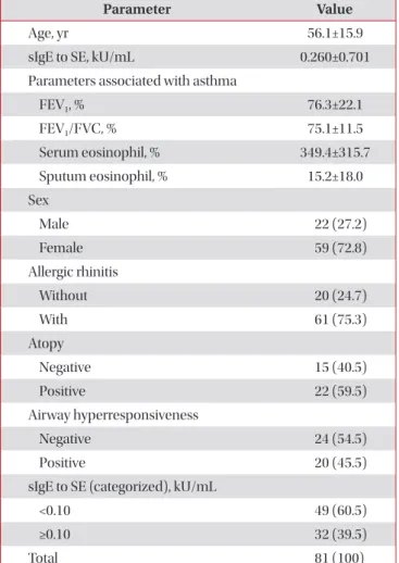
1. Demographics of subjects
We enrolled a total of 81 asthma patients. The mean age of subjects and the standard deviation from the mean was 56.1 years old. The mean value of sIgE to SE and the standard de- viation from the mean was 0.260 kU/mL. The FEV
1and FEV
1/ FVC were assessed by pulmonary function tests and repre- sented a mildly obstructive pattern. The percentage of sputum eosinophils and the standard deviation was elevated beyond the normal range. There was a predominance of females. The prevalence of allergic rhinitis and atopy was 75.3% and 59.5%, respectively. Airway hyperresponsiveness was evaluated in 44 patients, and the results were positive in 45.5%. We defined positive to SE-sIgE as more than 0.10 kU/mL, so that the posi- tivity rate is about 40% (39.5% exactly) (Table 1).
2. Correlation of sIgE to SE with parameters associated with asthma
We evaluated the correlation of SE-sIgE with parameters Table 1. Demographics of subjects
Parameter Value
Age, yr 56.1±15.9
sIgE to SE, kU/mL 0.260±0.701
Parameters associated with asthma
FEV
1, % 76.3±22.1
FEV
1/FVC, % 75.1±11.5
Serum eosinophil, % 349.4±315.7
Sputum eosinophil, % 15.2±18.0
Sex
Male 22 (27.2)
Female 59 (72.8)
Allergic rhinitis
Without 20 (24.7)
With 61 (75.3)
Atopy
Negative 15 (40.5)
Positive 22 (59.5)
Airway hyperresponsiveness
Negative 24 (54.5)
Positive 20 (45.5)
sIgE to SE (categorized), kU/mL
<0.10 49 (60.5)
≥0.10 32 (39.5)
Total 81 (100)
Values are presented as mean±standard deviation or number (%).
sIgE: specific immunoglobulin E; SE: staphylococcal enterotoxin;
FEV
1: forced expiratory volume in 1 second; FVC: forced vital capacity.
Table 2. Correlation of sIgE to SE with parameters associated with asthma
Parameter Pearson’s coefficient p-value
FEV
10.099 0.385
FEV
1/FVC 0.149 0.206
Serum eosinophil, % 0.091 0.417
Sputum eosinophil, % 0.027 0.815
sIgE: specific immunoglobulin E; SE: staphylococcal enterotoxin;
FEV
1: forced expiratory volume in 1 second; FVC: forced vital capacity.
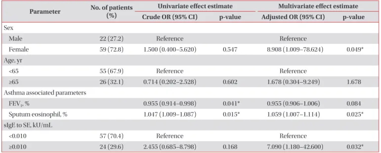
Figure 1. Comparison of specific immunoglobulin E (sIgE) to staphylococcal enterotoxin (SE) levels according to airway hyper- responsiveness (AHR) (p=0.034). *p-value<0.05 obtained by t-test.
slgEtoSE(kU/mL)
0 AHR negative
1.0 0.8 0.6 0.4 0.2
*
AHR positive