적양 추출물의 멜라닌 합성 저해효과
이 준 영⋅임 경 란⋅정 택 규⋅윤 경 섭
†(주)사임당화장품 기술연구소
(2013년 1월 14일 접수, 2013년 1월 21일 수정, 2013년 4월 28일 채택)
The Inhibitory Effects of Alnus Japonica Steud. Extract on Melanogenesis
Jun Young Lee, Kyung Ran Im, Taek Kyu Jung, and Kyung-Sup Yoon
†Saimdang Cosmetics Co. Ltd. R&D Center, 143, Yangcheongsongdae-gil, Ochang-eup, Chungcheongbuk-do 363-886, Korea (Received January 14, 2013; Revised January 21, 2013; Accepted April 28, 2013)
요 약: 본 연구에서는 새로운 미백소재를 개발하기 위해 적양 에탄올추출물을 효소처리 후 EtOAc 분획물(AJE) 을 준비하여 in vitro 상에서 이들의 tyrosinase 저해활성과 세포 수준에서의 멜라닌 합성 저해효과를 측정하였 다. AJE는 mushroom tyrosinase의 활성에는 영향을 미치지 않았으나 B16-F1 melanoma cell을 이용한 멜 라닌 합성 저해효과에 있어서 농도 의존적으로 멜라닌 합성을 저해하여, 40 µg/mL의 농도에서 52% 이상의 저해효과를 나타내었다. 이러한 멜라닌 합성 저해효과에 대한 작용 기전을 확인하기 위해 western blot을 통해 멜라닌 합성 경로에 관련된 단백질의 발현을 측정하였다. 그 결과 멜라닌 합성에 관여하는 효소인 tyrosinase related protein 1 (TRP-1)의 발현을 억제하였고, 이를 조절하는 전사인자인 microphthalmia associated transcription factor (MITF) 발현 역시 효과적으로 억제하였다. 또한 extracellular signal-regulated kinase (ERK) pathway를 활성화시킴으로써 phosphorylated extracellular signal-regulated kinase (p-ERK)의 발현을 상당히 증가시키는 것을 확인할 수 있었다. 이러한 결과는 AJE가 멜라닌 합성의 신호전달 경로 중 ERK pathway의 활성화를 통해 MITF의 분해를 촉진시키고 이로 인해 MITF의 발현을 감소시키며, 그 결과 멜라닌 합성에 관여하는 효소 중 TRP-1의 발현을 감소시킴으로써 멜라닌 합성을 저해하는 것으로 사료되며, 따라서 AJE는 미백용도의 기능성 원료로서의 가능성이 큰 것으로 판단된다.
Abstract: In order to develop new skin whitening agents, we prepared the EtOAc layer (AJE) after enzyme treatment of 75% EtOH extract of the Alnus Japonica Steud. We measured their tyrosinase inhibitory activity in vitro and melanin synthesis inhibitory activity in B16-F1 melanoma cells. They did not show inhibitory activity against mushroom ty- rosinase but showed melanin synthesis inhibitory activity in a dose-dependent manner. In a melanin synthesis inhibition assay, AJE suppressed melanin production up to 52% at a concentration of 40 µg/mL. To elucidate the mechanism of the inhibitory effects of AJE on melanogenesis, we measured expression of melanogenesis-related proteins by the west- ern blot assay. As a result, AJE suppressed the expression of tyrosinase related protein 1 (TRP-1) and microphthalmia associated transcription factor (MITF). Moreover, AJE increased the expression of phosphorylated extracellular sig- nal-regulated kinase ( p-ERK). These results conclude that ERK activation by AJE reduces melanin synthesis via MITF downregulation and is subsequent to the inhibition of TRP-1 expression. Therefore, we suggest that AJE could be used as active ingredients for skin whitening.
Keywords: Alnus Japonica Steud, melanogenesis, TRP-1, MITF, ERK activation 1)
† 주 저자 (e-mail: [email protected])
1. 서 론
멜라닌은 태양광선 중 유해한 자외선으로부터 생체 를 보호하는 중요한 방어수단이나 멜라닌이 과도하게 합성되거나 노화 등에 의해 피부의 생리기능이 떨어 지게 되면 멜라닌이 피부 표면에 침착되어 기미, 주근 깨 등 다양한 색소 침착을 유발하게 된다[1]. 멜라닌은 표피 기저층에 존재하는 멜라닌세포의 멜라노좀에서 합성되어 멜라닌세포의 수지상 돌기를 통해 피부 각 질층으로 이동된다[2].
멜라닌 합성 과정은 melanocyte specific enzymes인 tyrosinase와 tyrosinase- related proteins (TRPs) 등에 의 해 조절되며[3,4] 크게 두 가지 타입의 멜라닌이 만들 어지는데, 노란색에서 붉은색의 색소를 보이는 pheo- melanin과 검은색에서 갈색을 띠는 eumelanin이다[5].
멜라닌 합성에 필요한 tyrosinase는 tyrosine을 3,4-dihy- droxy phenyl-alanine (DOPA)으로 변환시키고, 이것이 DOPA quinone으로 산화되는 것을 촉진하며, 5,6-dihy- droxyindole (DHI)을 indole-5,6-quinone으로 산화하는 것을 촉진한다. 반면 TRPs는 그 뒤 단계인 eumelano- genesis에 관여한다[6,7]. 또한, 멜라닌 합성의 조절인 자로 잘 알려진 microphthalmia-associated transcription factor (MITF)는 helix-loop-helixleucine zipper 구조를 가진 transcription factor로서 멜라닌 합성을 통한 흑화 과정, 세포의 증식 및 생존을 조절함이 잘 알려져 있 다[8-11]. MITF는 tyrosinase 및 TRPs의 M-box se- quences에 결합하여 멜라닌 합성에 관여하는 효소들 의 발현을 조절하는 것으로 잘 알려져 있기 때문에 [12] 멜라닌 합성을 조절하는 skin-lightening agents를 개발하기 위한 주요 타깃으로서 여겨지고 있다[8, 13-16]. 또 다른 멜라닌 합성과정의 신호전달 기전으 로는 extracellular signal-regulated kinase (ERK) pathway 의 억제가 MITF와 tyrosinase의 활성을 증가시켜 결과 적으로 멜라닌의 합성을 증가시키는 한편 ERK의 활 성은 MITF의 ubiquitination (MITF-ⓟ-Ser73)으로 MITF 가 분해되어 멜라닌의 합성 억제를 유도한다는 연구 결과가 보고되기도 하였다[17-20]. 그 외에도 nitric ox- ide에 의한 cyclic guanosine monophosphate (cGMP)경 로, protein kinase C (PKC)의 경로 등이 알려져 있다.
자작나무과(Betulaceae)에 속하는 오리나무의 수피 는 한방에서 적양(Alnus japonica Steud)이라 부르고 청
열(淸熱), 강화(降火)하는 작용이 있어서 비출혈(鼻出 血), 설사, 외상출혈에 쓰였으며[21] 민간에서는 숙취 해소 등의 목적에도 응용한 것으로 나타나 있다[22].
함유 성분에 대한 보고로는 diarylheptanoid 계열이 주 성분이며 이를 비롯한 flavonoid, tannin 등 페놀성 화 합물이 많이 함유된 것으로 알려져 있다[23-34], 주요 성분인 diarylheptanoid류의 생리활성에 관한 연구로는 NO 및 COX-2 생성 억제효과, 항암효과, 항산화효과 등을 비롯한 다양한 생리활성이 알려져 있다[35-38].
Cho 등[39]은 같은 자작나무과의 물갬나무(Alnus hir- suta Turcz)로부터 분리한 diarylheptanoid 화합물의 멜 라닌 합성 억제효과를 보고한 바 있으나 그 작용기전 에 대해 연구한 보고는 없는 실정이다.
본 연구에서는 적양을 화장품의 새로운 미백소재로 이용하기 위해 in vitro 상의 tyrosinase 활성 억제효과 와 B16-F1 melanoma cell에서의 멜라닌 합성 억제효과 를 측정하여 미백효과를 평가하였고, 그 작용기전을 규명하고자 멜라닌 합성 과정에 관여하는 단백질의 발현을 확인하였다.
2. 재료 및 방법
2.1. 실험재료, 시약 및 기기
본 실험에 사용한 적양은 국내산으로 지유본초 (Korea)로부터 구입하여 사용하였다. 적양의 추출과정 및 분획에 사용된 용매들은 시약급을 사용하였다. 세 포배양에 필요한 배지 및 시약은 Hyclone (USA), Sigma (USA)로부터 구입하여 사용하였다. 흡광도 측정은 BIO- TEK Instruments (USA)의 EL800 microplate reader를 사용하였다. Western blot을 위해 Bio-rad (USA)의 west- ern blot kit 및 semi-dry transfer system을 사용하였고 image analysis system (Bio-rad, USA)으로 결과를 확인 하였다. 항체는 Santa Cruz Biotech (USA), Cell Signaling Technology (USA), Abcam Biochemicals (UK)로부터 구입하였다.
2.2. 적양 추출물의 제조
건조한 적양 100 g에 75% 에탄올수용액 500 mL을 가하여 3일 동안 상온 교반 후 여과한 다음 추출여액 을 회전식 증발 건조기로 40 ℃에서 감압 농축하여 적 양 조추출물(8.88 g)을 얻었다. 이것을 10% 에탄올수
용액으로 용해한 뒤 당분해 효소인 Viscozyme L을 처 리한 액을 50 ∼ 60 ℃에서 20 h 반응시킨 뒤 여과한 후, EtOAc로 분획하여 얻어진 상층을 농축하여 EtOAc 분획물(AJE) 0.54 g을 얻었다.
2.3. 미백효과 평가
2.3.1. Tyrosinase 저해활성 측정
Tyrosinase에 대한 활성은 각 농도별 시료 0.9 mL, 0.1 M 인산완충액(pH 6.8) 1.0 mL, 1.5 mM L-tyrosine 용액 1.0 mL을 넣은 후, 37 ℃에서 10 min 간 반응시 킨 뒤 mushroom tyrosinase (1,500 units/mL) 0.1 mL를 첨가하여 37 ℃에서 10 min 간 반응시킨 후 UV-Vis spectorphotometer를 사용하여 475 nm에서 흡광도를 측정하였다. Tyrosinase 저해활성은 다음 식에 따라 시 료용액의 첨가군과 무첨가군의 흡광도 감소율로 나타 내었다.
저해율 시료 첨가군의 흡광도무첨가군의 흡광도 ×
2.3.2. 세포주 및 세포배양
세포주는 마우스 흑색종 세포주인 B16-F1 melano- ma cell을 ATCC (American Type Culture Collection)에 서 분양받아 사용하였으며, 세포배양에 사용된 배지 (Dulbecco’s modified Eagle’s medium; DMEM)는 10%
fetal bovine serum (HyClone Lab., USA), 1% antibiotic antimycotic (100 U/mL penicillin and 50 µg/mL strepto- mycin, Life Tech Inc., USA)을 혼합한 배지를 사용하 여 37 ℃, 5% CO2 incubator에서 배양하여 70 ∼ 80%의 cell culture confluency를 보일 때 실험을 실시하였다.
2.3.3. 세포 생존율 시험
본 실험에서 B16-F1 melanoma cell에 대한 시료의 처리농도를 결정하기 위해 MTT [3-(4,5-dimethythia- zol-2-yl)-2,5-diphenytetrazolium bromide, Sigma] assay 를 Mosmann의 방법[40]을 변형하여 실시하였다. 이 분석법은 노란색의 수용성 기질인 MTT를 진청색의 비수용성 formazan으로 변환시키는 살아있는 세포의 mitochondria dehydrogenase의 능력을 이용한 방법이 다. 생성된 formazan의 양은 살아있는 세포 수에 비례
한다.
B16 melanoma cell을 5 × 10³ cells/well 농도로 96- well plate에 분주하여 37 ℃, 5% CO2 조건하에서 24 h 동안 배양하였다. 배양 후 배양액을 제거하고 시료를 농도별로 배지에 희석하여 교체한 후, 최종 200 nM α- MSH가 되도록 첨가하여 48 h 동안 더 배양하였다. 배 양 후 MTT assay를 통하여 세포 생존율을 확인하였다.
대조군은 시료를 처리하지 않은 배양액으로 설정한 후 흡광도를 측정하였다. 세포의 생존율은 다음의 식 에 따라 계산하였다.
세포 생존율
대조군의 흡광도
시료 첨가군의 흡광도 대조군의흡광도 ×
2.3.4. 멜라닌 함량 측정
멜라닌 함량 측정은 Oka의 방법[41]을 변형하여 사 용하였다. B16-F1 melanoma cell을 6-well plate에 1 × 10⁵ cells/well이 되게 준비한 후, 24 h 동안 37 ℃, CO2
항온기에서 배양하였다. 배양액을 제거하고 시료를 농도별로 배지에 희석하여 교체한 후, 최종 200 nM α -MSH가 되도록 첨가하여 48 h 동안 더 배양하였다.
대조군은 α-MSH만 첨가한 것으로 하였으며 양성 대 조군으로는 알부틴을 사용하였다. 배양 후 배양액을 제거하고 PBS로 세척한 후, 10% DMSO가 함유된 1 N NaOH를 첨가한 후 50 ℃ 항온조에서 세포 내 멜라닌 을 용해시켰다. 이 액을 ELISA reader를 이용하여 490 nm에서 흡광도를 측정하였으며, 총 단백질량으로 보 정하였다.
2.4. 미백 기전 평가
2.4.1. 단백질 발현 조사(Western Blot 분석)
B16-F1 melanoma cell을 10% FBS를 첨가한 DMEM 에 1 × 10⁶ cells의 밀도로 100 mm dish에 분주하여 37 ℃, 5% CO2 조건하에서 24 h 동안 배양하였다. 배양액을 제거하고 시료를 농도별로 배지에 희석하여 교체한 후, 최종 200 nM α-MSH가 되도록 첨가하여 48 h 동안 더 배양하였다. 배양한 세포를 PBS로 세척하여 1.5 mL microtube에 옮기고 세포침전물을 pro-prep protein ex- traction solution (Intron Biotechnology, Korea)을 첨가하
Figure 1. The effect of AJE on cell viability in B16-F1 melanoma cells by MTT assay. The cells were treated with various concentration of AJE for 48 h. The cell viability was measured by the MTT method. The results were expressed as the mean ± S.D. from the three independent experiments. ** p < 0.01 vs. control group.
Figure 2. Inhibitory effect of AJE on melanin synthesis in B16-F1 melanoma cells. The cells were treated with AJE at indicated concentration for 48 h. The absorbance was measured at 490 nm and corresponding total protein content was used to normalize absorbance. The results were expressed as the mean ± S.D. from the three independent experiments. ** p <
0.01 vs. control group.
여 세포를 파괴한 후 4 ℃에서 15,000 rpm으로 20 min 동안 원심분리하고 상층액을 취하여 단백질을 분리하 였다. 분리한 단백질은 BCA 방법에 따라 정량한 후 12% SDS-ployacrylamide gel을 사용하여 전기영동한 다음, PVDF membrane에 transfer시켰다. Membrane을 5% skim milk solution으로 1 h 동안 blocking한 뒤 pri- mary antibody로 상온에서 3 ∼ 4 h 동안 반응시키고, horseradish peroxidase가 결합된 secondary antibody로 처리하였다. Chemiluminescence kit을 이용하여 발색된 밴드의 강도를 확인하였다. β -actin은 각각의 시료에 동량의 단백질이 들어있는지 확인하기 위한 대조군으 로 사용하였다.
2.5. 통계처리
모든 실험 결과는 평균 ± 표준편차로 표기하였고 통계적 유의성은 SPSS를 이용한 Student’s t-test를 시 행하여 p-value를 구하였으며, p < 0.05인 경우 *, p <
0.01인 경우 **로 유의성이 있다고 표시하였다.
3. 결과 및 고찰
3.1. 미백효과
3.1.1. 세포독성
AJE의 실험에 사용할 농도범위를 결정하기 위해 MTT assay를 시행하였다. 세포 수준의 연구에 많이
이용되고 있는 MTT assay는 cell proliferation과 via- bility의 in vitro 분석에 매우 유용하게 사용되고 있다 [40]. AJE에 의한 B16-F1 melanoma cell의 생존율을 확 인한 결과(Figure 1), 40 µg/mL 이하의 농도로 처리시 세포생존율이 80% 이상으로 나타났지만 그 이상 즉 50 µg/mL 이상의 농도로 처리시 세포독성으로 인해 60% 이하로 급격히 생존율이 저하되었다. 따라서 40 µg/mL 농도 이하에서 멜라닌 합성 저해 및 단백질 발 현 실험을 진행하였다.
3.1.2. 멜라닌 합성 억제효과
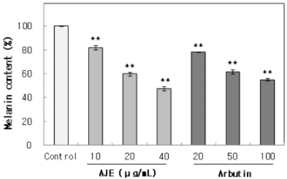
우선 in vitro 상에서 mushroom tyrosinase를 이용하 여 tyrosinase 활성에 미치는 영향을 측정한 결과, mushroom tyrosinase에 대한 저해 활성을 나타내지 않 았다(data 미첨부). 다음으로 멜라닌 합성에 미치는 영 향을 확인하기 위해 B16-F1 melanoma cell에 10, 20, 40 µg/mL의 농도로 처리하고 48 h이 지난 후 멜라닌 함량을 측정한 결과, 농도 의존적으로 멜라닌 합성이 저해됨을 확인하였다. Figure 2에서와 같이 최종 농도 인 40 µg/mL에서 53%의 멜라닌 합성 억제효과를 나 타내었다. 이는 100 µg/mL의 농도에서 45%의 멜라닌 합성 억제효과를 나타낸 양성대조군인 알부틴보다 뛰 어난 효과였다. 이러한 결과로 보아 AJE는 tyrosinase 의 활성을 직접적으로 저해하지 않고 멜라닌 합성 신 호전달 경로에 작용할 것으로 추정된다. 따라서 AJE
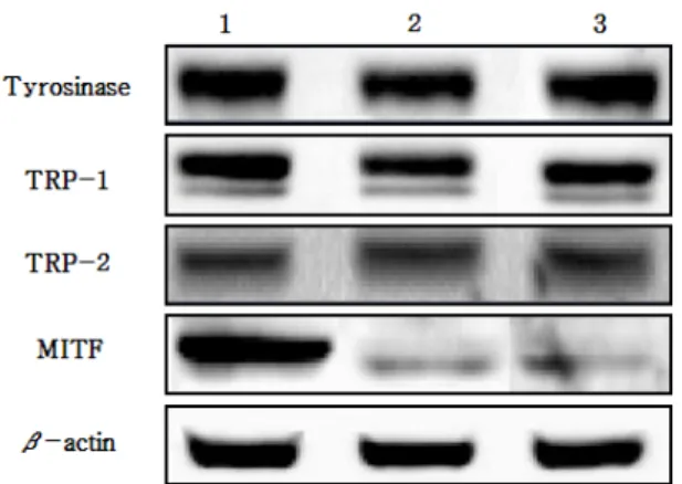
Figure 3. The effect of AJE on the melanogenic protein expression in B16-F1 melanoma cells. The cells were treated with AJE at indicated concentration for 48 h. lane 1, Control; lane 2, AJE 40 µg/mL; lane 3, Arbutin 100 µg/mL.
Figure 4. The effect of AJE on the ERK activation in B16-F1 melanoma cells. The cells were treated with AJE at indicated concentration for 48 h. lane 1, Control; lane 2, AJE 40 µg/mL; lane 3, Arbutin 100 µg/mL.
에 의한 멜라닌 합성 관련 단백질의 발현 양상을 분석 하였다.
3.1.3. 멜라닌 합성 관련 단백질 발현 저해효과
멜라닌 합성 신호전달 체계에는 매우 다양한 신호 전달물질이 관여하고 있다. 멜라닌은 몇 가지 세포 내 신호전달 기전을 통하여 합성되는데, 그 중 cAMP / PKA 경로가 멜라닌 합성의 주요 경로로서 UV에 피 부가 노출되었을 때 멜라닌세포의 cAMP가 증가되고 하류 신호전달 물질인 PKA를 활성화 시키며, CREB 을 거쳐 MITF의 발현을 증가시킨다. MITF는 멜라닌 합성 과정에서 중요한 전사 조절 인자로 tyrosinase, TRP-1, TRP-2의 전사를 촉진한다[42-44]. 본 연구에서 는 AJE의 미백효과 검증을 위해 멜라닌 합성 관련 단 백질인 MITF, tyrosinase, TRP-1, TRP-2의 발현 정도를 측정하였다. 40 µg/mL 농도의 AJE를 B16-F1 melano- ma cell에 처리한 후 48 h 배양한 뒤 단백질을 모아 western blot assay를 수행하였다. 이때 세포의 여러 조 건에서도 그 발현 정도의 차이가 거의 없는 house- keeping gene인 β -actin을 대조군으로 사용하였다. 그 결과 Figure 3에서와 같이 AJE를 처리한 군은 TRP-1 의 발현을 30% 정도 저해하였으며, tyrosinase와 TRP-2 의 발현에는 영향을 미치지 않는 것으로 확인되었다.
또한 64%의 MITF의 발현저해 효과를 확인할 수 있었 다. 한편 100 µg/mL의 알부틴을 처리한 군에서도 AJE 처리군과 유사한 경향을 보였는데 TRP-1의 발현을 18%, MITF의 발현을 54% 정도 저해하였으나 tyrosinase와
TRP-2의 발현에는 영향을 미치지 않는 것으로 확인되 었으며 이는 Park 등[45]의 결과와 유사한 경향을 나 타내었다.
3.1.4. ERK pathway activation에 미치는 효과
주로 세포의 증식, 분화에 관여하는 것으로 알려진 MAPK 경로에 속하는 ERK 신호는 MITF의 인산화를 유도하여 MITF의 ubiquitination이 이루어져 proteoso- mal degradation을 일으키게 됨으로써 멜라닌 합성을 감소시키는 것으로 알려져 있다[18,46]. AJE가 이러 한 ERK pathway 활성화에 영향을 미치는지 확인하고 자 p-ERK의 발현을 측정하였으며 그 결과 40 µg/mL 의 AJE를 처리한 경우 p-ERK가 증가되어(Figure 4) AJE가 ERK의 활성화에 영향을 미치는 것으로 확인 되었다. 한편 100 µg/mL의 알부틴을 처리한 군에서도 p-ERK의 발현이 증가됨을 확인하였다. 이러한 결과 로 보아 AJE의 멜라닌 합성 저해효과는 ERK pathway 활성화를 통해 MITF의 분해를 촉진함으로써 MITF 발현을 억제하고 그 결과 TRP-1의 발현 역시 억제되 었으며 최종적으로 멜라닌 합성이 억제된 것으로 사 료된다.
4. 결 론
본 연구에서는 적양을 화장품의 새로운 미백소재로 이용하고자 적양 에탄올추출물을 효소처리한 후 EtOAc 로 분획하여 EtOAc 분획물인 AJE를 얻어 이의 미백 활성을 측정하고, 그 작용기전에 대해 확인하였다.
AJE의 B16-F1 melanoma cell을 이용한 멜라닌 합성 억제효과 측정 결과 미백소재로 알려져 있는 알부틴 보다 우수한 미백 활성을 가지는 것으로 확인되었다.
그러나 in vitro 상의 mushroom tyrosinase의 활성에는 영향을 미치지 않는 것으로 나타나, tyrosinase의 활성 을 직접적으로 저해하지 않고 멜라닌 합성 신호전달 경로에 작용할 것으로 추정되어 멜라닌 합성 관련 단 백질의 발현 양상을 분석하였다. 그 결과 멜라닌 합성 에 관여하는 효소인 TRP-1의 발현을 억제하였고 이를 조절하는 전사인자인 MITF 발현 역시 효과적으로 억 제하였다. 또한 ERK pathway를 활성화시킴으로써 p-ERK의 발현을 상당히 증가시키는 것을 확인할 수 있었다.
이상의 결과를 종합해 볼 때 AJE는 멜라닌 합성의 신호전달 경로 중 ERK pathway의 활성화를 통해 MITF의 분해를 촉진시키고 이로 인해 MITF의 발현을 감소시키며, 그 결과 멜라닌 합성에 관여하는 효소 중 TRP-1의 발현을 감소시킴으로써 멜라닌 합성을 저해 하는 것으로 사료된다. 향후 활성 물질에 대한 분석과 인체실험을 통한 안정성 및 미백효능의 검증이 필요 하나 적양은 화장품의 새로운 미백 소재로서의 활용 가치가 큰 것으로 판단된다.
감사의 글
본 연구는 2011년 중소기업기술혁신개발사업(과제 번호 SA112778)의 연구비 일부에 의하여 이루어진 것 으로 지원에 감사드립니다.
Reference
1. H. Z. Hill, W. Li, P. Xin, and D. L. Michell, Melanin: a two edged sword?, Pigment Cell Res., 10, 158 (1998).
2. M. Seiberg, L. Babiarz, and C. B. Lin, IL-41 the PAR-2 pathway is differentially expressed in skin of color, Pigment Cell Res., 16, 591 (2003).
3. V. J. Hearing and M. Jimenez, Analysis of mamma- lian pigmentation at the molecular level, Pigment Cell Res., 2, 75 (1989).
4. V. J. Hearing and K. Tsukamoto, Enzymatic control of pigmentation in mammals, FASEB J., 5, 2902 (1991).
5. T. Kobayashi, W. D. Vieira, B. Potterf, C. Sakai, G.
Imokawa, and V. J. Hearing, Modulation of melano- genic protein expression during the switch from eu-to pheomelanogenesis, J. Cell Sci., 108, 2301 (1995).
6. T. Kobayashi, K. Urabe, A. Winder, C. Jimenez- Cervantes, G. Imokawa, T. Brewington, F. Solano, J.
C. Garcia-Borron, and V. J. Hearing, Tyrosinase re- lated protein 1 (TRP1) functions as a DHICA oxi- dase in melanin biosynthesis, EMBO J., 13, 5818 (1994).
7. K. Yokoyama, H. Suzuki, K. Yasumoto, Y. Tomita, and S. Shibahara, Molecular cloning and functional analysis of a cDNA coding for human DOPAchrome tautomerase/tyrosinase-related protein-2, Biochim. Bio- phys. Acta., 1217, 317 (1994).
8. I. Aksan and C. R. Goding, Targeting the micro- phthalmia basic helix-loop- helix-leucine zipper tran- scription factor to a subset of E-box elements in vi- tro and in vivo, Mol. Cell. Biol., 18, 6930 (1998).
9. C. A. Hodgkinson, K. J. Moore, A. Nakayama, E.
Steingrimsson, N. G. Copeland, N. A. Jenkins, and H. Arnheiter, Mutations at the mouse microphthalmia locus are associated with defects in a gene encoding a novel basic-helix-loop-helix-zipper protein, Cell, 74, 395 (1993).
10. E. Steingrimsson, K. J. Moore, M. L. Lamoreux, A.
R. Ferre-D’Amare, S. K. Burley, D. C. Zimring, L.
C. Skow, C. A. Hodgkinson, H. Arnheiter, N. G.
Copeland, and N. A. Jenkins, Molecular basis of mouse microphthalmia (mi) mutations helps explain their developmental and phenotypic consequences, Nat. Genet., 8, 256 (1994).
11. H. R. Widlund and D. E. Fisher, Microphthalamia associated transcription factor: a critical regulator of pigment cell development and survival, Oncogene, 22, 3035 (2003).
12. N. J. Bentley, T. Eisen, and C. R. Goding, Melano- cyte-specific expression of the human tyrosinase pro- moter: activation by the microphthalmia gene prod- uct and role of the initiator, Mol. Cell. Biol., 14, 7996 (1994).
13. C. Bertolotto, P. Abbe, T. J. Hemesath, K. Bille, D.
E. Fisher, J. P. Ortonne, and R. Ballotti, Microph- thalmia gene product as a signal transducer in cAMP-induced differentiation of melanocytes, J. Cell Biol., 142, 827 (1998).
14. C. Levy, M. Khaled, and D. E. Fisher, MITF: master regulator of melanocyte development and melanoma oncogene, Trends Mol. Med., 12, 406 (2006).
15. K. Yasumoto, K. Yokoyama, K. Takahashi, Y. Tomita, and S. Shibahara, Functional analysis of micro- phthalmia-associated transcription factor in pigment cell-specific transcription of the human tyrosinase family genes, J. Biol. Chem., 272, 503 (1997).
16. U. Yavuzer, E. Keenan, P. Lowings, J. Vachtenheim, G. Currie, and C. R. Goding, The microphthalmia gene product interacts with the retinoblastoma pro- tein in vitro and is a target for deregulation of mela- nocyte-specific transcription, Oncogene, 10, 123 (1995).
17. W. Englaro, C. Bertolotto, R. Busca, A. Brunet, G.
Pages, J. P. Ortonne, and R. Ballotti, Inhibition of the mitogen-activated protein kinase pathway triggers B16 melanoma cell differentiation. J. Biol. Chem., 273, 9966 (1998).
18. D. S. Kim, E. S. Hwang, J. E. Lee, S. Y. Kim, S.
B. Kwon, and K. C. Park, Sphingosine-1-phosphate decrease melanin synthesis via sustained ERK activa- tion and subsequent MITF degradation. J. Cell Sci., 116, 1699 (2003).
19. D. S. Kim, S. Y. Kim, J. H. Chung, K. H. Kim, H.
C. Eun, and K. C. Park, Delayed ERK activation by ceramide reduces melanin synthesis in human mela- nocytes. Cell. Signal., 14, 779 (2002).
20. W. Englaro, R. Rezzonico, M. Durand-Clement, D.
Lallemand, J. P. Ortonne, and R. Ballotti, Mitogen- activated protein kinase pathway and AP-1 are acti- vated during cAMP-induced melanogenesis in B-16 melanoma cells. J. Biol. Chem., 270, 24315 (1995).
21. S. J. Lee, Korean Folk Medicine, 3, Seoul National University, Seoul (1966).
22. 中藥大辭典, 3, 3042, 小學館, Tokyo (1985).
23. M. Terazawa, H. Okuyama, and M. Miyake, Isola-
tion of hirsutanonol and hirsutenone, two new diary- lheptanoids form the green bark of keyamahannoki, Alnus hirsuta Turcz, Mokuzai Gakkaishi, 19, 45 (1973).
24. J. J. Karches, M. L. Laever, D. F. Barofsky, and E.
Barofsky, Structure of oregonin, a natural diary- lheptanoid xyloside. J. Chem. Soc., Chem. Commun., 16, 649 (1974).
25. M. W. Lee, T. Tanaka, G. Nonaka, and I. Nishioka, Hirsunin, an ellagitannin with a diarylheptanoid moi- ety from Alnus hitsuta var. Microphylla, Phytochemistry, 31, 967 (1992).
26. M. Tori, A. Hashimoto, K. Hirose, and Y. Asakawa, Diarylheptanoids, flavonoids, stilbenoids, sesquiterpe- noids and a phenanthrene from Alnus maximowiczii, Phytochemistry, 40, 1263 (1995).
27. H. Wada, H. Tachibana, H. Fuchino, and N. Tanaka, Three new diarylheptanoid glycosides from Alnus ja- ponica, Chem. Pharm. Bull., 46, 1054 (1998).
28. R. F. Gonzalez-Laredo, J. Chem, Y. M. Karchesy, and J. J. Karchesy, Four new diarylheptanoid glyco- sides from Alnus Rubra Bark, Nat. Prod. Lett., 13, 75 (1999).
29. T. Suga, N. Iwata, and Y. Asakawa, Chemical con- stituents of the male flower of Alnus pendula (Betul- aceae), Bull. Chem. Soc. Jpn., 45, 2058 (1972).
30. Y. Asakawa, Chemical constituents of Alnus sie- boldiana (Betulaceae) Ⅱ. The isolation and structure of flavonoids and stilbenes, Bull. Chem. Soc. Jpn., 44, 2761 (1971).
31. V. A. Stikhin, A. I. Ban’kovskii, V. I. Glyzin, and I. A. Kir’yanova, Quercetin-3-sophoroside from the pollen of Alnus glutinosa and Fraxinus lanceolata, Chem. Nat. Comp., 10, 526 (1974).
32. M. W. Lee, D. W. Jeong, Y. A. Lee, M. S. Park, and S. H. Toh, Flavonoids from the Leaves of Alnus hir- suta, Yakhak Hoeji, 43, 547 (1999).
33. K. W. Ahn, S. H. Toh, D. W. Jeong, J. S. Kim, S.
M. Sho, and M. W. Lee, Flavonoids from the Leaves of Alnus maximowiczii Call, Yakhak Hoeji, 44, 41 (2000).
34. M. W. Lee, M. S. Park, D. W. Jeong, K. H. Kim,
and S. H. Toh, Diarylheptanoids from the Leaves of Alnus hirsuta Trucz, Arch. Pharm. Res., 23, 50 (2000).
35. M. W. Lee, M. S. Park, D. W. Jeong, K. H. Kim, S. H. Toh, D. R. Hahn, Y. C. Kim, and H. T.
Chung, Diarylheptanoids with in vitro inducible ni- tric oxide synthesis inhibitory activity from Alnus hirsuta, Planta Med., 66, 551 (2000).
36. M. W. Lee, J. H. Kim, D. W. Jeong, K. H. Ahn, S.
H. Toh, and Y. J. Surh, Inhibition of cyclooxy- genase-2 expression by diarylheptanoids from the bark of Alnus hirsuta var. sibirica, Biol. Pharm.
Bull., 23, 517 (2000).
37. D. I. Lee, J. K. Chang, M. W. Lee, and S. G. Hong, Effects of oregonin, diarylheptanoid derivative from plant on antitumor, Chung-Ang J. Pharm. Sci., 12, 50 (1998).
38. Y. A. Lee, K. H. Kim, J. S. Kim, S. M. Cho, S. W.
Kim, and M. W. Lee, Antioxidative effects of diary- lheptanoids from Alnus hirsuta, Yakhak Hoeji, 44, 193 (2000).
39. S. M. Cho, Y. M. Kwon, J. H. Lee, K. H. You, and M. W. Lee, Melanogenesis inhibitory activities of di- arylheptanoids from Alnus hirsuta Turcz in B16 mouse melanoma cell, Arch. Pharm. Res., 25, 885 (2002).
40. T. Mosmann, Rapid colorimetric assay for the cel- lular growth and survival: application to proliferation
and cytotoxic assay, J. Immun. Methods, 65, 55 (1983).
41. M. Oka, M. Ichihashi, and AK. Chakraborty, Enhanced expression of protein kinase C subspecies in melano- genic compartments in B16 melanoma cells by UVB or MSH. J. Invest. Dermatol., 106, 377 (1996).
42. R. Busca and R. Ballott, Cyclic AMP a key mes- senger in the regulation of skin pigmentation. Pigment Cell Res., 13, 60 (2000).
43. C. P. Sassone, Coupling gene expression to cAMP signalling: Role of CREB and CREM. Int. J.
Biochem. Cell B., 30, 27 (1998).
44. B, Saha, S. K. Singh, C. Sarkar, R. Bera, J. Ratha, D. J. Tobin, and R. Bhadra, Activation of the MITF promoter by lipid-stimulated activation of p38-stress signalling to CREB. Pigment Cell Res., 29, 595 (2006).
45. K. T. Park, J. K. Kim, D. H. Hwang, Y. M. Yoo, and Y. H. Lim, Inhibitory effect of mulberroside A and its derivatives on melanogenesis induced by ul- traviolet B irradiation. Food Chem. Toxicol., 49, 3038 (2011).
46. Xu, W., L. Gong, M. M. Haddad, O. Bischof, J.
Campisi, E. T. Yeh, and E. E. Medrano, Regulation of Microphthalmia-associated transcription factor MITF protein levels by association with the ubiq- uitin-conjugation enzyme hUBC9. Exp. Cell Res., 255, 135 (2000).