Effects of Ovary Status and In Vitro Maturation Condition on the Developmental Competence of Canine Oocytes
Su-Jin Cho1, Dong-Hoon Kim3, Chan-Sik Min4 and Il-Keun Kong1,2,*
1Division of Applied Life Science (BK21), Graduate School of Gyeongsang National University, Jinju 660-701, Republic of Korea
2Institute of Agriculture and Life Science, Jinju 660-701, Republic of Korea
3Animal Biotechnology Division, National Institute of Animal Science, Suwon 441-706, Republic of Korea
4Gyeongsangnam-do Agricultural Research and Extension Services, Jinju 660-985, Republic of Korea
ABSTRACT
In canine, oocytes are ovulated at the GV (germinal vesicle) stage and they have to fulfill maturation phase before reaching metaphase ΙΙ stage. The efficiency of in vitro maturation is still very low. Therefore, the aim of this study was to investigate the effect of in vitro maturation on nuclear changes of immature canine oocytes recovered from different reproductive stages ovaries and different culture conditions. The oocytes were cultured in TCM-199 with supplement at 5% CO2 and 38.5℃ for 72 h. The nuclear maturation of canine oocytes was evaluated with Hoechst 33342 stain under fluorescence microscope (Fig. 1). The results of this study detected differences in in vitro maturation rate between oocytes recovered from follicle status and non-follicle status ovaries. However, these differences were not significant as indicated in Table 1 and Fig. 2. In regard to the effect of culture condition with supplements, we did not found significant differences compared with control group (Table 2, Table 3). One of the reasons for this data could be the conditions that ovaries were exposed during slaughtering process or the long distant transportation of the ovaries. Although these data have not shown clearly significant differences results compared with control, furthermore the different reproductive status ovaries was beneficial for maturation of oocytes in vitro and can be a basic part of knowledge to improve in vitro maturation of canine oocytes.
(Key words : canine, oocytes, in vitro maturation, ovary, estrus cycle)
†This study was supported by a scholarship from the BK21 program and Rural Development Administration (Project No. PJ008975042012), Republic of Korea, and sabbatical year (2012) research of Gyeongsang National University.
*Correspondence : E-mail : [email protected]
INTRODUCTION
The developmental competence and quality of embryos pro- duced in vitro depends upon several factors in addition to the conditions of in vitro maturation, fertilization and culture. Espe- cially, In vitro maturation (IVM) is one of the most important steps that determine the developmental competence of the oocytes (Eppig et al., 1994). IVM is a reproductive technology that enables oocytes to be matured in vitro from ovaries that have received either no or low levels of gonadotrophin stimu- lation (Edwards, 1965). Furthermore, IVM is an important assisted reproductive technologies (ART) as it has the potential to capture the vast supply of oocytes within an ovary (Gil- christ et al., 2008). In domestic animals except canine, IVM success rates are relatively high and therefore are more widely accepted. However, the efficiency of IVM is still very low in
canine compared to other mammalian species. One of the rea- sons for low efficiency of IVM in this species may be due to its unique reproductive physiological characteristics (Kim et al., 2001). Unlike other species, canine oocytes are ovulated at pro- phase of the first meiotic division and undergo maturation in the distal part of the oviduct for at least 48 to 72 h (Lee et al., 2005; Songsasen and Wildt, 2007). Female dogs also have unusual characteristics including the follicular environment, extremely high lipid content representing a uniform dense, dark appearing cytoplasm, with highly compact, and unexpanded cumulus cells of ovulated oocytes (Hewitt and England, 1997).
Many researchers have examined the feasibility of IVM of canine immature oocytes (Yamada et al., 1992; Bogliolo et al., 2002; Bolamba et al., 2002; Songsasen et al., 2003; Hatoya et al., 2006; Saikhun et al., 2008; Apparicio et al., 2011; Lopes et al., 2011; Salavati et al., 2012; Songsasen et al., 2012).
Nevertheless, the rates of maturation of canine oocytes to me- taphase ΙΙ (MΙΙ) remain low, especially when compared with those of many other mammalian species. Thus, there is no in vitro system has been developed to increase the maturation rates of canine oocytes comparable to in vivo one. A short- coming that has been attributed to this low efficiency was the specific and highly complex requirements of in vivo canine oocytes maturation (B.A. Rodrigues JLR, 2010). Once research results improved, the reproductive techniques of in vitro matu- ration, in vitro fertilization, and embryo transfer promise to be useful tools for specific canine species conservation.
This study was conducted to evaluate the nuclear develop- ment of canine oocytes when collected from different repro- ductive stages ovaries or cultured with different supplements during in vitro maturation.
MATERIALS AND METHODS
All chemicals used in this study were purchased from Sigma Che- mical Company (Sigma, St. Louis, MO, USA), unless indicated.
1. Collection of Canine Ovaries and Cumulus-oocyte Complexes Ovaries were obtained from canine females of various breeds and at different ages with unknown health status that under- went ovariohysterectomy at the veterinary hospital nearby Jinju city, Republic of Korea. The cumulus-oocyte complexes (COC) were released by repeated slicing of the ovarian cortex with the scalpel blades in TCM-199 medium with 25 mM HEPES supplemented with 0.1% bovine serum albumin (BSA) and 1%
penicillin-streptomycin at 38.5℃.
2. In Vitro Maturation
The oocytes selected for this study had a perfectly spherical shape and an even, smooth, dark pigmented cytoplasm. The oocytes were cultured in TCM-199 supplemented with 1 μg/ml estradiol-17β, 10 μg/ml FSH, 0.6 mM cysteine, 0.2 mM Na- pyruvate, 10% FBS and 1% penicillin-streptomycin. The ma- turation was performed by culturing approximately 50~60 COCs in 500 μl of maturation medium in four-well dishes for 72 h at 5% CO2 and 38.5℃.
3. Removal of Cumulus Cells and Assessment of Oocytes Nuclear Maturation
At the end of the maturation period, COCs were transferred to D-PBS buffer containing 0.1% hyaluronidase and the cumu-
Fig. 1. Chromatin configuration of canine oocytes stained with Hoe- chst 33342. GV: germinal vesicle stage, GVBD: germinal vesicle breakdown stage, MΙ: metaphase Ι stage, MΙΙ: me- taphase ΙΙ stage, PB: first polar body. The bar is 50 μm.
lus cells were removed by gentle pipetting with glass pipette.
The denuded oocytes were washed several times in D-PBS with 0.1% polyvinyl alcohol (PVA) and transferred to 3.7% for- maldehyde solution at room temperature before mounting on a slide with an overlay of 1.9 μM Hoechst 33342 in glycerol.
The oocytes were evaluated under an inverted epifluorescence microscope with a UV light to determine the stage of meiosis, such as germinal vesicle (GV), germinal vesicle breakdown (GVBD), MΙ (metaphase Ι) stage, and MⅡ (metaphaseⅡ) stage (Fig. 1).
4. Experimental Design
For the effect of reproductive stage ovaries, the cumulus- oocyte complexes (COCs) from non-follicle status (n=163) and follicles status (n=224) ovaries were released by repeated sli- cing of the ovarian cortex with the scalpel blades in TCM-199 medium with 25 mM HEPES supplemented with 0.1% bovine serum albumin (BSA) and 1% penicillin-streptomycin at 38.5℃.
The oocytes were cultured in maturation medium at 38.5℃ of 5% CO2 for 72 h.
For the different culture conditions, the oocytes from ran- domly stage ovaries were cultured in maturation medium with (n=54)/without (n=49) PMSG (0.5 IU/ml) & hCG (1 IU/ml) at 38.5℃ of 5% CO2 for 72 h. The other group was cultured in maturation medium with (n=50)/without (n=20) MG132 (1 μM) at 38.5℃ of 5% CO2 for 72 h.
5. Statistical Analysis
Data were expressed as the mean ± SEM by t-test. Diffe- rences were considered significant at p<0.05.
RESULTS AND DISCUSSION
1. The Effect of Ovary Status on Maturation Rate of Canine Oocyte In domestic animals, the oocytes are recovered from slaugh-
tered animals that could be at different stages of estrus cycle and from different sizes of follicles. Even so, effects of ovary status on nuclear kinetics of canine oocytes were varied depending on in vitro maturation. The nuclear configuration of immature canine oocytes collected from different reproductive stage ovaries were evaluated after IVM as shown in Fig. 2.
Overall, this study found that there were differences but not significant in maturation rate between oocytes recovered from follicle status and non-follicle status ovaries collected from different reproductive stages (Table 1. 24.6 ± 18.5%; Fig. 2-A vs. 21.4 ± 18.1%; Fig. 2-B, C). Oocytes recovered from follicle
Fig. 2. Morphology evaluations of the canine ovaries. (A) follicular ovaries (red arrow), (B, C) non-follicular ovaries from diffe- rent reproductive stages; (B) anestrus and (C) corpus lu- teum stage.
Table 1. Meiotic progression of canine oocytes from follicle and non-follicle status ovaries
Ovary status
No. of oocytes developed to (%)
GV GVBD MΙ MΙΙ Non MΙ - ΙΙ
Follicle* 47 (21 ± 20.1) 115 (51.3 ± 18.5) 28 (12.5 ± 5.6) 27 (12.1 ± 12.9) 7 (3.1 ± 4.1) 55 (24.6 ± 18.5) Non-follicle** 9 (5.5 ± 1.4) 116 (71.2 ± 18.2) 25 (15.3 ± 11.2) 10 (6.1 ± 6.9) 3 (1.8 ± 2.0) 35 (21.4 ± 18.1)
* Follicle showed Fig. 2-A status, ** Non-follicle showed Fig. 2-B, Fig. 2-C status.
GV: germinal vesicle stage, GVBD: germinal vesicle breakdown stage, MΙ: metaphase Ι stage, MΙΙ:: metaphase ΙΙ stage, PB: first polar body, Non: Not detected nuclear.
status ovaries reached metaphase stage in higher rate than that of recovered from non-follicle ovaries (Table 1). One of the reasons behind this result could be that there was no available information in regard to the health, and age of animals. An- other possibility is that canine ovaries usually exposed to higher temperature in slaughterhouse, which could reduce the quality of the oocytes.
Oh et al. (2005) have reported that immature canine oocytes within follicles are exposed to high levels of E2 and P4 due to preovulatory luteinization of follicles which could influence their in vitro maturation. Following ovulation, canine oocytes are also exposed to high levels of P4 and E2 in the bursa and oviduct (Kim et al., 2005). In addition, size of the follicle is one of major factors influencing IVM success of the canine oocyte. The large follicles provide oocytes with higher meiotic competency compared to small ones. Songsasen and Wildt (2005) have reported also that nearly 80% of oocytes recovered from follicles > 2 mm have the capacity to achieve MΙΙ com- pared to that recovered from 1 to 2 mm (38%), from > 0.5 to 1 mm (26%) and from < 0.5 mm (17%) follicles. Therefore, oocytes collected from follicle status ovaries have a beneficial effect on the nuclear maturation in vitro.
2. The Effect of Culture Condition on Maturation RAte of Canine Oocyte
Oocytes aspirated from the follicle and exposed to an arti- ficial environment such as a maturation medium will experience changes associated with the action of compounds to which they were subjected. Moreover, spontaneous apoptosis occurs in cumulus oocyte complexes (COCs) during suboptimal culture condition (Ikeda et al., 2003; Yuan et al., 2005). Specific com- mercial culture media can generate reactive oxygen species (ROS) depending on its composition, and media additives also are playing a crucial role in ROS induction (Agarwal et al.,
2006). Therefore, media additives may interfere with the extent of either cell death or survival of the oocytes. Nevertheless, nobody has been yet established efficient in vitro maturation system of canine oocyte as the well-established one of other domestic animal species.
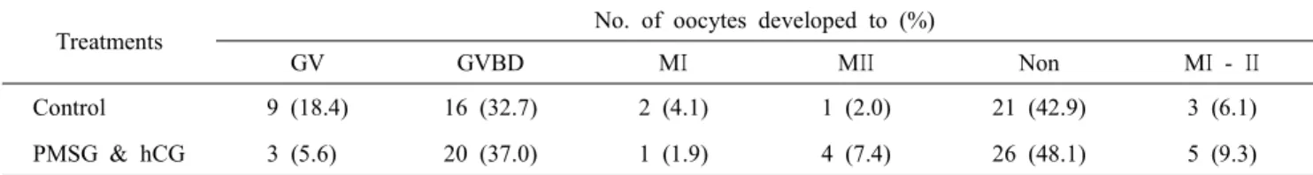
We also have demonstrated that the effect of culture con- ditions on the in vitro maturation competence of immature ca- nine oocytes when supplements were added. In this study, the various supplements specifically PMSG and hCG as hormone (Table 2) and in addition to MG132 as a proteosome inhibitor (Table 3) were added during in vitro maturation. The rates of maturation for the control, and hormone supplemented group (0.5 IU/ml PMSG and 1 IU/ml hCG) were 6.1% and 9.3%, respectively. The percentage of maturation to MⅡ stage was higher in the hormone treatment group that the control. But, there was no significant difference between control and hor- mone treatment groups. As the other study, the MG132 is a specific inhibitor of proteasome activity and prevents MPF inactivation by blocking cyclin B degeneration (Josefsberg et al., 2000; You et al., 2012). The rates of maturation for the control (non-treatment) and 1 μM MG132 treatment groups were 35% and 48%, respectively. The percentage of matura- tion to MⅡ stage was also higher in the MG132 treatment group than the control. But, there was no significant difference between control and MG132 treatment groups. Overall, no sig- nificant differences were observed on the in vitro maturation
Table 2. Effect of hormonal supplementation on in vitro maturation rate of canine oocytes
Treatments No. of oocytes developed to (%)
GV GVBD MΙ MΙΙ Non MΙ - ΙΙ
Control 9 (18.4) 16 (32.7) 2 (4.1) 1 (2.0) 21 (42.9) 3 (6.1)
PMSG & hCG 3 (5.6) 20 (37.0) 1 (1.9) 4 (7.4) 26 (48.1) 5 (9.3)
GV: germinal vesicle stage, GVBD: germinal vesicle breakdown stage, MΙ: metaphase Ι stage, MΙΙ: metaphase ΙΙ stage, PB: first polar body, Non: Not detected nuclear.
Table 3. Effect of MG132 treatment on in vitro maturation rate of canine oocytes
Treatments No. of oocytes developed to (%)
GV GVBD MΙ MΙΙ Non MΙ - ΙΙ
Control 9 (45.0) 3 (15.0) 6 (30.0) 1 (5.0) 1 (5.0) 7 (35.0)
MG132 15 (30.0) 6 (12.0) 21 (42.0) 3 (6.0) 5 (10.0) 24 (48.0)
GV: germinal vesicle stage, GVBD: germinal vesicle breakdown stage, MΙ: metaphase Ι stage, MΙΙ: metaphase ΙΙ stage, PB: first polar body, Non: Not detected nuclear.
rate among the different supplements. Furthermore, we need investigate effect of MG132 treatment during oocyte matu- ration in vitro on the MPF activity in canine oocytes.
Chemical supplement using glucose in the maturation me- dium, seems to enhance the rates of meiosis resumption and metaphase stage of in vitro matured canine oocytes (Silva et al., 2009). Glucose is the predominant energy substrate used by canine oocytes (Wesselowsky, 2008). It has also been su- ggested that retinoic acid may promote embryonic develop- ment by preventing oxidative stress (Deb et al., 2011), and may also regulates the expression of several growth factors con- trolling genes during maturation of bovine oocytes (Gomez et al., 2004). Liang et al. (2012) reported that the influence of different concentration of 9-cis RA on nuclear maturation of canine oocytes during IVM. In the previous stuy, five nM 9- cis RA in the IVM medium was beneficial to nuclear and cytoplasmic maturation of canine oocytes (5 nM 9-cis RA vs.
control: 18.3 ± 2.5 vs. 8.7 ± 1.5%) (Liang et al., 2012).
To date, several studies in canine oocytes have been per- formed to improve the rate of IVM by supplementation IVM medium with fetal bovine serum (Yamada et al., 1993), estrus bitch serum (Bogliolo et al., 2002), bovine serum albumin (Hewitt et al., 1998; Lee et al., 2003; Rodrigues and Rodrigues, 2003;
Willingham-Rocky et al., 2003) or medium without serum (Songsasen et al., 2002) as well as collecting oocytes at va- rious estrus phases (Yamada et al., 1993; Luvoni et al., 2001;
Willingham-Rocky et al., 2003). However the maturation rate has not been satisfactorily increased. In addition, the majority of in vitro matured canine oocytes have remained at the GV stage regardless of the culture conditions.
Likewise, many researchers have supplied the in vitro ma- turation media of canine oocyte with the several supplemen- tations. However, there is still progress is urgently needed to establish more efficient in vitro maturation system that could help to advance reproductive technologies based on the mass production of canine embryos. Furthermore, efforts in this field are also required to increase the rates of metaphase stages of canine oocytes to compare with other domestic animals. This goal could be achieved through further studies that mainly fo- cus on developing ideal in vitro culture condition suitable for canine oocytes.
REFERENCES
Agarwal A, Said TM, Bedaiwy MA, Banerjee J and Alvarez JG. 2006. Oxidative stress in an assisted reproductive tech- niques setting. Fertil Steril. 86:503-512.
Apparicio M, Alves AE, Pires-Butler EA, Ribeiro AP, Covizzi GJ and Vicente WR. 2011. Effects of hormonal supplemen- tation on nuclear maturation and cortical granules distri- bution of canine oocytes during various reproductive stages.
Reprod. Domest. Anim. 46:896-903.
B.A. Rodrigues JLR. 2010. In vitro maturation of canine oocytes:
a unique conundrum. Anim. Reprod. 7:3-15.
Bogliolo L, Zedda MT, Ledda S, Leoni G, Naitana S and Pau S.
2002. Influence of co-culture with oviductal epithelial cells on in vitro maturation of canine oocytes. Reprod. Nutr.
Dev. 42:265-273.
Bolamba D, Russ KD, Olson MA, Sandler JL and Durrant BS.
2002. In vitro maturation of bitch oocytes from advanced preantral follicles in synthetic oviduct fluid medium: serum is not essential. Theriogenology 58:1689-1703.
Deb GK, Dey SR, Bang JI, Cho SJ, Park HC, Lee JG and Kong IK. 2011. 9-cis retinoic acid improves developmental competence and embryo quality during in vitro maturation of bovine oocytes through the inhibition of oocyte tumor necrosis factor-alpha gene expression. J. Anim. Sci. 89:2759- 2767.
Edwards RG. 1965. Maturation in vitro of mouse, sheep, cow, pig, Rhesus monkey and human ovarian oocytes. Nature 208:
349-351.
Eppig JJ, Schultz RM, O'Brien M and Chesnel F. 1994. Rela- tionship between the developmental programs controlling nu- clear and cytoplasmic maturation of mouse oocytes. Dev.
Biol. 164:1-9.
Gilchrist RB, Lane M and Thompson JG. 2008. Oocyte-secreted factors: regulators of cumulus cell function and oocyte quality. Hum. Reprod. Update 14:159-177.
Gomez E, Rodriguez A, Goyache F, Diez C, Jose Royo L, Mo- reira PN, Nestor Caamano J, Moran E and Gutierrez-Adan A. 2004. Retinoid-dependent mRNA expression and poly- (A) contents in bovine oocytes meiotically arrested and/or matured in vitro. Mol. Reprod. Dev. 69:101-108.
Hatoya S, Sugiyama Y, Torii R, Wijewardana V, Kumagai D, Sugiura K, Kida K, Kawate N, Tamada H, Sawada T and Inaba T. 2006. Effect of co-culturing with embryonic fib- roblasts on IVM, IVF and IVC of canine oocytes. Therio- genology 66:1083-1090.
Hewitt DA and England GC. 1997. Effect of preovulatory en- docrine events upon maturation of oocytes of domestic bit- ches. J. Reprod. Fertil. Suppl. 51:83-91.
Hewitt DA, Watson PF and England GC. 1998. Nuclear staining and culture requirements for in vitro maturation of domes- tic bitch oocytes. Theriogenology 49:1083-1101.
Ikeda S, Imai H and Yamada M. 2003. Apoptosis in cumulus cells during in vitro maturation of bovine cumulus-enclosed oocytes. Reproduction 125:369-376.
Josefsberg LB, Galiani D, Dantes A, Amsterdam A and Dekel N. 2000. The proteasome is involved in the first metaphase- to-anaphase transition of meiosis in rat oocytes. Biol. Re- prod. 62:1270-1277.
Kim MK, Fibrianto YH, Oh HJ, Jang G, Kim HJ, Lee KS, Kang SK, Lee BC and Hwang WS. 2005. Effects of estradiol-17 beta and progesterone supplementation on in vitro nuclear maturation of canine oocytes. Theriogenology 63:1342-1353.
Kim MK, Kim HJ, Cho JK, Jang K, Lee KS, Kang SK, Lee BC and Hwang WS. 2001. In vitro nuclear maturation of canine oocytes obtained from differents stages of estrus cycle. Korean J. Emb. Trans. 17:145-151.
Lee HS, Lee YH, Yin XJ and Kong IK. 2005. Effect of meio- tic maturation of canine oocytes cultured in reproductive tract. Korean J. Emb. Trans. 20:63-69.
Lee HS, Yin XJ, Lee YH, Kang TY and Kong IK. 2003. Effect of IVM medium and protein source on in vitro maturation of canine oocytes. Korean J. Emb. Trans. 18:75-80.
Liang S, Kang J, Jin H, Liu X, Li J, Li S, Lu Y, Wang W and
Yin XJ. 2012. The influence of 9-cis-retinoic acid on nu- clear and cytoplasmic maturation and gene expression in canine oocytes during in vitro maturation. Theriogenology 77:1198-1205.
Lopes G, Alves MG, Carvalho RA, Luvoni GC and Rocha A.
2011. DNA fragmentation in canine oocytes after in vitro maturation in TCM-199 medium supplemented with diffe- rent proteins. Theriogenology 76:1304-1312.
Luvoni GC, Luciano AM, Modina S and Gandolfi F. 2001. In- fluence of different stages of the oestrous cycle on cumulus- oocyte communications in canine oocytes: effects on the efficiency of in vitro maturation. J Reprod. Fertil. Suppl.
57:141-146.
Oh HJ, Fibrianto YH, Kim MK, Jang G, Hossein MS, Kim HJ, Kang SK, Lee BC and Hwang WS. 2005. Effects of canine serum collected from dogs at different estrous cycle stages on in vitro nuclear maturation of canine oocytes. Zygote 13:227-232.
Rodrigues BA and Rodrigues JL. 2003. Meiotic response of in vitro matured canine oocytes under different proteins and he- terologous hormone supplementation. Reprod. Domest. Anim.
38:58-62.
Saikhun J, Sriussadaporn S, Thongtip N, Pinyopummin A and Kitiyanant Y. 2008. Nuclear maturation and development of IVM/IVF canine embryos in synthetic oviductal fluid or in co-culture with buffalo rat liver cells. Theriogenology 69:1104-1110.
Salavati M, Ghafari F, Zhang T and Fouladi-Nashta AA. 2012.
Effects of oxygen concentration on in vitro maturation of canine oocytes in a chemically defined serum-free medium.
Reproduction 144:547-556.
Silva AE, Rodriguez P, Cavalcante LF, Rodrigues BA and Ro- drigues JL. 2009. The influence of oxygen tension on cumu- lus cell viability of canine COCs matured in high-glucose medium. Reprod. Domest. Anim. 44 Suppl. 2:259-262.
Songsasen N and Wildt DE. 2005. Size of the donor follicle, but not stage of reproductive cycle or seasonality, influences meiotic competency of selected domestic dog oocytes. Mol.
Reprod. Dev. 72:113-119.
Songsasen N and Wildt DE. 2007. Oocyte biology and challenges
in developing in vitro maturation systems in the domestic dog. Anim. Reprod. Sci. 98:2-22.
Songsasen N, Wesselowski S, Carpenter JW and Wildt DE.
2012. The ability to achieve meiotic maturation in the dog oocyte is linked to glycolysis and glutamine oxidation.
Mol. Reprod. Dev. 79:186-196.
Songsasen N, Yu I and Leibo SP. 2002. Nuclear maturation of canine oocytes cultured in protein-free media. Mol. Reprod.
Dev. 62:407-415.
Songsasen N, Yu I, Gomez M and Leibo SP. 2003. Effects of meiosis-inhibiting agents and equine chorionic gonadotro- pin on nuclear maturation of canine oocytes. Mol. Reprod.
Dev. 65:435-445.
Wesselowsky S. 2008. Metabolic analysis of glucose, pyruvate and glutamine in dog oocytes collected from different sized follicles and matured in vitro. Manhattan: Kansas State Uni- versity.
Willingham-Rocky LA, Hinrichs K, Westhusin ME and Krae- mer DC. 2003. Effects of stage of oestrous cycle and pro- gesterone supplementation during culture on maturation of canine oocytes in vitro. Reproduction 126:501-508.
Yamada S, Shimazu Y, Kawaji H, Nakazawa M, Naito K and Toyoda Y. 1992. Maturation, fertilization, and development of dog oocytes in vitro. Biol. Reprod. 46:853-858.
Yamada S, Shimazu Y, Kawano Y, Nakazawa M, Naito K and Toyoda Y. 1993. In vitro maturation and fertilization of preovulatory dog oocytes. J. Reprod. Fertil. Suppl. 47:227- 229.
You J, Lee E, Bonilla L, Francis J, Koh J, Block J, Chen S and Hansen PJ. 2012. Treatment with the proteasome inhi- bitor MG132 during the end of oocyte maturation improves oocyte competence for development after fertilization in cattle. PLoS One. 7:e48613.
Yuan YQ, Van Soom A, Leroy JL, Dewulf J, Van Zeveren A, de Kruif A and Peelman LJ. 2005. Apoptosis in cumulus cells, but not in oocytes, may influence bovine embryonic developmental competence. Theriogenology 63:2147-2163.
(received: 2012. 10. 16 / revised: 2012. 10. 17 / accepted: 2012. 11. 12)