Veterinary Science
DOI: 10.4142/jvs.2011.12.1.75
Received: 09 Mar. 2010, Accepted: 10 Oct. 2010
Original Article
*Corresponding author
Tel: +32-9-264-75-61; Fax: +32-9-264-77-97 E-mail: [email protected]
†Present address: Reproductive Biology Unit, Department of Reproduction, Obstetrics and Herd Health, Faculty of Veterinary Medicine, Ghent University, Salisburylaan 133, B-9820 Merelbeke, Belgium
In vitro maturation and fertilization of prepubertal and pubertal black Bengal goat oocytes
Momena Khatun, Mohammad Musharraf Uddin Bhuiyan, Jalal Uddin Ahmed, Aminul Haque, Mohammad Bozlur Rahman*
,†, Mohammed Shamsuddin
Department of Surgery and Obstetrics, Bangladesh Agricultural University, Mymensingh 2202, Bangladesh
Oocytes retrieval, in vitro maturation (IVM) and fertilization (IVF) efficiency are inevitable steps towards
in vitro production of embryos. In the present study, theseparameters were investigated in the ovaries of prepubertal (n = 31) and pubertal (n = 61) black Bengal goats obtained from a slaughterhouse. Nuclear maturation was evaluated upon aspiration and following IVM in TCM-199 (Earle’s salt with L-glutamine and sodium bicarbonate) for 27 h at 39
oC under 5% CO
2in humidified air. The oocytes retrieval and efficiency (mean ± SD) per prepubertal and pubertal goats were 5.2 ± 0.6 and 6.8 ± 0.6, and 77.3 ± 0.1% and 80.5 ± 0.6%, respectively. Anaphase I - telophase I stages differed significantly (7.3 ± 0.8 vs. 2.6 ± 0.2, p < 0.05) between the two groups of goats. After IVM, the percentages of metaphase II were significantly higher (66.3 vs. 60.3, p < 0.05) in pubertal goats than in their prepubertal counterparts. The percentages of normal in
vitro fertilization (IVF) in Fert-Tyrode’s albumin lactatepyruvate of pubertal goat oocytes did not differ between Percoll and swim-up sperm separation methods (36.7 ± 0.9% vs. 32.7 ± 1.3%, p > 0.05). Furthermore, sperm capacitation by heparin alone or in combination with ionomycin did not lead to a significant increase in the normal fertilization rate (34.8 ± 1.7 vs. 32.2 ± 1.5%, respectively) in the oocytes of pubertal goats. In conclusion, the ovaries of pubertal black Bengal goats obtained from the slaughterhouse could be used for in
vitro embryo production. However, further optimization ofthe IVM and IVF techniques are necessary for satisfactory
in vitro embryo production.Keywords: black Bengal goat, IVF, IVM, nuclear status, oocyte retrieval
Introduction
The preliminary goal of assisted reproductive technologies (ART) such as multiple ovulation embryo transfer and in vitro production (IVP) of embryos is to rapidly gain genetic improvement of livestock [12,28]. More recently, ART has been used for the conservation of endangered species [32], stem cell research [27] and genetic improvement. In addition, the production of large numbers of IVP embryos in an animal can increase the understanding of basic biological processes such as the endocrine control of oocyte maturation and embryo implantation or the molecular switches and metabolic pathways regulating early embryo development [12].
The IVP comprises three inevitable processes, in vitro
maturation (IVM), in vitro fertilization (IVF) and in vitro
culture. Maturation of mammalian oocytes is defined as the
sequence of events occurring from the germinal vesicle
stage to completion of the second meiotic division with
formation of the first polar body [33]. The importance of
oocyte quality, one of the most important intrinsic factors
involved in the developmental competence of embryos,
might be more appropriate to determine the oocyte’s nuclear
and cytoplasmic maturation, which are attained during its
growth in the follicle [45]. A competent oocyte is by
definition able to sustain embryonic development to term
[13]. Nuclear maturation is characterized by the oocyte’s
ability to resume meiotic division up to metaphase II during
IVM. Nuclear maturation can be visualized by the extrusion
of the first polar body and the appearance of the metaphase
plate using a nuclear staining technique such as Hoechst
33342 [42] or orcein [26,47]. Cytoplasmic maturation is
dictated by the entire array of maternal mRNAs, proteins,
substrates, nutrients and mitochondrial accumulation in the
ooplasm during folliculogenesis [4,13,53]. Importantly,
cytoplasmic maturation enables the oocytes to control the
first cleavage divisions until the embryonic genome is
activated and takes over this responsibility [9]. Follicular
diameter [8,29,40], follicle status [49], oocyte diameter
[8,37], cumulus morphology [56] and reproductive status of animals [49] are some factors that have been linked to the maturational competence of oocytes and have therefore been proposed as selection criteria for oocyte quality. Good quality oocytes are indeed competent for fertilization and embryo production in vitro. In the quest for good quality oocytes Wright et al. [54] showed that pubertal ewe oocytes are superior to prepubertal oocytes for fertilization both in vivo and in vitro. Similarly, pubertal oocytes were found to be of superior quality when compared to prepubertal oocytes in cows [22,29].
However, data regarding oocyte retrieval, IVM and IVF are lacking in black Bengal goats in Bangladesh. To the best of our knowledge, only Islam et al. [18] conducted a qualitative assessment of ovaries from black Bengal goats obtained in a slaughterhouse and the oocytes recovery rate.
Therefore, we evaluated the oocyte retrieval rate, IVM and IVF potential of prepubertal and pubertal oocytes from black Bengal goats prior before implementing in vitro embryo production.
Different sperm preparation techniques are used in ART to separate spermatozoa with normal appearance and higher motility [5]. In cattle, it has been shown that sperm preparation techniques increased sperm motility [39] and fertilization rates [38]. Additionally, treatment of sperm with ionomycin at different concentrations increases the fertilization rate in goats [52]. Moreover, heparin is widely used for capacitation of spermatozoa in cattle IVF. This indicates the importance of capacitation agents for successful IVF in many species. Hence, in this study, we attempted to identify a suitable sperm separation method for preparation of spermatozoa and to select an agent for capacitation of spermatozoa to improve the potential for fertilization of black Bengal goats.
Materials and Methods Chemicals and reagents
All chemicals, reagents, media, biologics and media constituents were purchased from Sigma-Aldrich Chemicals, USA, unless otherwise stated. All maturation and fertilization media were filtered with membrane filter (0.22 μ m pore size; Millipore, Ireland) and routinely equilibrated at 39
oC under 5% CO
2in humidified air for at least 1 h prior to use.
Collection of ovaries and oocytes recovery
Goat ovaries were collected at a slaughterhouse and transported to the laboratory in a thermo flask containing warm physiological saline (35
oC, 0.9% sodium chloride solution, w/v) supplemented with penicillin-streptomycin (125 μ g/mL Streptopen; Renata Bangladesh, Bangladesh) within 2 h of slaughtering the animal. In the laboratory, the ovaries were rinsed 3 times in physiological saline at 35
oC.
Follicular fluid was then aspirated from all visible follicles using an 18-gauge needle (Terumo, China) attached to a 10-mL disposable plastic syringe (Steriopack Disposable Syringe; Opso Saline, Bangladesh). The retrieved follicular fluid was then transferred to a 60 mm Petri dish (Greiner Bio-One, Germany) and diluted with TL-HEPES, after which the samples were examined for cumulus-oocyte complexes (COCs) under a stereomicroscope (MZ6; Leica Microsystems, Germany). The TL-HEPES was prepared according to Sirard et al. [44]. The COCs were washed three times in fresh TL-HEPES and once in maturation medium prior to being placed into maturation drops.
In vitro maturation of oocytes
The basic medium for oocytes maturation was tissue culture medium-199 (TCM-199, Earle’s salt with L- glutamine and sodium bicarbonate; Invitrogen, USA). On the day of maturation, TCM-199 was supplemented with 0.25 mM sodium pyruvate, 10% (v/v) fetal bovine serum (Invitrogen, USA), 0.05 μ g/mL bovine follicle stimulating hormone (FSH; Sioux Biochemical, USA), 5 μ g/mL luteinizing hormone (LH; Soiux Biochemical, USA), 1 μ g/mL estradiol and 50 μ g/mL gentamycin. Next, 50 μ L droplets of maturation medium were prepared in a 35 mm Petri dish (Greiner Bio-One, Germany) and covered with mineral oil. Ten to fifteen oocytes were then placed in each drop and cultured in the incubator at 39
oC under 5% CO
2in humidified air for 27 h.
In vitro fertilization
Frozen semen from five bucks obtained from the BRAC Bull and Buck Station (Sombugunj, Mymensingh, Bangladesh) was used for IVF. One straw (0.25 mL straw, 120 × 10
6sperm / straw) from each batch was thawed at 37
oC for 30 sec. The thawed semen was layered onto a discontinuous Percoll gradient column in a 15 mL centrifuge tube (bottom layer 2 mL 90% and top layer 2 mL 45%). The tube was then centrifuged at 1,500 rpm for 10 min in a centrifuge (Centra-CL2; International Equipment, USA), and the supernatant was discarded after leaving 100∼200 μ L of the sperm pellet. The sperm concentration in the pellet was determined using a hemocytometer and then adjusted to 80 × 10
6/mL with capacitation-Tyrode’s albumin lactate pyruvate (CAP- TALP) supplemented with fatty acid free BSA (6 mg/mL). The diluted semen was then mixed with CAP-TALP (1:1, v/v) containing 20 μ g/mL heparin and 400 nM ionomycin (final concentrations in the IVF droplet
= 10 μ g/mL heparin and 200 nM ionomycin) and incubated for 15 min at 39
oC under 5% CO
2in a humidified air atmosphere.
In the swim-up method, 500 μ L of semen were layered
below 1 mL of CAP-TALP and then incubated at 39
oC for
60 min in a humidified air atmosphere under 5% CO
2.
Following incubation, 800 μ L of the supernatant was
recovered and centrifuged at 1,500 rpm for 10 min three times, with 700 μ L of supernatant being discarded and replaced with 700 μ L CAP-TALP after each round. The sperm concentration in the pellet was determined using a hemocytometer and adjusted to 80 × 10
6/mL with CAP- TALP. The diluted semen was then mixed with CAP-TALP (1:1, v/v) containing 20 μ g/mL heparin and 400 nM ionomycin at final concentrations of 10 μ g/mL and 200 nM ionomycin, respectively, after which the samples were incubated for 15 min at 39
oC under 5% CO
2in a humidified air atmosphere.
IVF-TALP was used for sperm-oocytes co-incubation.
Four to five droplets (38 μ L) of IVF-TALP were prepared in a 35 mm Petri dish, covered with mineral oil and allowed to equilibrate in the incubator for 1 h before sperm-oocytes co-incubation. After 27 h of maturation, the expanded COCs were removed from the IVM droplets, washed three times in washing TALP and then once in IVF-TALP. Next, five COCs in 5 μ L IVF-TALP were transferred to each droplet of IVF-TALP, after which 2 μ L penicillamine, hypotaurine and epinephrine (PHE; 20 μ M D-penicillamine, 10 μ M hypotaurine, and 1 μ M epinephrine) and 5 μ L capacitated sperm suspension were added to each fertilization droplet.
Spermatozoa and COCs were co- incubated for 18 h at 39
oC under 5% CO
2in a humidified air atmosphere.
Staining and examination of oocytes nuclei
Upon aspiration from the follicles, oocytes were recovered and denuded using 3% sodium citrate solution [36]. Conversely, in vitro matured oocytes were placed in 1.5 mL eppendorf tubes with minimum TL-HEPES and denuded by vortexing for 3 min. One drop of TL-HEPES was then placed on separate clean dry glass slides for each group of denuded oocytes. The oocytes were then picked up (5∼10 oocytes) by a mouth controlled pipette and kept in the drops. The drops were then covered with a clear glass cover slip (18 × 18 mm) after making a rail with a paraffin and Vaseline mixture. Next, the slides were submersed in acid-alcohol (acetic acid:ethanol = 1:3) overnight to fix the oocytes. On the day of evaluation, the oocytes were washed with 70% ethanol and then stained with 1% orcein for 10 min. The oocytes were subsequently cleaned with aceto-glycerol (glycerol:acetic acid:water = 1:1:3) and sealed with clear finger nail polish and examined under a differential interference contrast microscope (BX-51;
Olympus, Japan).
Experimental design
In experiment 1, the oocytes retrieval efficiency from prepubertal and pubertal black Bengal goat ovaries was determined. The goats were classified as prepubertal or pubertal based on the absence or presence of corpus luteum, hemorrhagicum or albicans on either of the ovaries [3]. Before aspiration, all visible follicles on the ovaries of
each goat were counted. Following aspiration of the follicular fluid, the number of COCs recovered from each goat was counted and the oocytes retrieval efficiency was calculated.
In experiment 2, the stages of oocyte nuclei upon aspiration (immature oocytes) with respect to prepubertal and pubertal goats were determined by nuclear staining. By observing nuclear stages, the nuclei were classified into germinal vesicle (GV), GV breakdown (GVBD), metaphase I (M I) and anaphase I-telophase I (AI-TI) stages.
In experiment 3, the metaphase II (M II) rate of oocytes was evaluated based on examination of the nuclei after staining at 27 h post-IVM. Oocytes containing M II nuclei with the 1st polar body were regarded as mature oocytes.
In experiment 4, the IVF rate of pubertal goat oocytes was evaluated by examination of the pronuclei formation after staining at 18 h post-insemination with respect to two sperm preparation procedures (Percoll separation vs.
swim-up). The 18 h post-insemination oocytes were then classified as fertilized normally, fertilized by more than one spermatozoa or activated oocytes on the basis of the presence of two pronuclei, more than two pronuclei or one pronucleus, respectively.
In experiment 5, the IVF rate of the goats of pubertal oocytes were examined on the basis of two sperm capacitation methods (heparin vs. heparin + ionomycin) using techniques similar to those employed in experiment 4.
Statistical analysis
All values were expressed as the mean ± SD. Differences between parameters were evaluated by a student’s unpaired t-test. All statistical analyses were conducted using SPSS (SPSS, USA). The difference between parameters was regarded as significant when the p value was less than 0.05.
Results Experiment 1
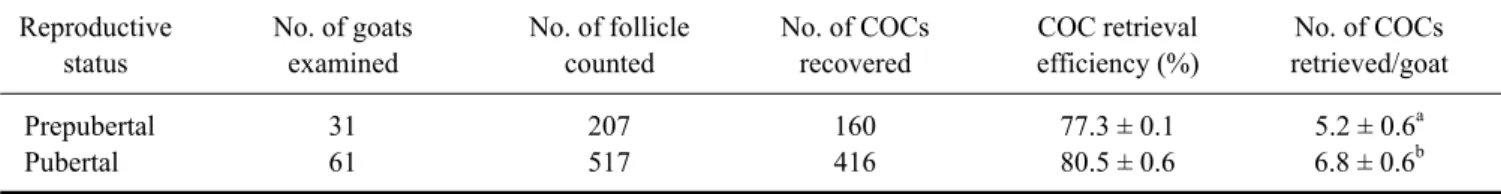
The number of oocytes retrieved from each prepubertal and pubertal goat was 5.2 ± 0.6 and 6.8 ± 0.6, respectively (Table 1). Among 62 ovaries from 31 prepubertal goats, 207 follicles were aspirated and 160 COCs were retrieved, giving an oocyte retrieval efficiency of 77.3 ± 0.1%. In contrast, 517 follicles were aspirated and 416 COCs were retrieved from 122 ovaries of 61 pubertal goats, giving an oocyte retrieval efficiency of 80.5 ± 0.5%.
Experiment 2
The results of the nuclear status of oocytes at the time of
collection are presented in Table 2. The percentages of
oocytes at GV stages did not different between prepubertal
and pubertal goats (63.6 ± 1.6 vs. 63.9 ± 1.2). Similarly, the
percentages of GVBD in the two groups did not differ
significantly (23.6 ± 1.1 vs. 27.7 ± 1.0, p > 0.05). GV with
Table 1. Oocyte recovery from black Bengal goats with respect to reproductive status
Reproductive No. of goats No. of follicle No. of COCs COC retrieval No. of COCs
status examined counted recovered efficiency (%) retrieved/goat
Prepubertal 31 207 160 77.3 ± 0.1 5.2 ± 0.6
aPubertal 61 517 416 80.5 ± 0.6 6.8 ± 0.6
ba,bValues within the same column did not differ significantly (p > 0.05). COCs: cumulus-oocyte complexes. Data represent mean ± SD.
Table 2. Nuclear status of immature oocytes of black Bengal goats with respect to reproductive status
Reproductive status No. of oocytes examined GV (%) GVBD (%) M I (%) AI-TI (%)
Prepubertal 65 63.6 ± 1.6 23.6 ± 1.1 5.5 ± 1.0 7.3 ± 0.8
bPubertal 173 63.9 ± 1.2 27.7 ± 1.0 5.8 ± 1.2 2.6 ± 0.2
aa,bValues within the same column differed significantly (p < 0.05). GV: germinal vesicle; GVBD: germinal vesicle breakdown; M I: metaphase I; AI-TI: anaphase I-telophase I. Data represent mean ± SD.
Fig. 1. Immature oocyte. (A) Germinal vesicle stage. (B) Germinal vesicle breakdown stage. 1% orcein stain and differential interference contrast (DIC) microscopy. ×400.
Fig. 2. (A) An in vitro matured oocyte with a polar body (black arrow) and metaphase II stage chromosomes (white arrow). (B) Normally fertilized oocyte. (C) Oocytes with one pronucle. (D) Polyspermic fertilization. 1% orcein stain and DIC microscopy,
×400.
a clear nuclear membrane and GVBD without a nuclear membrane are shown in Figs. 1A and B, respectively. The percentages of M I were 5.5 ± 1.0 and 5.8 ± 1.2, whereas the percentages of AI-TI were 7.3 ± 0.8 and 2.6 ± 0.2 in prepubertal and pubertal goats, respectively. The percentages of AI-TI oocytes in the two groups of goats differed significantly (p < 0.05).
Experiment 3
The rates of IVM are presented in Table 3. The percentage of mature oocytes was 60.3 ± 2.6 and 66.3 ± 8.4 in prepubertal and pubertal goats, respectively (p < 0.05). In contrast, the percentage of oocytes in the AI-TI/MI stage in pubertal goats was higher (19.5 ± 3.1 vs. 15.5 ± 0.6) than that of the prepubertal goat group, but this difference was not significant (p > 0.05). Although no significant differences were observed in the percentage of GV (5.0 ± 1.3 vs. 6.9 ± 1.5, p > 0.05) in pubertal and prepubertal goats, a significantly lower percentage of GVBD (9.3 ± 1.9 vs. 17.2 ± 0.5, p < 0.05) was observed in the pubertal goats
than in their prepubertal counterparts. An in vitro matured oocyte with a polar body and M II stage chromosome is shown in Fig. 2A.
Experiment 4
The rates of IVF following Percoll and swim-up separation
of spermatozoa are shown in the Table 4. The rates of single
pronucleus formation (46.9 ± 1.5 vs. 45.5 ± 1.3%), normal
Table 4. The effects of methods of sperm preparation on fertilization rates of pubertal black Bengal goat oocytes
Methods of No. of oocytes Percentage Percentage Percentage
sperm preparation examined of one pronulceus of two pronuclei of three pronuclei
Percoll 49 46.9 ± 1.5 (23) 36.7 ± 0.9 (18) 16.3 ± 0.9 (8)
Swim-up 55 45.5 ± 1.3 (25) 32.7 ± 1.3 (18) 21.8 ± 0.8 (12)
The values did not differ significantly between groups. Data represent mean ± SD (n).
Table 3. In vitro maturation rate of COCs of black Bengal goats with respect to reproductive status
Reproductive status No. of oocytes examined M II (%) AI-TI/M I (%) GVBD (%) GV (%)
Prepubertal 58 60.3 ± 2.6
a15.5 ± 0.6 17.2 ± 0.5
b6.9 ± 1.5
Pubertal 421 66.3 ± 8.4
b19.5 ± 3.1 9.3 ± 1.9
a5.0 ± 1.3
a,bValues within the same column differed significantly (p < 0.05). M II: metaphase II. Data represent mean ± SD.
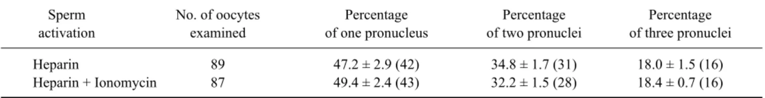
Table 5. The effects of sperm capacitation agents on fertilization rate of pubertal goat oocytes
Sperm No. of oocytes Percentage Percentage Percentage
activation examined of one pronucleus of two pronuclei of three pronuclei
Heparin 89 47.2 ± 2.9 (42) 34.8 ± 1.7 (31) 18.0 ± 1.5 (16)
Heparin + Ionomycin 87 49.4 ± 2.4 (43) 32.2 ± 1.5 (28) 18.4 ± 0.7 (16)
The values did not differ significantly between groups. Data represent mean ± SD (n).