201
Immune Network
Introduction
Prostaglandin E2 (PGE2), an arachidonic acid metabolite, exerts diverse effects on the immune response and the biological outcome in a variety of inflammatory diseases including rheumatoid arthritis (RA). It was initially believed to be a pro-inflamma- tory mediator causing vasodilation, hyperalgesia, and
fever (1,2), but has progressively been recognized for its immuno-regulatory and anti-inflammatory activ- ities (3-6). PGE2 dose-dependently inhibits IL-2 and IFN-γ production in stimulatedhuman peripheral blood lymphocytes, although IL-5 production is increased (3). These divergent actions of PGE2 on Th1 and Th2 cytokines were reproduced in other experiments reported by Katamura et al (4). They showed that nave CD4+ T cells from cord blood lost their ability to produce IFN-γ and IL-2 at the mRNA level after being treated with PGE2. PGE2 also strongly inhibited the LPS-induced production of IL-12, a critical cytokine of the Th1response (5). In contrast, PGE2 induced IL-10 approximately two fold in same condition. Wu et al also reported that
Prostaglandin E2 in Rheumatoid Synoviocytes
So-Youn Min, Young Ok Jung, Ju-Ho Do, So-Yang Kim, Jeong-Pyo Kim, Chul-Soo Cho, Wan-Uk Kim1
Department of Medicine, Division of Rheumatology, The Center for Rheumatic Diseases in Kang-Nam St. Mary's Hospital, 1St. Vincent Hospital, The Catholic University of Korea, Seoul, Korea
ABSTRACT
Objective: The role of prostaglandin E2 (PGE2) in the etiopathogenesis of immune and inflammatory diseases has become the subject of recent debate. To determine the role of PGE2 in rheumatoid arthritis (RA), we tested the effect of exogenous PGE2 on the production of cyclooxygenase-2 (COX-2) by rheumatoid synoviocytes. Methods:
Fibroblast-like synoviocytes (FLS) were prepared from the synovial tissues of RA patients, and cultured in the presence of PGE2. The COX-2 mRNA and protein expression levels were determined by RT-PCR and Western blot analysis, respectively.
The PGE2 receptor subtypes in the FLS were analyzed by RT-PCR. Electrophoretic mobility shift assay (EMSA) was used to measure the NF-κB binding activity for COX-2 transcription. The in vivoeffect of PGE2 on the development of arthritis was also tested in collagen induced arthritis (CIA) animals. Results: PGE2 (10-11 to 10-5 M) dose-dependently inhibited the expression of COX-2 mRNA and the COX-2 protein stimulated with IL-1β, but not COX-1 mRNA. NS-398, a selective COX-2 inhibitor, displayed an additive effect on PGE2-induced COX-2 downregulation. The FLS predominantly expressed the PGE2 receptor (EP) 2 and EP4, which mediated the COX-2 suppression by PGE2. Treatment with anti-IL-10 monoclonal antibodies partially reversed the PGE2-induced suppression of COX-2 mRNA, suggesting that IL-10 may be involved in modulating COX-2 by PGE2. Experiments using an inducer and an inhibitor of cyclic AMP (cAMP) suggest that cAMP is the major intracellular signal that mediates the regulatory effect of PGE2 on COX-2 expression. EMSA revealed that PGE2 inhibited the binding of NF-κB in the COX-2 promoter via a cAMP dependent pathway. In addition, a subcutaneous injection of PGE2 twice daily for 2 weeks significantly reduced the incidence and severity of CIA as well as the production of IgG antibodies to type II collagen. Conclusion: Our data suggest that overproduced PGE2 in the RA joints may function as an autocrine regulator of its own synthesis by inhibiting COX-2 production and may, in part, play an anti-inflammatory role in the arthritic joints. (Immune Network 2003;3(3):201-210)
Key Words: Prostaglandin E2, synoviocytes, COX-2, cAMP, NF-κB
Correspondence to: Dr. Wan-Uk Kim, Division of Rheumatology, Department of Internal Medicine, School of Medicine, The Catholic University of Korea, St. Vincent's Hospital, 93 Chi-dong, Suwon 442-060, Korea. (Fax) 82-31-253-8898, (E-mail) [email protected] Supported by a grant from the Ministry of Health and Welfare of the Republic of Korea (No 03-PJ1-PG10-20500-0021).
the surface expression of IL-12Rβ1, IL-12Rβ2 mRNA, and the receptor binding of IL-12 were diminished in PBMC after a treatment with PGE2 (6). This inhibitory effect of PGE2 is probably due to its cyclic AMP (cAMP) inducing capacity because other cAMP inducers or cAMP analogues mimic its action (4-6).
The regulatory function of PGE2 on the target cells is exerted through receptor-coupling events.
There are at least four subtypes of G-protein-coupled cell surface receptors, termed EP1, EP2, EP3, and EP4 receptors that bind (7,8). However, the conse- quence of PGE2 binding differs according to the receptor subtypes. For example, the EP3 receptor couples to Gi and inhibits adenylate cyclase. In contrast, EP2 or EP4 receptor couples to Gs and activates adenylate cyclase, resulting in an elevation of intracellular cAMP (9,10). Elevated cAMP subse- quently induces the transcriptional activation of anti- inflammatory cytokines such as IL-10 via the activa- tion of the cAMP responsive element-binding pro- teins (CREB) (11,12), whereas inhibits NF-κB-medi- ated transcription (13,14). It has been documented that PGE2 can block the DNA binding activity of NF-κB to the IL-2 transcriptional starting site (15).
The rate limiting step of PGE2 synthesis is cy- clooxygenase (COX) expression. This enzyme cata- lyzes the conversion of arachidonic acid to PGH2, a precursor of the biologically active prostaglandins.
Two COX isoforms have been described, COX-1 and COX-2. COX-1 is a constitutive isoform that is expressed in most human cells and tissues (16).
COX-2 is an inducible form of the COX enzyme, which is induced by stimulation by cytokines, mito- gens, and bacterial endotoxins (16-18). In particular, the influence of multiple cytokines on COX-2 expres- sion has been reported in many in vitro studies.
Proinflammatory cytokines including TNF-α (19,20), IL-1α (20), and IFN-γ (21) have been demonstrated to induce COX-2 expression, whereas anti-inflamma- tory cytokines such as IL-4 (22) and IL-10 (23) have been reported to inhibit COX-2 induction. Therefore, it is possible that COX-2 expression may be finely regulated by a number of cytokines within the RA joints. However, it is unclear whether PGE2, which is highly expressed in RA joints (24,25), can suppress COX-2 expression in RA as effective as it inhibit the production of Th1 and proinflammatory cytokines.
Here, it is shown that exogenous PGE2 strongly inhibits COX-2 expression in fibroblasts-like synov- iocytes (FLS) of RA via the EP2 and EP4 receptors.
The inhibition of COX-2 transcription appears to be affected by an increase in IL-10, at least in part. The effects of PGE2, including the reduced NF-κB binding activity to a COX-2 promoter, can be mimi-
cked by a cAMP inducer but completely abrogated by a cAMP inhibitor. Moreover, PGE injection re- duces the incidence and severity of collagen (CII)- induced arthritis (CIA). Collectively, these results suggest that PGE2 strongly inhibits NF-κB-mediated activation of COX-2 gene transcription in FLS via a cAMP-dependent pathway and has the potential to suppress RA inflammation.
Materials and Methods
Reagents and antibodies. PGE2 (Dinoprostone), 3-isobutyl- 1-methylxanthine (IBMX), and 2'-3'-dideoxyadenosine (DDA) were purchased from Sigma (St. Louis, MO).
The LPS was also obtained from Sigma. NS-398, a COX-2 inhibitor, was obtained from Calbiochem (La Jolla, CA). The EP2/EP4 agonist, 11-deoxy-PGE2, and the EP1/EP3 agonist, sulprostone, were pur- chased from Cayman Chemical (Ann Arbor, MI). The recombinant IL-1β and IL-10 were purchased from R & D systems (Minneapolis, MN), respectively. [γ-
32P] ATP was purchased from Amersham Pharmacia (Uppsala, Sweden). Reagents used for the culture contained <200 pg/ml of the endotoxin, as deter- mined by a Limulus amebocyte cell lysate assay.
Isolation of synoviocytes. The FLS cell lines were pre- pared from the synovial tissues of 6 RA patients undergoing a total joint replacement surgeryaccording to the procedure described previously (26). The mean age of the patients (5 females and 1 male) was 52.6 years. The mean disease duration was 81.4 months.
Five out of six patients had a positive rheumatoid factor. All had erosions as indicated by hand X-rays.
The cells between the forth and the eight passages were used, during which time they comprised a FLS homogenous population (<2.5% CD 14, <1% CD3, and<1% CD19 positive by flow cytometry analysis).
Culture conditions. The FLS cells were seeded in 24 well plates at a concentration of 6×10⁴ cells per well in 1 ml DMEM/5% FCS, and incubated at 37oC for 24 h in the presence of varying PGE2 concentrations, ranging from 10-11 to 10-5 M. To induce COX-2 synthesis in FLS, 10 ng/ml of the IL-β1 was added to the wells at the onset of culturing with PGE2. In some experiments, NS-398, a selective COX-2 inhibi- tor, was simultaneously treated to the wells in the presence of PGE2 and IL-1β. To investigate the effect of endogenous IL-10 on COX-2 expression, IL-10 was neutralized by adding monoclonal antibod- ies (mAbs) to IL-10. Various concentrations of a cAMP inducer, IBMX, were used to determine whether they could mimic the effect of PGE2. A cAMP inhibitor, DDA, was used to block cAMP accumulation. Both IBMX and DDA were added to each well at the onset of the culture. After 24 h of incubation, unless otherwise stated, the cell-free me-
dium was collected and stored at -20oC until assayed. All cultures were set up in either triplicate or quadruplicate.
RNA isolation and RT-PCR for COX-2 mRNA. The FLS were incubated with various concentrations of IL-1β. After 6 h of incubation, which was optimal for COX-2 induction, the mRNA was extracted using RNAzol B according to the manufacturer's instruc- tion (Biotex Laboratoryies, Houston, TX). The rever- se transcription of 5μg total mRNA was carried out at 42oC using the SuperscriptTM reverse transcription system (Life Technologies). PCR amplification of the cDNA aliquots was performed by adding 2.5 mM dNTPs, 2.5 U Taq DNA polymerase (TaKaRa SHUZO Co., Shiga, Japan) and 0.25μM each of the sense and antisense primers. The reaction was done in a PCR buffer (1.5 mM MgCl2, 50 mM KCl, 10 mM Tris HCl, pH 8.3) in a total volume of 25μl. The following sense and antisense primers for each molecules were used (all written in 5' → 3' direction): COX-2 sense GCAGTTGTTCCAGACAAGCA, COX-2 antisense C- AGGATACAGCTCCACAGCA; GAPDH sense CGAT- GCTGGGCGTGAGTAC, GAPDH antisense CGTT- CAGTCCA-GGGATGACC. The reactionswere proc- essed in a DNA thermal cycler (Perkin-Elmer, Foster City, CA) through cycles of 5 min denaturation at 94oC, 1 min annealing at 55oC for GAPDH and 45 seconds at 55oC for COX-2, and 30 seconds elonga- tion at 72oC. The PCR rounds were repeated for 25 cycles for GAPDH and 27 cycles for COX-2, which were determined to fall within the exponential phase of amplification for each molecule. PCR products were run on a 1.5% agarose gel and stained with eth- idium bromide. The mRNA expression level was presented as a ratio of the cytokine PCR product over the GAPDH product.
Western blot analysis for COX-2 protein. The FLS (5×
105 cells) were incubated for 12 h in the presence or absence of IL-β1. After incubation, the cells were harvested and lysed in 150μl of solubilization buffer (1% Tween 20, 10 mmol/L phenylmethylsulfonyl flu- oride, and 50 mmol/L Tris-HCl, pH 8.0). The pro- tein extracts (25μg) were then separated on a 12%
SDS-polyacrlamide gel electrophoresis (SDS-PAGE) and transferred onto a polyvinylidene difluoride (PVDF) membrane. The membrane was blocked with PBS containing 5% skim milk and 0.1% Tween 20, and then incubated with 0.25μg/ml of rabbit anti-human COX-2 polyclonal Abs in the blocking buffer at 25oC for 2 h. The membrane was subse- quently incubated with peroxidase- conjugate anti- rabbit IgG (1:1000 dilution) and analyzed using an Amersham enhanced chemiluminescence (ECL) sys- tem (Amersham, Arlington Heights, IL). Fuji X-omat AR film (Fuji Photi Film Co, Tokyo, Japan) with
cassette closure times of 5 to 10 minutes was used to obtain adequate exposure and to visualize the bands.
RT-PCR for subtypes of prostaglandin E2 receptors. The total RNA was extracted from synoviocytes of RA patients using the TRIzol reagent according to the manufacturer's instruction (Life Technologies). The RT-PCR was performed with 2μg of RNA in the first strand synthesis in a total volume of 20μl containing 80 pmol of the random hexamer primers (TaKaRa SHUZO Co.), 10 mM DTT solution, 0.5 mM dNTPs, 40 U of RNase inhibitor (TaKaRa SHUZOCo.), and 200 U of Superscript II reverse transcriptase (Invitrogen, Groningen, Netherland).
One microliter of the product was for PCR in a total volume of 25μl with 10 pmol each of the appropri- ate forward and reverse primers, 200μM dNTPs, and 0.5 U DNA Taq Polymerase (TaKaRa SHUZOCo.) in 10×buffer. The PCR amplification was carried out in a PCR thermal cycler (Perkin-Elmer Cetus) with an initial denaturation at 94oC for 10 minutes, follow- ed by 35 cycles of denaturation for 30 seconds at 94oC, annealing for 30 seconds at 56oC, and exten- sion time of 30 seconds at 72oC, with a final exten- sion time of 10 minutes at 72oC. The upstream and downstream primers (5`→3`) were TTGTCGGTAT- CATGGTGGTG and GGCCTCTGGTTGTGCTT- AGA for EP1, CAAGCCTGCTGTCTTGTGAT and TCCTCCCCCAGAAATAGGAT for EP2, AACTG- GGGCAACCTTTTCTT and TTTCTGCTTCTCC- GTGTGTG for EP3, and TGTGAACCCCATCC- TAGACC and GCACACCTGGAAGCAAATTC for EP4. The PCR products were analyzed on a 1.5%
agarose gel.
EMSA (Electrophoresis Mobility Shift Assay). The FLS nuclear extract was prepared as previously described (27). For the induction of NF-κB activity, the cells were pretreated with 10 ng/ml of IL-1β for 1 h in the presence or absence of PGE2. A double-stranded oligonucleotide probe containing the NF-κB recog- nition site (underlined) of human COX-2 promoter (5'-GGAGAGGGGATTCCCTGCGCC-3') was gen- erated by a 5'-end labeling of the sense strand with [γ -32P] dATP using T4 polynucleotide kinase (TaKaRa SHUZO Co.), and purified by NucTrap columns (Stratagene, La Jolla, CA). The NF-κB binding reactions were performed with 2μg of the nuclear extract in 10μl of the binding buffer containing 400,000 cpm of the labeled oligonucleotide for 30 min at room temperature. A 100×excess of the unlabeled oligonucleotide was used as a competitor.
The DNA-protein complex was analyzed on a 5%
polyacrylamide gel that was electrophoresed in a TBE buffer. The gels were then dried and exposed to BioMax-MR film (Eastman Kodak Co, Rochester, NY) at -70oC at 24 h.
The supershift assay was performed to verify the identity of the bound factors using specific antibodies to p65, p50, and c-Rel proteins. Briefly, 100× of Abs against p65, p50 and c-rel (Santa Cruz Laboratory, Santa Cruz CA) were added to the binding reaction prior to addition of the probe, and then incubated on ice for 30 min.
Determination of in vivo effect of PGE2 in collagen-induced arthritis. Male DBA/1 mice, which were obtained from Jackson Laboratories (Bar Harbor, ME) were maintained in groups of three to five in polycar- bonate cages and fed standard mouse chow (Ralston Purina, St. Louis, MO) and water ad libitum. The environment was made specifically pathogen-free for the mice. Neonatal mice were obtained by breeding the mice in our facility. The mice were immunized with native CII, a generous gift from Dr. Andrew H Kang (University of Tennessee, Memphis, TN), at 8 to 12 weeks of age. For the injection, ClI was dis- solved in 0.1 N acetic acid at 2 mg/ml, and emul- sified (1:1 ratio) with complete Freund's adjuvant (CFA) at 4oC (28). The mice received 0.1 ml of the emulsion containing 100μg of CII in the base of the tail as a primary immunization. Booster injections were given into the footpad with 50μg of CII similarly dissolved and emulsified 1:1 with CFA 14 days after the primary immunization. From 3 weeks after the primary immunization, 10 to 20μg of PGE2 dissolved in PBS was injected subcutaneously in the CIA animals twice daily during 2 weeks. The control mice received PBS alone. The incidence and severity of arthritis in the two groups of mice were deter- mined using a visual inspection. The mice were ob- served two to three times a week for the onset, duration, and severity of joint inflammation over a period of 10 weeks after the primary immunization.
Each limb was assessed on a 0- to 4-point scale, as described earlier (28). The hindfoot that received the booster immunization was excluded from the evalu- ation. Therefore, the maximum arthritis score possi- ble was 12. The mean arthritic index and incidence of arthritis (%) were used for data comparison among the experimental groups.
Assay for IgG antibodies to type II collagen. Sera were collected from each group of mice on day 35 after the primary immunization and stored at -20oC until the assay. The IgG anti-CII levels in the sera were determined by a commercial enzyme-linked immuno- sorbent assays kit (Chondrex, Redmond, WA), as previously described (28). The optical density of the standard serum, which was serially diluted 2-fold, is expressed as 100, 50, 25, 12.5, 6.25, 3.125 arbitrary units, respectively. The relationship of the optical density measured in the standard serum diluted serially and arbitrary units showed a good linear correlation in
all determinations (r>0.98, data not shown). The IgG anti-CII concentrations in the sera diluted 1:4,000 are presented as relative values (arbitrary units) compared to the optical density of the standard sera.
Statistical analysis. The data is expressed as a mean ± the standard deviation (SD). Statistical analysis was performed using the Student's t-test for matched pairs. Differences with a confidence level of 95% or higher were considered to be statistically significant (P<0.05).
Results
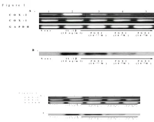
PGE2 inhibits IL-1-induced COX-2 expression in rheuma- toid synoviocytes. As shown in Fig. 1A, the unstimulated FLS expressed COX-2 mRNA slightly, which was strongly upregulated by 10 ng/ml of IL-1β. Incuba- tion of the cells with PGE2 (10-11 to 10-5 M) re- sulted in a dose-dependent decrease in IL-1β induced COX-2 mRNA expression to their spontaneous levels. The increase in the COX-2 protein levels induced by IL-1β was also dose- dependently inhibi-
Figure 1. Repression of IL-1β-induced cyclooxygenase 2 ex- pression by prostaglandin E2 (PGE2). (A) Messenger RNA ex- pression of the cyclooxygenases in the fibroblasts-like synovi- ocytes (FLS). The FLS were incubated with PGE2 in the presence or absence of IL-1β (10 ng/ml). After 6 h of incubation, which is optimal for COX-2, the FLS were analyzed for COX-2 mRNA expression by semi-quantitative RT-PCR. COX-2 mRNA expression after treatment without (lane 1) or with IL-1β (lane 2), IL-1β plus 10-11 M PGE2 (lane 3), IL-1β plus 10-8 M PGE2 (lane 4), IL-1β plus 10-5 M PGE2 (lane 5). COX-1 amplifica- tion was used as a negative control. The mRNA levels are correc- ted for the levels of the GAPDH mRNA signal. Representative data from three independent experiments is shown. (B) Western blot analysis of the COX-2 protein. FLS were incubated for 12 hr with various PGE2 concentrations in the presence of IL-1β (10 ng/ml). The whole cell lysate of the FLS (5×10⁵ cells) was subjected to immunoblot analysis using polyclonal antibodies against the purified COX-2 proteins. The other culture conditions were similar to those used for RT-PCR.
C O X- 2
C O X- 1
G AP D H
No n e I L - 1β
( 1 0 n g / ml ) P GE 2 P GE 2 P GE 2 ( 1 0- 1 1 M) ( 1 0- 8M ) ( 1 0- 5M )
B . A . F i g u r e 1
No n e I L - 1β
( 1 0 n g / ml ) P GE 2 P GE 2 P GE 2 ( 1 0- 1 1 M ) ( 1 0- 8M ) ( 1 0- 5M ) 1 2 3 4 5
CO X -2
CO X -1
G APD H
None IL-1β
(10 ng/ ml ) PG E2 PGE2 PGE2
(10-11 M ) (10-8M ) (10-5M)
B .
A .
Fi gure 1
None IL-1β
(10 ng/ m l ) PGE2 PG E2 PGE2
(10-11 M ) (10-8M ) (10-5M )
1 2 3 4 5
ted by the treatment with PGE2 (Fig. 1B). However, the COX-1 mRNA levels were not changed by either IL-1β or PGE2. These results suggest that PGE2 could regulate its own synthesis by blocking COX-2 expression.
It is well known that non-steroidal anti-inflam- matory drugs (NSAIDs) inhibit the COX-2 produc- tion, a key enzyme for the endogenousPGE2 func- tion. Moreover, NSAIDs such as aspirin and salicylic acid repress the NF-κB-responsive genes in mono- cytes (29). Assays performed in the presence of a COX-2 specific inhibitor NS-398 (1μM and 10μM), showed that this agent dose-dependently decreased COX-2 mRNA expression (Fig. 2A). Moreover, NS-398 potentiated the downregulation of COX-2 mRNA when added together with PGE2 (Fig. 2B). This suggests that the blockade of endogenous PGE2 by NSAIDs did not attenuatethe downregulatory effect of exogenous PGE2 on COX-2 expression.
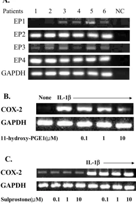
EP2 and EP4 receptors are involved in COX-2 inhibition in response to exogenous PGE2. PGE2 functions through the receptor-coupling events (7-10). To investigate which type of EP receptors predominantly mediates PGE2-induced COX-2 suppression, we examined the
mRNA expression of EP four subtypes (EP1, EP2, EP3, EP4) in the FLS obtained from 6 RA patients.
In all 6 RA patients, FLS strongly expressed two subtypes of the PGE2 receptor, EP2 and EP4, as revealed by RT-PCR (Fig. 3A). EP1 and EP3 were also detected in 4 to 5 FLS out of the 6 cell lines, but the banddensities of the PCR products were faint compared to the EP2 and EP4 under similarPCR conditions. Moreover, 1-hydroxy-PGE1 dose-depen- dently inhibited the COX-2 expression stimulated with IL-1β when the FLS were treated with 1-hy- droxy-PGE1, which has been shown to have high a affinity for EP2 and EP4. In contrast, sulprostone,
Figure 2. Effect of NS-398 on PGE2-mediated COX-2 regula- tion. (A) Dose dependent decrease in the COX-2 mRNA by NS-398. NS-398 (1 or 10μM) was added to fibroblasts-like synoviocytes (FLS) in the presence or absence of IL-1β (10 ng/ml). The COX-2 mRNA expression level was determined by semi-quantitative RT-PCR. (B) Additive effect of NS-398 on PGE2-mediated COX-2 down-regulation. Ten μM of NS-398 was simultaneously added to the FLS with 10-8 M or 10-5 M of PGE2 in the presence of IL-1β. The boxes indicate the co-treatment of NS-398 with PGE2.
N S 3 9 8 (μ Μ ) 1 1 0
C O X -2 G A P D H
N o n e IL -1β
A .
C O X - 2 G A P D H
N o n e IL -1β
P G E 2 (M ) 1 0- 8 1 0- 8 1 0-5 1 0- 5 N S 3 9 8 (μ Μ ) ( -) 1 0 ( -) 1 0
B .
Figure 3. Involvement of prostaglandin receptor (EP) 2 and EP4 subtypes in COX-2 inhibition in response to exogenous PGE2 (A) Analysis of the EP subtypes in the synovial fibroblasts. The expressionof the EP subtypes (EP1, EP2, EP3, and EP4) was determined by RT-PCR in the FLS from six different patients with rheumatoid arthritis. GAPDH amplification was used as internal control. NC indicates the negative control without cDNA. (B and C) Effect of hydroxy-PGE1 (EP2/EP4 agonist) and sulprostone (EP1/EP3 agonist) on the IL-1β-induced COX-2 expression.
Various concentrations of hydroxy-PGE1 and sulprostone, ran- ging from 0.1 to 1μM, were added to the FLS stimulated with 10 ng/ml of IL-1β. COX-2 mRNA expression was determ- inedby RT-PCR. Representative data from three independent experiments is shown.
EP1 EP2 EP3 EP4 GAPDH
Patients 1 2 3 4 5 6 NC
A.
COX-2 GAPDH
None IL-1β
11-hydroxy-PGE1(μM) 0.1 1 10
B.
Sulprostone(μM) 0.1 1 10 0.1 1 10 COX-2
GAPDH
IL-1β
C.
an EP1/EP3 agonist, had no significant effect on the basal and IL-1-β-induced COX-2 expression levels (Fig. 3B). These results suggest that the EP2 and EP4 receptor may be predominantly involved in COX-2 inhibition by exogenous PGE2.
Suppression of COX-2 by PGE2 was partially mediated by the increase of IL-10. IL-10 is a central regulator of COX-2 expression (30). We demonstrated previously that PGE2 increased IL-10 production from FLS.
Consequently, we tested whether or not increased IL-10 by PGE2 acted as an intermediate in COX-2 suppression. As shown in Fig. 4, incubation of FLS with neutralizing anti-IL-10 mAbs in the presence of 10-5 M of PGE2 resulted in the partial recovery of COX-2 mRNA stimulated by IL-1β. In this exper- iment, the equivalent concentration of the isotype control Abs (IgG1; R & D systems) did not show any significant effect (data not shown). Theses obser- vations indicate that the suppression of COX-2 by PGE2 is partially dependent on the enhancement of IL-10 production.
PGE2 inhibits NF-κB-mediated COX-2 transcription via a cAMP dependent pathway. The activation of adenylate cyclase and the resultant accumulation of cAMP are
downstream pathways activated by PGE2 receptor signaling (4-6). The agents known to enhance cAMP were analyzed to determine if they could mimic
Figure 4. IL-10 dependency of PGE2-mediated COX-2 suppre- ssion. FLS were cultured with 10-5 M of PGE2 in the presence or absence of neutralizing antibodies (Abs) to IL-10. COX-2 mRNA expression after treatment without (lane 1) or with IL-1β (lane 2), IL-1β plus 10-5 M of PGE2 (lane 3), and IL-1β plus 10-5 M of PGE2 in the presence of 10μg/ml of anti-IL-10 (lane 4). The levels of mRNA are expressed as the fold increase relative to the mRNA from the unstimulated control cells, and corrected for the levels ofthe GAPDH mRNA signal. The data represents the results from one of three similar experiments.
C O X -2
Relative level of COX-2 mRNA
0 5 1 0 1 5 2 0
2 5 a n ti-IL -1 0
N o n e IL -1β
P G E 2 (1 0-5M ) 1 2 3 4
G A P D H
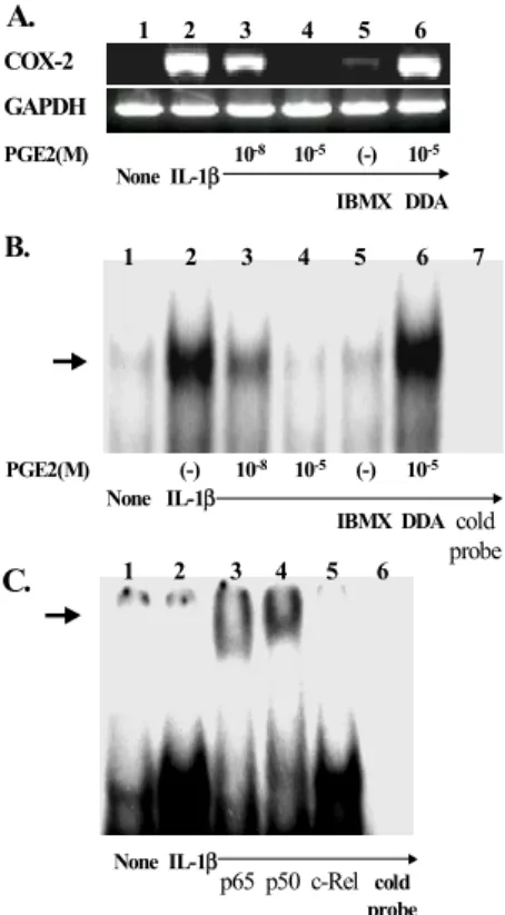
Figure 5. PGE2 inhibits NF-κB-mediated COX-2 transcription via a cAMP dependent pathway. The IL-1β-stimulated FLS were incubated with the cyclic AMP (cAMP) agonists, 3-isobutyl-1- methylxanthine (IBMX) or c-AMP antagonist, 2'-3'-dideoxya- denosine (DDA) with in the presence or absence of PGE2. (A) cAMP dependency of COX-2 mRNA suppression by PGE2.
COX-2mRNA expression after treatment without (lane 1) or with IL-1β (lane 2), IL-1β plus 10-8 M PGE2 (lane 3), IL-1β plus 10-5 M PGE2 (lane 4), IL-1β plus IBMX 500μM (lane 5), and IL-1β plus 10-5 M PGE2 in the presence of DDA 500μM (lane 6). (B) Effect of PGE2 and the cAMP inducer on NF-κB complex in the nuclear extracts of FLS. The cells were pretreated with 10 ng/ml of IL-1β for the induction of NF-κB activity for 1 h prior to the analysis. The NF-κB binding activity in the nuclear extract was determined by EMSA, as described in Material and Methods”. The abrogation of NF-κB activity by an excess of unlabeled oligonucleotide in the IL-1β-stimulated cells (lane 7), the NF-κB activity in the absence (lane 1) or presence IL-1β (lane 2), IL-1β plus 10-8 M PGE2 (lane 3), IL-1β plus 10-5 M PGE2 (lane 4), IL-1β plus IBMX 500μM (lane 5), and IL-1β plus 10-5 M PGE2 in the presence of DDA 500 μM (lane 6). The arrow on the left denotes the labeled oli- gonucleotide band shifted upon NF-κB binding. A representa- tive result from three independent experiments using the cell lines of different RA patients is shown. (C) Supershift assay of the NF- κB site using antibodies against p65 (lane 3), p50 (lane 4), and c-Rel (lane 5). The cells in all lanes, except lane 1 (medium alone), were pre-stimulated with IL-1β. The arrow on the left denotes the labeled oligonucleotide band shifted upon NF-κB binding.
A.
None IL-1β
p65 p50 c-Rel
C.
None IL-1β
PGE2(M) (-) 10-8 10-5 (-) 10-5
IBMX DDA
B. 1 2 3 4 5 6 7
cold probe
PGE2(M) 10-8 10-5 (-) 10-5
COX-2 GAPDH
None IL-1β
IBMX DDA 1 2 3 4 5 6
1 2 3 4 5 6
cold probe
PGE2 action on COX-2 expression. IBMX, a pho- sphodiesterase inhibitor, suppressed the expression of COX-2 mRNA stimulated by IL-1β, whereas DDA, an adenylate cyclase inhibitor, completely abrogated the effect of PGE2 (Fig. 5A), suggesting that the COX-2 regulation by PGE2 is mainly mediated via a cAMP-dependent pathway.
IL-1β has been associated with the induction of the nuclear factor-κB (NF-κB) and increased COX-2 transcription (31), on the basis of the existence of two putative NF-κB-binding motifs on human COX-2 gene (32).We tested if the accumulation of cAMP by PGE2 is related to the down-regulation of COX-2 promoter activation, by a mobility shift assay of the NF-κB site. Fig. 5B shows that when compared to unstimulated cells (lane 1), the incubation of the FLS with IL-1β (10 ng/ml) strongly induced the DNA-binding activity of NF-κB (lane 2). The shifting of the radiolabeled NF-κB element was
chased by a competition with an excess amount of the unlabeled oligonucleotide (lane 7). The addition of PGE2 to the IL-1β-stimulated FLS cultures resul- ted in a dose-dependent reduction in NF-κB binding (lane 3 and 4). Treatment with IBMX has a similar result to that of PGE2 10-5 M (lane 5), whereas DDA completely restored the NF-κB activity (lane 6). These results suggest that PGE2 inhibits COX-2 mRNA production via the elevation of cAMP, which in turn blocks NF-κB-signaling through the COX-2 promoter. A supershift assay using antibodies to p65, p50, and c-relwas performed to verify the identity of binding NF-κB isoforms (Fig. 5C). It appears that the factors binding to the oligonucleotide include p65 and p50, but not c-rel.
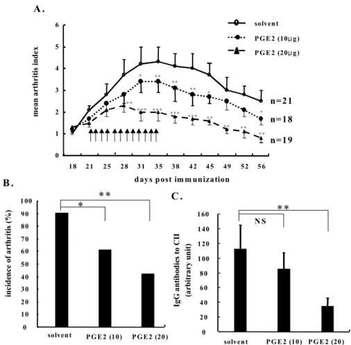
Suppression of collagen-induced arthritis by PGE2. Finally, we investigated the in vivo effect of exogenous PGE2 on the progression of arthritis. As shown in Fig. 6, a subcutaneous injection of PGE2 (10 to 20μg)twice
Figure 6. Suppression of collagen induced arthritis (CIA) by PGE2.
From 3 weeks after the primary immunization, 10 (closed circles) or 20 μg (closed triangle) of PGE2 dissolved in PBS was injected in the CIA animals twice daily for 2 weeks. The control mice (open cir- cles) were receivedPBS alone. The severity and incidence of arthritis were determined in the three groups of mice by a visual inspec- tion. The arrows denote a sub- cutaneous injection with PGE2 or the solvent. (A) Decrease in the arthritis index in the PGE2-injected mice during the course of CIA. The data is presented as the mean (±
SD) values. (B) Reduction in the incidence of arthritis in the PGE2- treated mice, as determined 5 weeks after immunization. (C) The levels of circulating IgG antibodies to CII in PGE2 versus the solvent-treated mice, as determined 5 weeks after primary immunization. Data are presented as the mean (±SD) titers of the circulating IgG anti-CII anti- bodies. *, P<0.05; **, P<0.01; ***, P<0.001 versus solvent alone.
0 10 20 30 40 50 60 70 80 90 100
solvent P G E 2 (10) P G E 2 (20)
incidence of arthritis (%) 160
0 20 40 60 80 100 120 140
solven t P G E 2 (10) P G E 2 (20) IgGantibodies to CII (arbitrary unit)
0 1 2 3 4 5 6
18 21 25 28 31 35 38 42 45 49 52 56
days post im m unization
mean arthritis index
solven t P G E 2 (10μ g) P G E 2 (20μ g)
n=21 n=18 n=19 A .
B .
C .
**
*** ***
*** *** **
** **
*
* **
**
** *
**
* **
N S
**
daily for 2 weeks reduced the incidence and severity of CIA in a dose-dependent manner (Fig. 6A and 6B). In addition, the serum levels of the IgG antibodies to CII were significantly lower in the mice treated with PGE2 than in the mice treated with PBS alone, as determined 5 weeks after a primary immun- ization (Fig. 6C). This clearly shows that exogenous PGE2 has strong anti-inflammatory activity in vivo.
On histological examination of the joints, it was also found that the paws of the PGE2-treated (20μg twice) mice exhibited a lower degree of inflammation, cartilage destruction, and synovial proliferation com- pared to the PBS-treated mice, as determined on day 35 after immunization (data not shown).
Discussion
Enhanced COX-2 production has been associated with the pathogenesis of RA (33-36). In RA synov- ium, both COX-1 and -2 are highly expressed, with COX-2 expression being elevated in relation to the de- gree of the inflammation in the synovial tissue (33,34).
Blocking COX-2 activity with a selective inhibitor pre- vented the progression of arthritis and the production of the pro-inflammatory cytokines such as TNF-α and IL-6, which are key mediators of rheumatoid inflammation (35). Recently, Myers et al. reported that CIA was prevented in genetically COX-2 depleted mice whereas COX-1 depleted mice could not be protected from developing CIA (36), thereby demonstrating the crucial role of COX-2 in RA inflammation.
We have previously demonstrated that PGE2 strong- ly suppresses IL-15 production, whereas it induces IL-10 production by FLS (37), suggesting that PGE2 may display anti-inflammatory activity. In this study, PGE2 dose dependently inhibited COX-2 mRNA and protein expression in FLS, an important enzyme in mediating autoimmune arthritis. The PGE2 concentration (10-5 M) at which regulatory effects were clearly evident is much higher than the tissue level found in RA (24,25). However, the action of PGE2 was also observed at concentrations as low as 10-8 M, which approximates the physiological concen- trationof PGE2 in RA joints. Furthermore, in an inflammatory response, PGE2 is mainly produced by monocytes and macrophages through the direct cell-to-cell contact. Therefore, it is possible that during the interaction of the macrophages with the synovial fibroblasts or T cells, sufficient amount of PGE2 to result in a functional modulation of FLS may be secreted in vivo, as shown in this paper.
A negative feedback exerted by the COX-2 end products on COX-2 expression itself may provide an important autocrine regulatory mechanism in several cell types (38-41). For example, in porcine aortic smooth-
muscle cells, endogenous and exogenous PGE2 inhibits COX-2 expression following activation by the fibroblast growth factor-2 (38). PGE2 also inhibits the LPS- or IL-1β-induced COX-2 expression in J774 macrop- hages and endothelial cells, respectively (39,40). The inhibition of COX-2 expression by 15-deoxy-PGJ2, a PGD2 metabolite, has been reported in FLS (41). In this respect, the results in this study support the negative feedback role of PGE2 in limiting COX-2 expression and thereby its own synthesis. Overall, overproduced PGE2 may have the potential to quench the ongoing inflammation within the joints by inhibiting COX-2 production and its end products mediating RA inflammation.
On the other hand, there are contrary reports showing PGE2 increasing COX-2 expression (42-44).
In the airway smooth-musclecells and microglial cells, PGE2 up-regulated COX-2 expression (42,43). More- over, in human FLS, PGE2 enhanced the COX-2 stability and translation (44). One potential explana- tion for this discrepancy would be the differential cellular effects of PGE2 according to the PGE2 receptor (EP) subtypes. For example, depending on the receptors, the consequence of ligand binding to these receptors can be increased cAMP, decreased cAMP, or a phosphoinositide response (7-10). In ad- dition, most experiments of the latter study (44) were performed in the presence of an endogenous PGE2 inhibitor, NS-398. In fact, NS-398 could alter the expression of the EP3 and EP4 receptors and thus change the sensitivities to exogenous PGE2 (45).
Therefore, the feedback modulation of COX-2 ex- pression by PGE2 appears to be influenced by mul- tiple factors including the cell types, the EP receptor subtypes, and the use of an endogenous COX-2 blocker.
In this study, all FLS cell lines obtained from the six differentpatients strongly expressed the EP2 and EP4 receptors relative to EP1 and EP3. An agonist of the EP2/EP4 receptor, 1-hydroxy-PGE1, mimick- ed the PGE2-mediated COX-2 down-regulation, whe- reas an EP1/EP3 agonist sulprostone did not. It is documented that PGE2 rapidly triggers cAMP for- mation in FLS (37). In this study, experiments using a cAMP analogue and inhibitor showed that the cAMP pathway was the major pathway responsible for the down-regulation of COX-2 by PGE2. To- gether, these data suggest that PGE2 may trigger cAMP accumulation through the activation of EP2/
EP4 receptor on FLS, which subsequently suppress IL-1β-stimulated COX-2 production. Again, this scenariois consistent with previous studies showing- that PGE2 blocked the production of pro-inflamma- tory cytokines and matrix metalloproteases in rheu- matoid synoviocytes via cAMP elevating mechanism
(37,46).
In RA synoviocytes, signaling via NF-κB is in- volved in regulating the COX-2 expression induced by IL-1β (47). In this study, both PGE2 and the cAMP inducer caused a dramatic reduction in the IL-1β-induced NF-κB-binding activity for COX-2 transcription, while a treatment with DDA comple- tely reversed the IL-1β-induced NF-κB-binding activity. These observations suggest that the down- regulation of the COX-2 gene by PGE2 may result from the inhibitory actions of cAMP on NF-κB activity. Considering that the anti-IL-10 Abs partially reversed the PGE2-mediated COX-2 down-regula- tion, PGE2 may regulate NF-κB activity through either indirect or direct means; e.g. the activation of intracellular cAMP signaling via the PGE2/PGE4 receptors, and its associated increase in IL-10, which is a well-known regulator of COX-2 expression and NF-κB activity (30,48). Still, it is possible thatthe signal transduction cascades after PGE2 stimulation may adopt different pathways for cAMP formation and IL-10 regulation.
It is quite evident that inducible COX-2 is a key enzyme that increases PGE2 in many inflammatory conditions and the selective COX-2 inhibitor is now an established treatment for RA. Nevertheless, the in vivonet effect of PGE2 on RA inflammation should be clarified because the role of PGE2 in the etiopath- ogenesis of RA has become the subject of recent debate. This study demonstrated that PGE2 dose- dependently reduced the incidence and severity of CIA and anti-CII Abs formation, which supports our data demonstrating the PGE2-mediated suppression of COX-2 and proinflammatory cytokines in the FLS (37). In this respect, overproduced PGE2 may play an anti-inflammatory role in vivo, rather than being a simple mediator of RA inflammation.
In conclusion, our data shows a novel mechanism for the action of PGE2 in RA in which PGE2 functions as an autocrine regulator of its own synthe- sis by inhibiting COX-2 production. PGE2 modula- tion of COX-2 production was mimicked by a EP2/
EP4 receptor agonist, and primarily mediated by a cAMP signal. Anti-inflammatory activity of PGE2 in RA was confirmed in animal models of RA. These results offer a new possibility of manipulating the function of the FLS by modulating the PGE2 recep- tors and cAMP.
Acknowledgements
The authors wish to acknowledge Dr. Ju-Young Kim for her advice on PGE2 receptor analysis.
References
1. Salmon JA, Higgs GA: Prostaglandins and leukotrienes as
inflammatory mediators. Br Med Bull 43;285-296, 1987.
2. Portanova JP, Zhang Y, Anderson GD, Hauser SD, Masferrer JL, Seibert K, et al: Selective neutralization of prostaglandin E2 blocks inflammation, hyperalgesia, and interleukin 6 production in vivo. J Exp Med 184;883-891, 1996
3. Snijdewint FG, Kalinski P, Wierenga EA, Bos JD, Kapsen- berg ML: Prostaglandin E2 differentially modulate cytokine secretion profiles of human T helper lymphocytes. J Imm- unol 150;5321-5329, 1993
4. Katamura K, Shintaku N, Yamauchi Y, Fukui T, Ohshima Y, Mayumi M, et al: Prostaglandin E2 at priming of naive CD4+ T cells inhibits acquisition of ability to produce IFN- γ and IL-2, but not IL-4 and IL-5. J Immunol 155;4604- 4612, 1995
5. van der Pouw Kraan TC, Boeije LC, Smeenk RJ, Wijdenes J, Aarden LA: Prostaglandin-E2 is a potent inhibitor of human interleukin 12 production. J Exp Med 181;775-779, 1995
6. Wu CY, Wang K, McDyer JF, Seder RA: Prostaglandin E2 and dexamethasone inhibit IL-12 receptor expression and IL-12 responsiveness. J Immunol 161;2723-2730, 1998 7. Coleman RA, Smith WL, Narumiya S: International Union
of Pharmacology classification of prostanoid receptors:
properties, distribution, and structure of the receptors and their subtypes. Pharmacol Rev 46;205-229, 1994
8. Negishi M, Sugimoto Y, Ichikawa A: Molecular mechanisms of diverse actions of prostanoid receptors. Biochim Biophys Acta 1259;109-119, 1995
9. Katsuyama M, Ikegami R, Karahashi H, Amano F, Sugimoto Y, Ichikawa A:Characterization of the LPS-stimulated expres- sion of EP2 and EP4 prostaglandin E receptors in mouse macrophage-like cell line, J774.1. Biochem Biophys Res Commun 251;727-731, 1998
10. Arakawa T, Laneuville O, Miller CA, Lakkides KM, Wingerd BA, DeWitt DL, et al: Prostanoid receptors of murine NIH 3T3 and RAW 264.7 cells. Structure and expression of the murine prostaglandin EP4 receptor gene. J Biol Chem 271;29569-29575, 1996
11. Eigler A, Siegmund B, Emmerich U, Baumann KH, Hartmann G, Endres S: Anti-inflammatory activities of cAMP-elevating agents: enhancement of IL-10 synthesis and concurrent suppres- sion of TNF production. J Leukoc Biol 63;101-107, 1998 12. Platzer C, Fritsch E, Elsner T, Lehmann MH, Volk HD,
Prosch S: Cyclic adenosine monophosphate-responsive ele- ments are involved in the transcriptional activation of the human IL-10 gene in monocytic cells. Eur J Immunol 29;
3098-3104, 1999
13. Ollivier V, Parry GC, CobbRR, de Prost D, Mackman N:
Elevated cyclic AMP inhibits NF-κB-mediated transcription in human monocytic cells and endothelial cells. J Biol Chem 271; 20828-20835, 1996
14. Parry GC, Mackman N: Role of cyclic AMP response ele- ment-binding protein in cyclic AMP inhibition of NF-κ B-mediated transcription. J Immunol 159;5450-5456, 1997 15. Chen D, Rothenberg EV: Interleukin 2 transcription factors
as molecular targets of cAMP inhibition: delayed inhibition kinetics and combinatorial transcription roles. J Exp Med 179;931-942, 1994
16. Smith WL, Garavito RM, DeWitt DL: Prostaglandin endope- roxide H synthases (cyclooxygenases)-1 and -2. J Biol Chem 271;33157-33160, 1996
17. Endo T, Ogushi F, Sone S: LPS-dependent cyclooxygenase-2 induction in human monocytes is down-regulated by IL-13, but not by IFN-γ. J Immunol 156;2240-2206, 1996 18. DuBois RN, Tsujii M, Bishop P, Awad JA, Makita K,
Lanahan A: Cloning and characterization of a growth factor-
inducible cyclooxygenase gene from rat intestinal epithelial cells. Am J Physiol 266;G822-827, 1994
19. Minghetti L, Walsh DT, Levi G, Perry VH: In vivo ex- pression of cyclooxygenase-2 in rat brain following in- traparenchymal injection of bacterial endotoxin and inflam- matory cytokines. J Neuropathol Exp Neurol 58;1184-1191, 1999
20. Vlahos R, Stewart A: Interleukin-1α and tumour necrosis factor-modulate airway smooth muscle DNA synthesis by induction of cyclooxygenase-2: inhibition by dexamethasone and fluticasone propionate. Br J Pharmacol 126;1315-1324, 1999
21. Barrios-Rodiles M, Chadee K: Novel regulation of cycloox- ygenase-2 expression and prostaglandin E2 production by IFN- γ in human macrophages. J Immunol 161;2441-2448, 1998 22. Niiro H, Otsuka T, Izuhara K, Yamaoka K, Ohshima K,
Tanabe T, et al: Regulation by interleukin-10 and interleu- kin-4 of cyclooxygenase-2 expression in human neutrophils.
Blood 89;1621-1628, 1997
23. Niiro H, Otsuka T, Tanabe T, Hara S, Kuga S, Nemoto Y, et al: Inhibition by interleukin-10 of inducible cyclooxygenase expression in lipopolysaccharide-stimulated monocytes: its underlying mechanism in comparison with interleukin-4.
Blood 85;3736-3745, 1995
24. Robinson DR, Dayer JM, Krane SM: Prostaglandins and their regulation in rheumatoid inflammation. Ann N Y Acad Sci 332;279-294, 1979
25. Salmon JA, Higgs GA, Vane JR, Bitensky L, Chayen J, Hen- derson B, et al: Synthesis of arachidonate cyclo-o xygenase products by rheumatoid and nonrheumatoid synovial lining in nonproliferative organ culture. Ann Rheum Dis 42;36-39, 1983 26. Cho ML, Kim WU, Min SY, Min DJ, Min JK, Lee SH, et
al: Cyclosporine differentially regulates interleukin-10, inte- rleukin-15, and tumor necrosis factor-α production by rheu- matoid synoviocytes. Arthritis Rheum 46;42-51, 2002 27. Jeong JY, Jue DM: Chloroquine inhibits processing of tumor
necrosis factor in lipopolysaccharide-stimulated RAW 264.7 macrophages. J Immunol 158;4901-4907, 1997
28. Kim WU, Lee WK, Ryoo JW, Kim SH, Kim J, Youn J, et al:
Suppression of collagen-induced arthritis by single adm- inistration of poly (lactic- co-glycolic acid) nanoparticles ent- rapping type II collagen: a novel treatment strategy for induc- tion of oral tolerance. Arthritis Rheum 46;1109-1120, 2002 29. Stevenson MA, Zhao MJ, Asea A, Coleman CN, Calderwood
SK: Salicylic acid and aspirin inhibit the activity of RSK2 kinase and repress RSK2- dependent transcription of cyclic AMP response element binding protein- and NF-κB-respon- sive genes. J Immunol 163;5608-5616, 1999
30. Berg DJ, Zhang J, Lauricella DM, Moore SA: IL-10 is a central regulator of cyclooxygenase-2 expression and prostaglandin production. J Immunol 166;2674-2680, 2001 31. Newton R, Kuitert LM, Bergmann M, Adcock IM, Barnes
PJ: Evidence for involvement of NF-κB in the transcrip- tional control of COX-2 gene expression by IL-1β. Biochem Biophys Res Commun 237;28-32, 1997
32. Appleby SB, Ristimaki A, Neilson K, Narko K, Hla T:
Structure of the human cyclo-oxygenase-2 gene. Biochem J 302;723-727, 1994
33. Crofford LJ, Wilder RL, Ristimaki AP, Sano H, Remmers EF, Epps HR, et al: Cyclooxygenase-1 and -2 expression in rheumatoid synovial tissues. Effects of interleukin-1β, phorbol ester, and corticosteroids. J Clin Invest 93;1095-1101, 1994 34. Siegle I, Klein T, Backman JT, Saal JG, Nusing RM, Fritz
P: Expression of cyclooxygenase 1 and cyclooxygenase 2 in human synovial tissue: differential elevation of cyclooxy-
genase 2 in inflammatory joint diseases. Arthritis Rheum 41;122-129, 1998
35. Anderson GD, Hauser SD, McGarity KL, Bremer ME, Isakson PC, Gregory SA: Selective inhibition of cyclooxy- genase (COX)-2 reverses inflammation and expression of COX-2 and interleukin 6 in rat adjuvant arthritis. J Clin Invest 97;2672-2679, 1996
36. Myers LK, Kang AH, Postlethwaite AE, Rosloniec EF, Morham SG, Shlopov BV, et al: The genetic ablation of cyclooxygenase 2 prevents the development of autoimmune arthritis. Arthritis Rheum 43;2687-2693, 2000
37. Min SY, Kim WU, Cho ML, Hwang SY, Park SH, Cho CS, et al: Prostaglandin E2 suppresses nuclear factor-κB mediated interleukin 15 production in rheumatoid synoviocy- tes. J Rheumatol 29;1366-1376, 2002
38. Karim S, Berrou E, Levy-Toledano S, Bryckaert M, Maclouf J: Regulatory role of prostaglandin E2 in induction of cyclo- oxygenase-2 by a thromboxane A2 analogue (U46619) and basic fibroblast growth factor in porcine aortic smooth- muscle cells. Biochem J 326;593-599, 1997
39. Pang L, Hoult JR: Repression of inducible nitric oxide syn- thaseand cyclooxygenase-2 by prostaglandin E2 and other cyclic AMP stimulants in J774 macrophages. Biochem Phar- macol 53;493-500, 1997
40. Akarasereenont P, Techatrisak K, Chotewuttakorn S, Tha- worn A: The induction of cyclooxygenase-2 in IL-1beta- trea- ted endothelial cells is inhibited by prostaglandin E2 through cAMP. Mediators Inflamm 8;287-294, 1999
41. Tsubouchi Y, Kawahito Y, Kohno M, Inoue K, Hla T, Sano H: Feedback control of the arachidonate cascade in rheu- matoid synoviocytes by 15-deoxy-Δ12,14-prostaglandin J2. Bio- chem Biophys Res Commun 283;750-755, 2001
42. Bonazzi A, Bolla M, Buccellati C, Hernandez A, Zarini S, Vigano T, et al: Effect of endogenous and exogenous prostaglandin E2 on interleukin- 1-induced cyclooxygenase-2 expression in human airway smooth-muscle cells. Am J Respir Crit Care Med 162;2272-2277, 2000
43. Minghetti L, Polazzi E, Nicolini A, Creminon C, Levi G:
Up-regulation of cyclooxygenase-2 expression in cultured microglia by prostaglandin E2, cyclic AMP and non-steroidal anti-inflammatory drugs. Eur J Neurosci 9;934-940, 1997 44. Faour WH, He Y, He QW, de Ladurantaye M, Quintero M,
Mancini A, et al: Prostaglandin E2 regulates the level and stability of cyclooxygenase-2 mRNA through activation of p38 mitogen-activated protein kinase in interleukin-1 beta-treated human synovial fibroblasts. J Biol Chem 276;
31720-31731, 2001
45. Nasrallah R, Laneuville O, Ferguson S, Hebert RL: Effect of COX-2 inhibitor NS-398 on expression of PGE2 receptor subtypes in M-1 mouse CCD cells. Am J Physiol Renal Phys- iol 281;F123-132, 2001
46. DiBattista JA, Martel-Pelletier J, Fujimoto N, Obata K, Zafarullah M, Pelletier JP: Prostaglandins E2 and E1 inhibit cytokine-induced metalloprotease expression in human sy- novial fibroblasts. Mediation by cyclic-AMP signaling pa- thway. Lab Invest 71:270-278, 1994
47. Crofford LJ, Tan B, McCarthy CJ, Hla T: Involvement of nuclear factor κB in the regulation of cyclooxygenase-2 ex- pression by interleukin-1 in rheumatoid synoviocytes. Ar- thritis Rheum 40;226-236, 1997
48. Schottelius AJ, Mayo MW, Sartor RB, Baldwin AS Jr:
Interleukin-10 signaling blocks inhibitor of κB kinase acti- vity and nuclear factor κB DNA binding. J Biol Chem 274;31868-31874, 1999