Infection &
Chemotherapy
http://dx.doi.org/10.3947/ic.2014.46.1.50 Infect Chemother 2014;46(1):50-53 pISSN 2093-2340 · eISSN 2092-6448
Received: May 28, 2013 Revised: July 11, 2013 Accepted: July 23, 2013 Corresponding Author : Won Suk Choi, MD, PhD
Division of Infectious Diseases, Department of Internal Medicine, Korea University Ansan Hospital, 123 Jeokgeum-ro, Danwon-gu, Ansan 152-703, Korea
Tel: +82-31-412-4271, Fax: +82-31-412-5984 E-mail: [email protected]
This is an Open Access article distributed under the terms of the Creative Commons Attribution Non-Commercial License (http://creativecommons.org/licenses/by-nc/3.0) which permits unrestricted non-commercial use, distribution, and repro- duction in any medium, provided the original work is properly cited.
Copyrights © 2014 by The Korean Society of Infectious Diseases | Korean Society for Chemotherapy
www.icjournal.org
Simultaneous Chylothorax and Chylous Ascites Due to Tuberculosis
Kyeong Jin Kim, Dae Won Park, and Won Suk Choi
Division of Infectious Diseases, Department of Internal Medicine, Korea University College of Medicine, Seoul, Korea
Chylothorax or chylous ascites is rare manifestation of tuberculosis. We report a case of simultaneous chylothorax and chylous ascites due to tuberculosis. A 17-year-old girl was admitted with fever, abdominal distention and dyspnea. Chest and abdominal computed tomography revealed bilateral pleural effusion, multifocal nodular consolidation on both lung fields and copious ascites and multiple necrotic lymphadenopathy in the abdominal cavity. Mycobacterium tuberculosis was isolated from sputum and pleu- ral fluid. The patient was treated with anti-tuberculosis medication. Pleural effusion and ascites improved with the medication.
Key Words: Chylothorax; Chylous ascites; Tuberculosis
Case Report
Introduction
Chylothorax and chylous ascites, which is characterized by the presence of chyle in the pleural and peritoneal cavities, is an uncommon condition [1, 2]. Chylothorax or chylous ascites result from trauma or a malignant condition such as lympho- ma. Tuberculosis (TB) can cause cylothorax or chylous ascites [3-5], but the concurrent occurrence of both manifestations associated with tuberculosis is extremely rare. We report a case of chylothorax and chylous ascites due to TB.
Case Report
A 17-year-old girl was admitted to the Korean University
Ansan Hospital with a one-month history of fever, abdominal distention and dyspnea. The patient had been treated in an- other hospital for 10 days but the cause of ascites and pleural effusion had not been discovered. She was transferred to our hospital for further care. On admission, body temperature was 38.6oC, heart rate was 146/min and body weight was 56 kg. On chest auscultation, breathing sound was decreased on both lower lung fields. Abdominal examination revealed shifting dullness and the lower extremities were severely edematous.
A chest radiograph taken on admission showed diffuse fine nodular opacities in both lungs and bilateral pleural effusion.
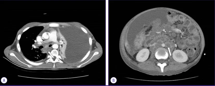
Chest computed tomography (CT) showed pleural effusion of both sides, more serious on the left side, and pericardial effu- sion and multifocal nodular consolidation on both lung fields (Fig. 1A). Abdominal CT showed a large amount of ascites
http://dx.doi.org/10.3947/ic.2014.46.1.50 • Infect Chemother 2014;46(1):50-53
www.icjournal.org 51
with diffuse peritoneal thickening and multiple necrotic lymphadenopathy in the abdominal cavity, and enhanced wall thickening in the distal ileum and ascending colon (Fig.
1B). Initial laboratory results were as follows: white blood cell count 6,670/mm3, hemoglobin 6.7 g/dL, serum total protein 4.6 g/dL and albumin 2.2 g/dL. Thoracentesis and paracente- sis were performed to verify the nature of fluid apparent in the chest and abdominal CT images.
Pleural fluid (Fig. 2A) was turbid and whitish-colored, with
pH 8.0, protein level 3.1 g/dL, lactate dehydrogenase of 5,855 IU/L, triglyceride 133 mg/dL and adenosine deaminase 121.6 IU/L. The lactate dehydrogenase ratio of pleural fluid versus serum was 15, consistent with exudate. Ascites (Fig. 2B) was milk-like and turbid in appearance, pH 8.0, albumin level 0.5 g/
dL, triglyceride 864 mg/dL and adenosine deaminase 14.8 IU/
L. We diagnosed chylothorax and chylous ascites because the triglyceride level of pleural fluid and ascites were higher than 110 mg/dL and 200 mg/dL, respectively. Direct smear of pleu- ral fluid and ascites for acid fast bacilli (AFB) was positive only in pleural fluid. So, finally we diagnosed the patient as having TB associated with chylothrax and chylous ascites on the basis of positive AFB stain and abdomen CT. Initially, the patient re- ceived anti-TB treatment with quadritherapy including isonia- zid 300 mg/day, rifampin 600 mg/day, pirazinamide 1,500 mg/
day and etambutol 1,200 mg/day. The patient also was put on a high protein and low fat diet with medium chain triglyceride.
Mycobacterium tuberculosis was cultured from sputum and pleural fluid on 28th day from admission and the isolate was susceptible to all anti-TB medications. The patient improved continuously after anti-tuberculosis medication, and pleural ef- fusion and ascites were slightly decreased on follow-up chest and abdomen X-ray. The patient was discharged on day 60 of hospitalization with ongoing anti-tuberculosis treatment.
Discussion
Chlyothorax and chylous ascites is characterized by chyle in Figure 1. Contrast-enhanced computed tomography (CT) scan images. (A) Chest CT scan showed left side dominant pleural effusion and multifocal mediastinal lymph node enlargement. (B) Abdominal CT scan showed large amount of ascites with diffuse peritoneal thickening and multiple necrotic lymphadenopathy in the abdomi- nal cavity suggesting tuberculous peritonitis with lymphadenopathy.
A B
Figure 2. Appearance of pleural fluid and ascites. (A) Pleural fluid drained by diagnostic thoracentesis, milk-like appearance. (B) Ascties drained by diagnos- tic paracentesis, whitish or opalescent looking rich in triglyceride.
B A
Kim KJ, et al. • Chylothorax and chylous ascites due to tuberculosis www.icjournal.org
52
the pleural and peritoneal cavities produced by obstruction and disruption of the lymphatic channel [2]. The reported in- cidence of the combined occurrence of chylothorax and chy- lous ascites has varied from 9% to 55% of chylous effusion [5, 6]. The etiologies of chylothorax and chylous ascites can be nontraumatic and traumatic. The most common cause of nontraumatic chylous effusion is a malignancy, such as lym- phoma or metastatic carcinoma [3, 4]. Other causes of non- traumatic chylous effusion include idiopathic, congenital anomaly, protein-losing enteropathy and TB [7, 8]. One case of systemic lupus erythematosus [7] and one case of Henoch- Schönlein purpura [9] accompanied by chylothorax and chy- lous ascites at a same time have been reported.
Our case did not have any other cause of chylothorax or chy- lous ascites except TB. The patient denied any history of trau- ma, chest and abdominal CT scan did not show any evidence of malignancy and repeated cytologic examinations of pleural effusion and ascites did not reveal malignant cells. Constric- tive pericarditis, possible cause of chylothorax and chylous as- cites, was not noted on the result of echocardiography. M. tu- berculosis was isolated from sputum and pleural fluid, but not from ascites. Despite the low adenosine deaminase level in ascites and a negative reaction for TB upon polymerase chain reaction examination, an abdominal CT scan showed multi- ple necrotic lymphadenopathy in the abdominal cavity, which was suggestive of TB infection. As a result, we diagnosed chy- lothoax and chylous ascites due to TB infection. The mecha- nism of TB for development of chylothorax and chylous asci- tes may be related to the enlarged lymph node obstructing lymphatic duct or direct invasion to lymphtic system inducing inflammation [10-13].
The mainstay of the treatment of chylothorax or chylous as- cites is conservative measures and correcting the underlying causes. The patient received anti-TB medication and nutrition replacement with high protein and low fat meal with medium chain triglycerides.
During the first two weeks after this therapy, the patient still complained of dyspnea and abdominal discomfort, and re- petitive paracentesis and thoracentesis was done to decom- press the pleural and peritoneal space. After one month, a fol- low-up chest X-ray revealed decreased pleural effusion and abdominal distension was also improved. Although some case reports have described the use of octreotide in the man- agement of chylothorax or chylous ascites [14-19], our patient improved with a regimen of anti-TB medication, diet control and supportive care; octreotide was not used.
The concurrent occurrence of both manifestations associat-
ed with tuberculosis is extremely rare. With considering high prevalence of TB in Korea, however, TB should be considered in the differential diagnosis of chylothorax and chylous ascites.
References
1. Cárdenas A, Chopra S. Chylous ascites. Am J Gastroenterol 2002;97:1896-900.
2. Aalami OO, Allen DB, Organ CH Jr. Chylous ascites: a col- lective review. Surgery 2000;128:761-78.
3. Valentine VG, Raffin TA. The management of chylothorax.
Chest 1992;102:586-91.
4. Press OW, Press NO, Kaufman SD. Evaluation and manage- ment of chylous ascites. Ann Intern Med 1982;96:358-64.
5. Romero S, Martín C, Hernandez L, Verdu J, Trigo C, Perez- Mateo M, Alemany L. Chylothorax in cirrhosis of the liver:
analysis of its frequency and clinical characteristics. Chest 1998;114:154-9.
6. Nix JT, Albert M, Dugas JE, Wendt DL. Chylothorax and chylous ascites; a study of 302 selected cases. Am J Gastro- enterol 1957;28:40-53; discussion, 53-5.
7. Lee CK, Han JM, Lee KN, Lee EY, Shin JH, Cho YS, Koh Y, Yoo B, Moon HB. Concurrent occurrence of chylothorax, chylous ascites, and protein-losing enteropathy in systemic lupus erythematosus. J Rheumatol 2002;29:1330-3.
8. Cakir E, Gocmen B, Uyan ZS, Oktem S, Kiyan G, Karakoc F, Ersu R, Karadag B, Dagli T, Dagli E. An unusual case of chy- lothorax complicating childhood tuberculosis. Pediatr Pul- monol 2008;43:611-4.
9. Lee TH, Lee EY, Cho YS, Yoo B, Moon HB, Lee CK. Concur- rent occurrence of chylothorax and chylous ascites in a pa- tient with Henoch-Sch?nlein purpura. Scand J Rheumatol 2003;32:378-9.
10. Doerr CH, Allen MS, Nichols FC 3rd, Ryu JH. Etiology of chylothorax in 203 patients. Mayo Clin Proc 2005;80:867-70.
11. Antón PA, Rubio J, Casán P, Franquet T. Chylothorax due to Mycobacterium tuberculosis. Thorax 1995;50:1019.
12. Vennera MC, Moreno R, Cot J, Marin A, Sanchez-Lloret J, Picado C, Agusti-Vidal A. Chylothorax and tuberculosis.
Thorax 1983;38:694-5.
13. Bond SJ, Guzzetta PC, Snyder ML, Randolph JG. Manage- ment of pediatric postoperative chylothorax. Ann Thorac Surg 1993;56:469-72; discussion 472-3.
14. Shah D, Sinn JK. Octreotide as therapeutic option for con- genital idiopathic chylothorax: a case series. Acta Paediatr 2012;101:e151-5.
http://dx.doi.org/10.3947/ic.2014.46.1.50 • Infect Chemother 2014;46(1):50-53
www.icjournal.org 53
15. Foo NH, Hwang YS, Lin CC, Tsai WH. Congenital chylotho- rax in a late preterm infant and successful treatment with octreotide. Pediatr Neonatol 2011;52:297-301.
16. Zhou DX, Zhou HB, Wang Q, Zou SS, Wang H, Hu HP. The effectiveness of the treatment of octreotide on chylous asci- tes after liver cirrhosis. Dig Dis Sci 2009;54:1783-8.
17. Ijichi H, Soejima Y, Taketomi A, Yoshizumi T, Uchiyama H, Harada N, Yonemura Y, Maehara Y. Successful manage- ment of chylous ascites after living donor liver transplanta-
tion with somatostatin. Liver Int 2008;28:143-5.
18. Baran M, Cakir M, Yüksekkaya HA, Arikan C, Aydin U, Ay- dogdu S, Kilic M. Chylous ascites after living related liver transplantation treated with somatostatin analog and par- enteral nutrition. Transplant Proc 2008;40:320-1.
19. Berzigotti A, Magalotti D, Cocci C, Angeloni L, Pironi L, Zoli M. Octreotide in the outpatient therapy of cirrhotic chylous ascites: a case report. Dig Liver Dis 2006;38:138-42.