Case Report pISSN 2383-9309❚eISSN 2383-9317 J Dent Anesth Pain Med 2021;21(3):261-268❚https://doi.org/10.17245/jdapm.2021.21.3.261
An anesthetic management of head and neck cancer reconstructive surgery in a patient having
hemophilia A: a case report
Seung-Hwa Ryoo
1, Dohyun Kwon
2, Jong-Ho Lee
2, Kwang-Suk Seo
1, Hyun Jeong Kim
1, Myong-Hwan Karm
11Department of Dental Anesthesiology, School of Dentistry, Seoul National University, Seoul, Republic of Korea
2Department of Oral and Maxillofacial Surgery, School of Dentistry, Seoul National University, Seoul, Republic of Korea
Hemophilia A is a hemorrhagic disease caused by coagulation factor VIII deficiency. In head and neck cancer surgery, especially during a reconstructive one, complications can occur. These include hematomas due to bleeding which can then lead to flap ischemia, necrosis, and impaired wound healing. There are fewer cases of reconstructive surgery in patients with hemophilia A. Here in we report, a reconstructive surgery that involved mass resection, partial glossectomy (right), selective neck dissection (right, Levels I, II, III, IV), and reconstruction at the lateral arm free flap (left) in a 25-year-old man with hemophilia A. The surgery was successfully performed without any complications after pretreatment with Factor VIII concentrate, which has not been reported earlier.
Keywords: Factor VIII; Hemophilia A; Reconstructive Surgical Procedures; Squamous Cell Carcinoma of Head and Neck.
This is an Open Access article distributed under the terms of the Creative Commons Attribution Non-Commercial License (http://creativecommons.org/licenses/by-nc/4.0/) which permits unrestricted non-commercial use, distribution, and reproduction in any medium, provided the original work is properly cited.
Received: April 27, 2021•Revised: May 18, 2021•Accepted: May 23, 2021
Corresponding Author: Myong-Hwan Karm, Department of Dental Anesthesiology, Seoul National University Dental Hospital, 101, Daehakro, Jongno-gu, Seoul 03080, Korea
Tel: +82-2-2072-3847 Fax: +82-2-766-9427 Email: [email protected] Copyrightⓒ 2021 Journal of Dental Anesthesia and Pain Medicine
INTRODUCTION
Hemophilia A, a congenital blood coagulation defi- ciency, is a hemorrhagic disease caused by factor VIII deficiency amongst other coagulation factors. Hemophilia A and B account for more than 95% of hemophilia cases, of which A accounts for nearly 80%. The incidence is approximately 1 in 5,000 males. Hemophilia A is six times more frequent than B [1]. In patients with hemophilia, symptoms vary depending on the degree of deficiency of blood coagulation factors. If dental procedure or surgery proceeds without treatment of hemophilia, it can lead to complications. As a result, the surgery may not be successful and chances of fatality can
increase [1,2].
To date, the most popular technique for head and neck reconstructive surgery is vascularized free flap tissue transfer. We performed partial glossectomy, selective neck dissection (SND) and reconstruction with a lateral arm free flap in a patient with hemophilia A. Herein, we present a case with a preoperative treatment approach, discuss the detailed procedure, and report the success of the surgery.
CASE REPORT
A 25-year-old male with American Society of Anesthesiologists class III physical status and body mass
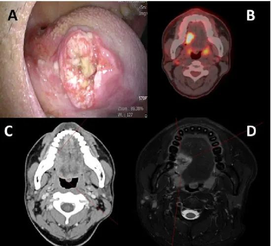
Fig. 1. Preoperative images of patient with hemophilia A. (A) Image of patient’s tongue lesion taken during first clinical visit. (B) Preoperative PET CT showing intense hypermetabolic lesion on right side of tongue, and suggestive of metastatic LNs at right level IIa. (C) Preoperative CT image showing the lesion on right tongue. (D) Preoperative MRI showing invasion of 19 mm depth. CT, computed tomography; MRI, magnetic resonance imaging; PET CT, positron emission tomography computed tomography; LN, lymph node.
index (height 167 cm, weight 65 kg) 23.3, was admitted for surgery for squamous cell carcinoma (SCC) of tongue.
The patient had been diagnosed with hemophilia 15 y ago. The previous medical history had no other abnormalities. Three months before being admitted, the patient had visited another hospital with complaints of intense pain on right tongue lesion. The patient had severe hemophilia A and required red blood cell transfusion because of bleeding after a swap test and incisional biopsy. The bleeding stopped within 30 min following an injection of factor VIII concentrate. Histological examination of the swab specimen revealed an atypical squamous cell carcinoma. The patient was then trans- ferred to the Oral and Maxillofacial Surgery Department, Seoul National University Dental Hospital for surgery.
On examination using CT and MRI, an ulcerative
lesion of 2.9 x 1.5 cm sized was seen on the right mid posterolateral border of tongue (Fig. 1). On clinical examination, the tumor showed endophytic growth, and the depth of the ulcer was >10 mm. Enhanced computational tomography (CT) image (Fig. 1) revealed the depth of invasion to be 19 mm, and a suspicious metastatic lymph node was observed on the right level IIa. Due to swab test, there was prolonged bleeding and oozing on the swab site. The Human papillomavirus (HPV) infection status was tested using intraoral mucosal swab and the result showed HPV16 (-), HPV 18 (-), other high risk (-). After the swab test at the outpatient department, as hemostasis was not achieved the patient was moved to the day-surgery unit. Hemostasis was then achieved by application of bipolar electrocauterization, SurgicelⓇ (Ethicon SARL, Puits-Godet 20, Swizerland)
Anesthetic management of head and neck cancer reconstructive surgery for hemophilia a patient
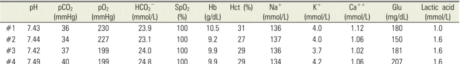
Table 1. Values of intraoperative arterial blood gas analysis
pH pCO2
(mmHg) pO2
(mmHg)
HCO3-
(mmol/L) SpO2
(%) Hb (g/dL)
Hct (%) Na+ (mmol/L)
K+ (mmol/L)
Ca++
(mmol/L) Glu (mg/dL)
Lactic acid (mmol/L)
#1 7.43 36 230 23.9 100 10.5 31 136 4.0 1.12 180 1.0
#2 7.44 34 227 23.1 100 9.2 27 137 4.0 1.06 150 1.6
#3 7.42 37 199 24.0 100 9.9 29 136 3.7 1.02 181 1.6
#4 7.49 40 199 24.8 100 9.9 29 134 4.2 1.06 207 1.6
The ABGA values were collected every 2 h during the operation. ABGA, arterial blood gas analysis; Ca, calcium; Hb, hemoglobin; HCO3-, bicarbonate;
Hct, hematocrit; K, potassium; Na, sodium; pCO2, partial pressure of carbon dioxide; pO2, partial pressure of oxygen; SpO2, arterial oxygen saturation.
(oxidized regenerated cellulose); gauze biting was also performed. After discharge, factor VIII concentrate injection was needed again due to the swab site bleeding.
The patient was scheduled to undergo partial glossec- tomy, selective neck dissection, and reconstruction with a lateral arm free flap.
A preoperative electrocardiogram (ECG) showed normal sinus rhythm and the chest radiography was unre- markable. Preoperative hemoglobin (Hb), hematocrit (Hct), and platelet count was 10.5 g/dl, 32.1, and 401
× 103/㎕ respectively. The coagulation profile showed that the prothrombin time (PT) international normalized ratio (INR) was 0.95, which was within the normal range of 0.8 to 1.2, and the activated partial thromboplastin time (aPTT) was 48.3 s, which was longer than the normal range of 26.7 to 36.6 s. The activity of coagulation factor VIII was reduced by 11%.
According to the answers of the consultation with the Hematology Department at Seoul National University Hospital, the patient was preoperatively injected with monoclonal antibody-purified factor VIII concentrate (GreenMonoⓇ, GC Pharma, Yongin, Republic of Korea, 2892 International Unit [IVS]) 1 h before the surgery.
Twelve hours after the initial injection, another injection of the same amount of GreenMonoⓇ was administered.
Thereafter, the injection was administered at every 12 h interval. Considering the extent of reconstructive surgery, the administration was recommended for 7 to 14 d so as to achieve sufficient hemostasis. In case of insufficient hemostasis after the injection of GreenMonoⓇ, 500 mg of tranexamic acid was recommended every 8 h.
General anesthesia was induced after routine monitoring (pulse oximetry, end-tidal CO2, ECG lead II with conti-
nuous ST-segment analysis, and noninvasive blood pressure monitoring) in the patient. His preoperative blood pressure (BP) was 121/65 mmHg, heart rate (HR) was 102 beats/min, body temperature was 36.1°C, and oxygen saturation (SpO2) was 100%. After sufficient preoxygenation, anesthesia was induced with lidocaine (30 mg), propofol (120 mg), and cisatracurium (16 mg).
Nasotracheal intubation with an endotracheal tube (right angle endotracheal tubes, cuffed, 7.0 mm) was performed using fiberoptic bronchoscopy. Anesthesia was maintained with an O2/air mixture (fraction of inspired oxygen 50%) and adjusted for desflurane (4.5-8% vol).
The patient was maintained on volume-controlled mechanical ventilation with a mean arterial pressure of 60-80 mmHg for a normal and regular heart rate. An additional neuromuscular blocker was administered, as needed. The tidal volume was 6-8 ml/kg, and positive end-expiratory pressure (5 cmH2O) was utilized. The respiratory rate was adjusted to maintain the partial pressure of end-tidal CO2 at 30-35 mmHg. Invasive BP monitoring and arterial blood gas analysis (ABGA) were performed with the catheter in the left dorsalis pedis artery.
ABGA was performed every two hours after the surgery began (Table 1).
Non-invasive continuous monitoring of total hemo- globin (SpHb; Masimo, Irvine, CA, USA) was also performed. Though this is not as accurate as the laboratory hemoglobin, it is used to identify relative numerical changes due to the serial measurements obtained. The initial value was 11.1, and 10.1 was the lowest value measured.
Central catheterization of the right femoral vein was performed to gain venous access because of the known
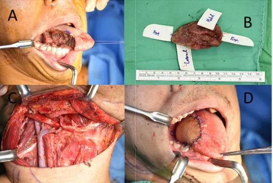
Fig. 2. Preoperative images of patient with hemophilia A. (A) Intraoperative image after right partial glossectomy. (B) Tumor specimen after resection.
(C) Reconstruction with lateral arm free flap. (D) Intraoperative image after completion of selective neck dissection, showing complete hemostasis.
bleeding tendency. During the reconstruction procedure, when the systolic blood pressure dropped to less than 120 mmHg, ephedrine 5-10 mg was injected twice, followed by dopamine, which was continuously injected for 275 min at a rate of 3.2-10 mcg/kg/min. This permitted maintaining the systolic blood pressure of 130-150 mmHg.
The routine surgical field was draped with povidone- iodine in the orofacial and neck areas. The tongue lesion was grossly marked with a surgical marking pen. Partial glossectomy was performed for a 1 cm safety margin (Figs. 2A and B). Muscular bleeding during partial glossectomy was controlled using bipolar electrocautery.
Frozen biopsies were performed around the marked margin. While waiting for the biopsy result, selective neck dissection was initiated. The internal jugular vein, carotid artery, vagus nerve, and spinal accessory nerves were isolated and preserved. Right levels I, II, III, and IV lymphatic chains were resected (Fig. 2C). Meticulous bleeding control was performed using electrocautery, vascular tying, and collagen sponge. The right side of the retromandibular vein branch and facial artery were
isolated for vessel anastomosis. After confirmation of negative frozen biopsy results, lateral arm fascio- cutaneous free flap harvesting was initiated. Skin incision and subcutaneous dissection were performed up to the junction of the radial collateral arteries. The branch and veins of the distal part of the posterior radial collateral artery were clamped and cut. The lateral arm fasciocutaneous free flap was transferred to the tongue to fit the defective region. The flap inset was closed with 4-0 Dafilon. Vessel anastomosis was performed using a microscope. The anastomosis of the posterior radial collateral artery with the facial artery and that of vena comitans with the retromandibular vein branch was performed via the end-to-end mode (Fig. 2D). All surgical sites were closed layer by layer, and the surgery was completed by suction drain and pressure dressing.
The surgery was uneventful. At the end of the surgery, 125 mg of methylprednisolone mixed with 50 ml of normal saline was injected. The total anesthesia time was 535 min, and the total surgery lasted for 500 min. During the operation, 5500 ml of crystalloid was administered and the total urine output was 2350 mL. The estimated
Anesthetic management of head and neck cancer reconstructive surgery for hemophilia a patient
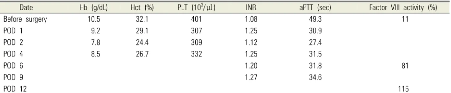
Table 2. Preoperative and postoperative laboratory data
Date Hb (g/dL) Hct (%) PLT (103/㎕) INR aPTT (sec) Factor VIII activity (%)
Before surgery 10.5 32.1 401 1.08 49.3 11
POD 1 9.2 29.1 307 1.25 30.9
POD 2 7.8 24.4 309 1.12 27.4
POD 4 8.5 26.7 332 1.25 31.5
POD 6 1.20 31.8 81
POD 9 1.27 34.6
POD 12 115
Normal range: Hb (13-17), Hct (39-52), PLT (130-400), INR (–0.8-1.2), aPTT (–26.7-36.6), and factor VIII activity (52-190). aPTT, activated partial thromboplastin time; Hb, hemoglobin; Hct, hematocrit; INR, international normalized ratio; PLT, platelet; POD, postoperative day.
blood loss was 400 ml. Following recovery at recover room for 40 minutes in the intubated state, the patient was transferred to the intensive care unit (ICU) and after 11 h, was transferred to the recovery room for extubation.
The oxygen was supplied as 5 L/min through the T-piece.
Oral and endotracheal tube suction was performed with routine monitoring. During the leakage test, the oxygen saturation was maintained at 99%. After extubation, the patient was monitored, and oxygen support by a reservoir bag mask was provided.
Monoclonal-antibody-purified Factor VIII Concentrate (GreenMonoⓇ) 6000IU (3000IU Bis in die, bid) was used on the day of surgery. From the first postoperative day (POD) and up to the next 15 days, 3000 U (1500 U bid, 8:30am, 8:30pm) were administered daily and then discontinued. On POD 18, a total of 1500 IU of GreenMonoⓇ was used before and after dental scaling,
#26 distal caries resin restoration, #28, and #38 surgical extraction. No additional hemostatic agents, such as tranexamic acid, were used. Following administration of GreenMonoⓇ after the surgery (Table 2), the aPTT level was rarely out of the normal range, and there were no bleeding-related symptoms or complications. The patient was discharged without any postoperative complications on POD 25.
DISCUSSION
The severity of symptoms associated with hemophilia depend on the deficiency degree of the clotting factor.
In case of mild deficiency (5%–40% of normal value), bleeding occurs mainly when there is a trauma, and is diagnosed since bleeding does not stop well after surgery or dental procedure. Thus, in case of such patients, the diagnosis is often delayed. Moderate deficiency (5%) is rare, but bleeding can occur without any cause, and starts even with a very small trauma. In case of severe deficiency (<1%), spontaneous bleeding is more common, enabling an earlier diagnosis (within a year after birth) [3,4]. Intracranial bleeding at the time of delivery, subcutaneous hematoma, intra-articular and muscle bleeding owing to movement around 12 months after birth, etc.
aid early diagnosis [4]. In addition, bleeding can occur within the brain, in the gastrointestinal tract, and in the urinary tract too. If the bleeding is recurrent, it results in severe pain even after a small wound or dental procedure, and the bleeding may not stop leading to dangerous complication. Hence, the prevention and treatment of severe hemorrhagic disease are very important [5-7].
In general, the goal of the treatment is to raise factor VIII concentrations to 80%–100% just before surgery, and a level of at least 50% should be maintained 5–14 d postoperatively [8,9]. Since the present case was a head and neck cancer reconstructive surgery, we set the target of factor VIII to 80%–100%.
Hemophilia can be diagnosed by measuring the level of coagulation factors through a detailed medical history and blood tests. In blood coagulation test, bleeding time, PT, and platelet count may be in the normal range, and only (aPTT) may show a delay depending on the degree
of deficiency [10-12]. Treatment involves transfusion of plasma containing insufficient clotting factors; at times desmopressin is used to treat mild symptoms [13]. It is recommended to diagnose hemophilia early, and prevent the damage of body function and minimize complications arising due to bleeding throughout life span. Hemophilia A is the most common type of hemophilia. In the present case study, the patient was already diagnosed with hemophilia A, and serum factor VIII activation was measured to be around 11%. Hence, in the present case, pre-surgical preparations were made to handle any complications arising due to moderate hemophilia [3,14,15].
When a patient with hemophilia has a lot of bleeding, and a lengthy operation such as cancer surgery is scheduled, preparations need to be made to take care of any situation before and after surgery. During the reconstructive procedure, declamping after arteriovenous anastomosis results in perfusion to the implanted site, and hypoperfusion can be prevented with appropriate anesthetic techniques. Therefore, in this case, we maintained normal body temperature, and provided adequate fluid therapy. We also used vasoactive drugs to promote vasodilation and adequate arterial pressure, which affected the flap blood flow thereby maintaining proper rate of blood flow [16]. Hemophilia may cause significant bleeding during surgery, which may become more severe in case blood coagulation factors get diluted due to the large amount of crystalloid and colloid solutions injected during surgery. As a result of hematoma , complications such as delayed healing and necrosis may occur due to compression of nerves and blood vessels. Thus, preparations are very important to prevent and control bleeding. In case of other surgical procedures for hemophilia patients, if several surgeries are to be performed together, there are cases where they are divided over different time spans. In a special situation such as cancer surgery, which has to be performed at once, we need to be more prepared for bleeding [17]. Partial glossectomy, SND, and recon- struction with lateral arm free flaps are the procedures
that are expected to have a high risk for hemophilia patients. As it is not a commonly performed procedure, only few have been reported.
<Preoperative treatment protocol for patients with hemophilia A>
One international unit (IU) of coagulation factor VIII per/kg of body weight can increase coagulation factor VIII activity by 2%. The required quantity of coagulation factor VIII was calculated as follows:
Factor VIII dose (IU) = desired factor activity (%) × weight (kg) × 0.5.
In this case, 2892.5 (IU) = (100% - 11%) × 65 (kg)
× 0.5.
Because of the wide dissection area, the dose administered given to the patient was calculated according to the medical hematology consult answer.
In adults, intravenous injection of factor VIII increases plasma levels by 2 IU/dl by 1 U/kg [3], with a half-life is 8-12 h. Dosage calculation can be performed at 12 h intervals, taking into account the patient's weight × target factor value × 0.5 half-life. The target factor value can be determined by considering the type of surgery and the expected bleeding level. In the presented case, we set the target value of the factor VIII to be close to 100%
[3,14,15,17]. We expected that the risk of intraoperative bleeding was higher than that of other surgeries considering the extent of the expected surgery. Hence appropriate procedures to minimize the amount of bleeding, and maintain adequate blood pressure (around 130-150 mmHg) were undertaken to ensure smooth arterial blood flow during and after the reconstructive surgery [16,18-21].
The surgery for head and neck cancer and recon- struction in hemophilia A patient is a situation in which specialists of various departments must cooperate to sufficiently prepare for the risk of bleeding and provide maximum safety. In this case, due to the use of factor VIII before surgery, the operation was uneventful and the patient did not require any blood transfusions either
Anesthetic management of head and neck cancer reconstructive surgery for hemophilia a patient
during or after the surgery.
AUTHOR ORCIDs
Seung-Hwa Ryoo: https://orcid.org/0000-0002-7442-8531 Dohyun Kwon: https://orcid.org/0000-0001-8450-5513 Jong-Ho Lee: https://orcid.org/0000-0002-8843-545X Kwang-Suk Seo: https://orcid.org/0000-0001-5906-0639 Hyun Jeong Kim: https://orcid.org/0000-0002-9265-7549 Myong-Hwan Karm: https://orcid.org/0000-0002-7494-4747
AUTHOR CONTRIBUTIONS
Seung-Hwa Ryoo: Writing – original draft Dohyun Kwon: Writing – original draft Jong-Ho Lee: Conceptualization Kwang-Suk Seo: Conceptualization Hyun Jeong Kim: Conceptualization
Myong-Hwan Karm: Writing – review & editing
DECLARATION OF CONFLICTS OF INTEREST: The authors declare no conflicts of interest.
CONSENT: The informed consent was obtained from the patient in this case report.
REFERENCES
1. Castaman G, Matino D. Hemophilia A and B: molecular and clinical similarities and differences. Haematologica 2019; 104: 1702-9.
2. Hoyer LW. Hemophilia a. N Engl J Med 1994; 330: 38-47.
3. Srivastava A, Santagostino E, Dougall A, Kitchen S, Sutherland M, Pipe SW, et al. WFH guidelines for the management of hemophilia, 3rd edition. Haemophilia 2020;
26: 1-158.
4. Shastry SP, Kaul R, Baroudi K, Umar D. Hemophilia A:
dental considerations and management. J Int Soc Prev Community Dent 2014; 4: S147-52.
5. Evatt BL. The natural evolution of haemophilia care:
developing and sustaining comprehensive care globally.
Haemophilia 2006; 12: 13-21.
6. Manco-Johnson MJ, Abshire TC, Shapiro AD, Riske B, Hacker MR, Kilcoyne R, et al. Prophylaxis versus episodic
treatment to prevent joint disease in boys with severe hemophilia. N Engl J Med 2007; 357: 535-44.
7. Gringeri A, Lundin B, von Mackensen S, Mantovani L, Mannucci PM. A randomized clinical trial of prophylaxis in children with hemophilia A (the esprit study). J Thromb Haemost 2011; 9: 700-10.
8. Zimmerman B, Valentino LA. Hemophilia: in review.
Pediatr Rev 2013; 34: 289-94.
9. Ljung RC, Knobe K. How to manage invasive procedures in children with haemophilia. Br J Haematol 2012; 157:
519-28.
10. Winter WE, Flax SD, Harris NS. Coagulation testing in the core laboratory. Lab Med 2017; 48: 295-313.
11. Mansouritorghabeh H. Clinical and laboratory approaches to hemophilia A. Iran J Med Sci 2015; 40: 194-205.
12. Peyvandi F, Kenet G, Pekrul I, Pruthi RK, Ramge P, Spannagl M. Laboratory testing in hemophilia: impact of factor and non-factor replacement therapy on coagulation assays. J Thromb Haemost 2020; 18: 1242-55.
13. Franchini M, Zaffanello M, Lippi G. The use of desmopressin in mild hemophilia A. Blood Coagul Fibrinolysis 2010; 21: 615-9.
14. White GC 2nd, Rosendaal F, Aledort LM, Lusher JM, Rothschild C, Ingerslev J. Definitions in hemophilia.
Recommendation of the scientific subcommittee on factor VIII and factor IX of the scientific and standardization committee of the international society on thrombosis and haemostasis. Thromb Haemost 2001; 85: 560.
15. Collins PW, Björkman S, Fischer K, Blanchette V, Oh M, Schroth P, et al. Factor VIII requirement to maintain a target plasma level in the prophylactic treatment of severe hemophilia A: influences of variance in pharmacokinetics and treatment regimens. J Thromb Haemost 2010; 8:
269-75.
16. Pereira CM, Figueiredo ME, Carvalho R, Catre D, Assunção JP. Anesthesia and surgical microvascular flaps.
Rev Bras Anestesiol 2012; 62: 563-79.
17. Pollei TR, Hinni ML, Moore EJ, Hayden RE, Olsen KD, Casler JD, et al. Analysis of postoperative bleeding and risk factors in transoral surgery of the oropharynx. JAMA Otolaryngol Head Neck Surg 2013; 139: 1212-8.
18. Kerawala CJ, Heliotos M. Prevention of complications in neck dissection. Head Neck Oncol 2009; 1: 35.
19. Malgonde M, Kumar M. Complications after neck dissection. Medical Journal of Dr DY Patil University 2015;
8: 458-62.
20. Laino L, Cicciù M, Fiorillo L, Crimi S, Bianchi A, Amoroso G, et al. Surgical risk on patients with coagulopathies:
guidelines on hemophiliac patients for oro-maxillofacial surgery. Int J Environ Res Public Health 2019; ; 16: 1386.
21. Wang KY, Yang KC, Su FY, Chen YC, Hsieh YH, Huang SL, et al. Association between blood pressure and postoperative hematomas in the patients undergoing head and neck cancer reconstruction. Head Neck 2019; 41:
3241-6.