Vol. 67, No. 3, March 2017, pp. 295∼300 http://dx.doi.org/10.3938/NPSM.67.295
Characterization of Iron-Oxide Nanoparticles Synthesized by Using the Sonochemical Method for Applications to Magnetic Hyperthermia
Hyewon Chung · Da-Ae Lee · Hongsub Bae · Ilsu Rhee
∗Department of Physics, Kyungpook National University, Daegu 41566, Korea
Sungwook Hong
Department of Physics Education, Daegu University, Gyeongsan 38453, Korea (Received 23 November 2016 : revised 21 December 2016 : accepted 23 December 2016)
Iron-oxide nanoparticles were synthesized by using the sonochemical method. Observation of the nanoparticles by using transmission electron microscopy revealed that they were spherical with an average diameter of 15 nm; further, the X-ray diffraction patterns of the nanoparticles showed that they possessed cubic spinel structures. The optimum concentration of the nanoparticles in an aqueous solution was found to be 0.62 mg/mL. The saturation temperature of this dispersion at a field strength of 5.5 kA/m and a frequency of 260 kHz was 42◦C, which is the desired temperature for applications of nanoparticles to magnetic hyperthermia. The field-strength dependence of the specific absorption rate (SAR) of the 2.88 mg/mL sample was also observed, and the SAR was found to depend on the square of the field intensity. These iron-oxide nanoparticles possessed all the physical properties suitable for their application to magnetic hyperthermia.
PACS numbers: 75.20.Ck, 81.05.-t, 61.46.Hk
Keywords: Iron-oxide nanoparticle, Sonochemical method, Magnetic hyperthermia
I. INTRODUCTION
Iron oxide nanoparticles are widely used in various fields, especially in medical applications such as targeted nano-drug delivery [1], MRI contrast agents [2–4], cell labeling [5], and magnetic hyperthermia [6]. The wide applicability of iron oxide is attributed to the facts that it is chemically stable and iron oxide nanoparticles can be easily synthesized by various methods such as the re- verse micelle method [7,8], coprecipitation [9,10], the Stöber method [11,12], and the sonochemical method [13]. Hyperthermia is one of the cancer therapies for killing cancer cells by heating them to up to 42◦C. This therapy is based on the fact that cancer cells are killed at the temperature of 42 ◦C whereas normal cells are able to sustain temperatures of up to 47◦C. The application of iron oxide nanoparticles to magnetic hyperthermia is
∗E-mail: [email protected]
based on the fact that magnetic nanoparticles can gener- ate heat by hysteresis and Brownian relaxation processes in an alternating magnetic field. At present, there is no way to administer the magnetic hyperthermia treatment directly to the human body. This is because difficulties faced in targeting nanoparticles only to the area with cancer cells and the effects of an alternating magnetic field on the human body have not yet been researched or understood thoroughly. Research works to overcome var- ious obstacles in the practical administration of magnetic hyperthermia therapy to the human body are currently in progress [14–17] via laboratory-based experiments and animal experimentation.
The sonochemical method is widely used to synthesize nanoparticles dispersed in liquid. The chemicals for the synthesis of nanoparticles and surfactants are mixed in liquid (water), and ultrasonic energy is applied to this mixture. The application of ultrasonic energy results in the formation of nanosized bubbles in which a very
This is an Open Access article distributed under the terms of the Creative Commons Attribution Non-Commercial License (http://creativecommons.org/licenses/by-nc/3.0) which permits unrestricted non-commercial use, distribution, and reproduction in any medium, provided the original work is properly cited.
system. The dependence of the saturation temperature on both the concentration of nanoparticles in aqueous solution and the field strength was intensively examined to obtain the optimal values of these parameters for prac- tical application of iron oxide nanoparticles to magnetic hyperthermia.
II. EXPERIMENTAL
First, 250 mL of distilled water was placed in a beaker and deoxygenated by bubbling argon gas through it for 30 min. Afterward, 1.90 g of FeCl2·4H2O and 4.87 g of FeCl3·6H2O were added to the beaker. The pH of the mixture was fixed at 7 by adding HCl solution. The con- tent was then sonicated with a high-density ultrasonic probe (CV3340, SONICS) at maximum power for 2 h.
Argon gas was bubbled through the mixture throughout the sonication process. The color of the mixture changed from milky to dark brown to black as the synthesis of the nanoparticles ended. After sonication, the beaker was placed on a permanent magnet and the content was al- lowed to cool to room temperature. After cooling of the content, the supernatant was removed and the particles were washed with acetone, alcohol, and distilled water in sequence. These washing processes were repeated until the supernatant was clear. Then, the particles were sep- arated using a centrifuge (CF-10, Wisd) at 13,000 rpm.
Finally, the particles were dried in an oven to obtain their powder sample for characterization.
The nanoparticles were characterized by TEM (H- 7600, Hitachi, Ltd.) and XRD (X’ pert PRO, PANa- lytical). The crystalline structure, shape, and size dis- tribution of the nanoparticles were observed by these
eral dispersion samples with different concentrations of nanoparticles were prepared by diluting the aqueous so- lution. The concentration of nanoparticles in the disper- sion was measured using an ICP spectrometer (IRISAP, Thermo Jarrell Ash). The heating effect of nanoparticles in the aqueous sample was observed using an induction heating system (OSH-120, Osung Tech). The tempera- ture of the sample was measured with an infrared ther- mometer (Pyro USB CF, Calex Electronics, Ltd.).
III. RESULTS AND DISCUSSION
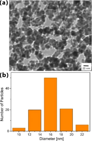
The TEM image and size distribution of iron oxide nanoparticles are shown in Fig. 1. As shown in Fig. 1(a), iron oxide nanoparticles are spherical in shape, with an average diameter of 15 nm. Fig. 1(b) shows the size distribution of 100 nanoparticles out of those observed in the TEM image in Fig. 1(a).
The XRD patterns of the powder sample of the iron ox- ide nanoparticles are shown in Fig. 2. The lattice indices for the peaks in this figure correspond to those of an in- verse spinel structure, which is one of the characteristics of iron oxide. Most ferrites have the chemical formula AB2O4, where A and B represent various metal cations and typically include those of iron. Ferrites usually have cubic close-packed oxides (O2−), with A cations occu- pying one-eighth of the tetrahedral sites and B cations occupying one-half of the octahedral sites; this is termed a spinel structure. On the other hand, if one-eighth of the tetrahedral sites are occupied by B cations, then one- fourth of the octahedral sites are occupied by A cations and the remaining one-fourth of the octahedral sites are occupied by B cations; this is termed the inverse spinel
Fig. 1. (Color online) (a) TEM image of iron oxide nanoparticles, (b) size distribution of 100 particles out of those observed in TEM image.
structure. Iron oxide (FeFe2O4 or Fe3O4) has the in- verse spinel structure [13]. The XRD data showed all lattice indices identical to those of iron oxide (JCPDS No. 01-089-2854). The lattice constant of the iron oxide nanoparticles was determined to be 8.34 Å from the data for the (440) peak.
The hysteresis of the iron oxide nanoparticles was ob- served using a VSM to confirm their superparamagnetic behavior. Fig. 3 shows the hysteresis curve, which re- veals a negligible coercive force; this is evidence of the superparamagnetic behavior of the nanoparticles.
When a magnetic system is subjected to an alternat- ing magnetic field, heat is generated owing to loss mech- anisms, which can be classified as hysteresis loss and re- laxation loss [6,15]. The latter can be further divided into Néel and Brown losses. Since our superparamag- netic nanoparticles show negligible hysteresis, we can ig- nore the hysteresis losses. The ferromagnetic resonance loss can also be neglected in the present study, because
Fig. 2. (Color online) XRD patterns of powder sample of iron oxide nanoparticles.
Fig. 3. (Color online) Hysteresis curve of nanoparticles at room temperature. The inset shows that there is a negligible coercive force, which confirms the superpara- magnetic behavior of the nanoparticles.
it occurs only in the gigahertz frequency range, which is much higher than the range of 200 – 300 kHz used in this study. Thus, the remaining heating mechanisms for our nanoparticles are the Néel loss and the Brown loss. We estimated the background heating effects due to pure water and the sample container and observed that the effect of these backgrounds was negligible.
The heat generation ability of iron oxide nanoparticles in liquid (water) was tested using an induction heating system, which is described elsewhere [15]. The powder sample of the nanoparticles was dispersed in distilled wa- ter and then diluted to obtain dispersion samples with five different concentrations of nanoparticles. The con- centration of nanoparticles in water was measured using
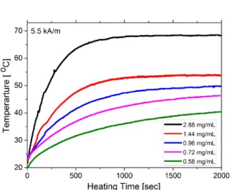
Fig. 4. (Color online) Temperature rise as a function of heating time for five samples of different particle concen- tration in the dispersion.
an ICP spectrometer. The iron concentrations in the five samples were 2.88, 1.44, 0.96, 0.72, and 0.58 mg/mL.
The dispersion sample was placed inside an induction coil to apply the alternating magnetic field to it. The field strength was 5.5 kA/m at a frequency of 260 kHz.
The temperature of the sample was measured with an infrared thermometer. Fig. 4 shows the temperature rise of the 2.88, 1.44, 0.96, 0.72, and 0.58 mg/mL dispersion samples as a function of heating time. The tempera- ture of the 2.88, 1.44, and 0.95 samples reached 42 ◦C within 200 – 500 s after the application of the alternat- ing magnetic field. The saturation temperatures of these three samples were 68, 54 and 49◦C, respectively. The 0.72 mg/mL sample showed slower temperature rise than above three samples, but crossed 42◦C at around 1,000 s and finally reached to the saturation temperature of 46 ◦C. On the other hand, the temperature of the 0.58 mg/mL sample did not reach 42◦C which is the desired temperature for hyperthermia therapy; instead, the sat- uration temperature of this sample was merely 39 ◦C.
This reveals that the concentration of nanoparticles in the 0.58 mg/mL sample is insufficient for employing it in hyperthermia. In contrast to this, the concentrations of nanoparticles in the other three samples were too high to employ them in hyperthermia. The optimum concen- tration of nanoparticles to heat the dispersion to up to 42 ◦C could be obtained from the graph of the concen- tration versus saturation temperature, which is depicted in Fig. 5. From this figure, the optimum concentration was determined to be about 0.62 mg/mL.
Fig. 5. (Color online) Dependences of saturation tem- perature and SAR on concentration of nanoparticles in aqueous solution.
The iron oxide nanoparticles in the alternating mag- netic field generate heat by hysteresis and Brownian re- laxation processes to increase the temperature of all con- stituents in the aqueous sample. This is expressed as
∆Q = mwcw∆T + mF ecF e∆T (1)
where cw and cF e are the specific heats of water and iron, respectively, and mw and mF e are the masses of water and iron, respectively. The specific absorption rate (SAR) is a measure of the heat generation ability of magnetic nanoparticles, which is defined as the heat generated in a unit time by a unit mass of iron. Thus, the SAR is expressed as
SAR = ∆Q/∆t mF e ≈ cw
[mw
mF e ]∆T
∆t, (2)
where cw = 4.2 J/g◦C and cF e = 0.45 J/g◦C. In ob- taining the last expression in Eq. (2), the second term in Eq. (1) was ignored, since the specific heat of water is much higher than that of iron and also since the quantity of iron in the sample is much smaller than that of water.
The rate of temperature rise in the first 100 s was used for estimating ∆T /∆t. The concentration dependence of the SAR is also depicted in Fig. 5. The SAR of our bare iron oxide nanoparticles has the value in the order of 100 comparable to others’ results [18,19]. The decrease in SAR with increasing particle concentration is due to the increase in the dipolar magnetic moment according to the increase in particle concentration, which affects the Néel relaxation time.
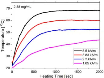
Fig. 6. (Color online) Magnetic heating curves for the 2.88 mg/mL sample at various field strengths.
The power loss density of nanoparticles is expressed as [20,21]
P = 1
2µoχ′′ωHo2 (3) where Ho and ω are the strength and frequency, respec- tively, of the alternating magnetic field and χ′′ is the magnetic susceptibility of nanoparticles, which is not re- lated to the field strength but is rather dependent on the relaxation times of nanoparticles. The SAR is di- rectly dependent on the power loss density of magnetic nanoparticles and is thus proportional to the square of the magnetic field strength, as shown in Eq. (3). The field strength dependence of the magnetic heating for the 2.88 mg/mL sample is shown in Fig. 6. From these data, we obtained the field dependence of the heating effect; the plot of the SAR versus the square of the field strength is shown in Fig. 7. The linear dependence of the SAR on the square of the field strength as given in Eq. (3) can be confirmed from this figure.
IV. CONCLUSION
Iron oxide nanoparticles were synthesized by the sono- chemical method. The particles were spherical in shape, with an average diameter of 15 nm; they also showed the inverse spinel crystal structure. The heat generation ability of nanoparticles dispersed in water was confirmed using an induction heating system. Nanoparticles in an alternating magnetic field generated heat by relaxation
Fig. 7. (Color online) Dependence of SAR of the 2.88 mg/mL sample on square of field strength.
processes to increase the temperature of the constituents of an aqueous solution of nanoparticles. The concentra- tion dependence of the heating effect was tested. The heating effect was also found to be influenced by the intensity of the magnetic field. The optimum concentra- tion of nanoparticles in the dispersion was determined to be 0.62 mg/mL. For this concentration, the saturation temperature of the aqueous sample at a field strength of 5.5 kA/m was 42 ◦C, which is the desired tempera- ture for magnetic hyperthermia. We also observed the field strength dependence of the heating effect and SAR for the 2.88 mg/mL sample. The SAR was observed to be linearly dependent on the square of the magnetic field intensity. Judging from these physical properties of the synthesized nanoparticles, we can conclude that iron oxide nanoparticles synthesized by the sonochemi- cal method can be applied to magnetic hyperthermia.
ACKNOWLEDGEMENTS
This work was supported by the National Research Foundation of Korea (2016R1A2B1006449).
REFERENCES
[1] M. Liong, J. Lu, M. Kovochih, T. Xia and S. G Ruehm et al., ACS Nano 2, 889 (2008).
[2] L. Li, W. Jiang, K. Luo, H. Song and F. Lan et al., Theranostics 3, 595 (2013).
Coll. Interface Sci. 128, 5 (2006).
[8] T. Ahmad, H. Bae, I. Rhee, Y. Chang and J. Lee et al., Curr. Appl. Phys. 12, 969 (2012).
[9] S. Hong and I. Rhee, Int. J. MRI 1, 15 (2007).
[10] I. Rhee and C. Kim, J. Magn. Magn. Mater. 261, 410 (2003).
[11] Y. Iqbal, I. Rhee, H. Bae, J. Kim and J. Jang et al., New Phys.: Sae Mulli 64, 339 (2014).
[12] L. M. Rossi, L. Shi, F. H. Quina and Z. Rosenzweig, Langmuir 21, 4277 (2005).
[13] J. H. Bang and K. S. Suslick, Adv. Mater. 22, 1039 (2010).
et al., Nano. Res. Lett. 6, 383 (2011).
[19] E. Garaio, J. M. Collantes, J. A. Garcia, S. Morner and F. Couill et al., J. Magn. Magn. Mater. 368, 432 (2014).
[20] T. Ahmad, H. Bae, Y. Iqbal, I. Rhee and S. Hong et al., J. Magn. Magn. Mater. 381, 151 (2015).
[21] F. Shubitidze, K. Kekalo, R. Stigliano and I. Baker, J. Appl. Phys. 117, 094302 (2015).
[22] R. Hergt, S. Dutz, R. Müller and M. Zeisberger, J.
Phys.: Condens. Matter 18, S2919 (2006).