Prepancreatic postduodenal portal vein(PPPV)은 1972년 에 Brook와 Gardner(1)에 의해서 처음으로 보고된 이후, 지 금까지 10예 미만이 보고된 매우 드문 문맥기형이다(2). 이러 한 문맥기형의 영상소견을 인지하고 있는 것은 수술이나 문맥 계 중재시술등에서 발생될 수 있는 원치 않는 문맥결찰이나 절 단, 출혈 등의 합병증을 예방하는데 중요하다. 2일간의 우상복 부 통증을 주소로 내원한 28세 여자환자가 담낭의 선근종증 (adenomyomatosis)으로 담낭적출술 시행하기전 시행된 CT에 서 췌장두부의 앞쪽과 십이지장의 뒤쪽으로 지나는 비정상적 인 형태의 문맥을 관찰할 수 있었다. 그리고 이어서 시행한 문 맥조영술상에서 omega 또는 arc 형태의 문맥기형의 소견을 보 여 PPPV로 진단하였으며 문헌고찰과 함께 보고한다.
증례 보고
28세 여자환자로 2일 동안의 상복부 동통을 주소로 외부병 원 방문하여 시행한 초음파에서 담낭의 선근종증이 의심되어 본원으로 전원되었다. 내원당시 이하학적 검사상에서 경증의 우상복부 압통이 관찰되었으며 과거력상 특이 소견은 없었다.
검사실 소견에서 혈색소가 11.8 g/dl, 백혈구수는 5800 103/uL, 혈소판은 249000 103/uL으로 정상이었으며 간기능 검사상에 서도 비정상적인 소견은 없었다. 본원에서 전산화단층촬영(Mx 8000 IDT; Philips Medical System, Best, the Netherland)을 시행하였고, 영상은 조영제 주입전 사진과 조영제(Optiray 320,
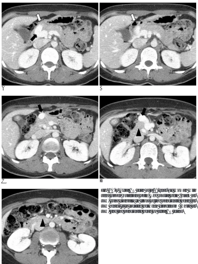
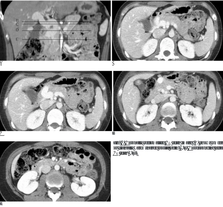
Mallinckrodt, U.S.A.) 140 cc를 자동주입기를 이용하여 초당 3 cc 속도로 주입후 40초와 80초에 각각 동맥기, 문맥기 영상 을 얻어 5 mm두께로 재구성하였다. 전산화단층촬영 소견상 담 낭의 기저부(fundus)에 국소적으로 담낭벽이 두꺼워져 있었으 며, 우연히 췌장 체부의 앞쪽으로부터 췌장두부의 앞쪽과 십이 지장의 뒤쪽을 지나는 비정상적인 형태의 문맥이 관찰되었다 (Fig. 1A-E). 우측 주문맥 주위로 경한 해면상 변형이 관찰되 었으나 담낭, 총담관, 췌장, 위장관, 비장에 다른 기형은 관찰 되지 않았다. 이어 시행한 관상면 재구성영상(Fig. 2A-E)과 수술전 간동맥의 변형을 확인하기 위해서 시행한 간동맥 및 상 장간막 조영술과 문맥조영술상(Fig. 3)에서 비정상적인 주행 경로를 보이는 omega 또는 arc 형태의 문맥이 관찰되었다. 환 자는 담낭선근종증이 의심되어 복강경유도하 담낭 적출술을 시 행하였으며, 수술 중에 술전 CT와 문맥조영술에서 관찰되었던 PPPV를 확인할 수 있었으며 수술 후 병리학적으로 담낭선근 종증으로 확진되었다.
고 찰
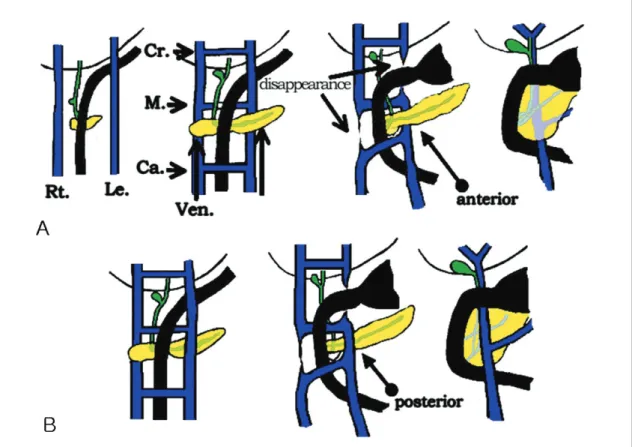
정상적인 문맥의 발생학적인 발달단계는 태생초기(5 mm 배 아시기)에 전장(foregut)으로부터의 정맥혈류는 평행하게 놓 인 두개의 난황 정맥(vitelline vein)에 의해서 배출되고 이 두 개의 난황 정맥은 간 내부에서 상부 문합 (cra nial anastomosis), 십이지장 뒤쪽에서 중앙문합 (middle anastomosis), 십이지장 앞쪽에서 하부문합 (caudal anastomosis )등 3개의 문합을 가지게 된다. 시간이 지나면서 (9 mm 배아시기) 하부 문합, 우측 난황정맥의 하부, 좌측 난
주문맥의 선천적 기형: 췌장두부의 앞쪽과 십이지장 후면사이의 비정상적 주행: 1예 보고1
정영진・이상진・양승부・박원규2・장재천2・김재운2・장한원2・이재교2
Prepancreatic postduodenal portal vein(PPPV)는 문맥이 췌장 두부의 앞쪽과 십이지장 후 면을 지나는 드문 문맥기형이다. 이러한 문맥기형을 이해하는 것은 수술중 발생할 수 있는 원 치않는 문맥결찰, 절제, 출혈등의 불필요한 합병증을 예방하는데 중요하다. 저자들은 2일간의 우상복부 통증을 주소로 내원한 28세 여자환자가 담낭의 선근종증으로 담낭적출술 시행하기전 검사중 우연히 발견된 췌장 두부의 앞쪽과 십이자장 후면으로 주행하는 비정상적인 형태의 문 맥을 경험하였기에 전산화단층촬영과 관상면 재구성 영상, 문맥조영술의 소견을 보고하고자 한 다.
1순천향대학교 구미병원 영상의학과학교실
2영남대학교 의과대학 영상의학과학교실
이 논문은 2005년 6월 3일 접수하여 2005년 8월 16일에 채택되었음.
A B
C D
Fig. 1A-E. Axial CT scan of a 28-year-old woman with right upper quadrant pain. The portal vein (black ar- row) runs cranially in front of the pancreatic head (ar- row head). it is located between duodenum (white ar- row) and the pancreatic head on axial CT scans.
E
(5, 6)에 따르면 발생학적으로 두개의 난황정맥을 연결하는 3 개의 문합중 상부와 중앙문합의 비정상적인 퇴화에 의해서 십 이지장의 앞쪽으로 지나는 하부문합만이 남게 되어 문맥을 형 성하는 것으로 생각된다(Fig. 4). 소아에서는 장회전이상, 담 도폐쇄증, 윤상췌장과 같은 내장기형과 높은 상관관계가 있으 며 성인에서는 무증상인 경우가 많지만 일부에서 담도결석과 연관이 있다는 보고가 있다.
반면에 PPPV은 1972년에 Brook and Gardner등(1)에 의해 서 처음으로 발표된 이후로 10 예 미만으로 보고되고 있으며, 이들 논문에 따르면 다른 복부 기형과는 연관성이 없는 것으 로 알려져 있으며 성인에서 담도결석, 악성질환과 동반된 경우 가 보고되고 있다(2).
Matsumoto 등(7)의 가설에 의하면 PPPV의 경우에는 PDPV 와는 다르게 앞에서 언급한 정상적인 문맥의 발달단계를 거쳐 서 문맥이 형성되지만 정상적으로 배측 췌장 원시배아(dorsal
A B
C D
Fig. 2. Coronally reformatted CT scan image (A) shows omega or cranially ‘arc’shaped portal vein. B-E. Corresponding axial CT scan(B-E).
E
Fig. 3. AP view of indirect SMA portography. Normal shape of superior mesenteric vein (white arrow) is demonstrated, and is continuous to omega or arc shaped portal vein (black arrow).
pancreatic bud)가 좌측 난황 정맥의 앞쪽에 위치해야 하는데 PPPV에서는 좌측 난황 정맥의 뒤쪽에 위치하여 췌장 앞쪽에 위치하는 문맥을 형성한다. 이후로는 정상적으로 회전을 하기 때문에 다른 장 회전이상, 십이지장, 담도 또는 췌장의 선천성 기형과 연관성이 드문 것으로 생각하였다.
Inoue 등(2)의 보고에 따르면 PPPV의 모양은 L shape 또 는 inverted L shape이며, 총담관의 앞쪽 또는 평행하게 주행 하며, 장 회전이상, 십이지장, 담도 또는 췌장 기형과 연관성은 없다고 보고하였다. 정상적으로는 주췌관이 문맥의 앞쪽에 위 치하고 총담관은 주췌관과 연결되기 때문에 총담관은 문맥의 앞쪽에 위치하게 된다. 그러나 PPPV에서는 문맥이 복측 췌장 원시배아(dorsal pacnreatic bud)의 앞쪽에서 주행하기 때문에 총담관은 췌장두부주위에서 문맥의 뒤쪽에서 주행하게 된다.
췌장 두부가 발달하게 되면서 PPPV의 모양이 L shape 또는 inverted L shape의 형태를 보이는 것으로 생각된다. 후천적 인 원인으로 문맥 폐색이 발생하고 이에 따른 주변에 굵어진 측부혈관(collateral vessel)이 비정상적인 문맥의 형태로 관찰 될 수 있을 것으로도 생각되나, 본 증례의 경우에 있어서는 다 른 측부혈관 및 문맥폐색을 시사하는 소견들을 관찰할 수가 없 었으며 하장간막정맥으로부터 주문맥까지 이어지는 비정상적 인 형태의 문맥이 명확하게 관찰되어 후천적인 문맥폐색에 따 른 굵어진 측부혈관이 아닌 선천적인 원인의 PPPV로 생각하
였다. 축상면 전산화단층촬영 영상에서 일부 관찰되었던 해면 상변형은 문맥폐색에 의해서 발생하였다가 보다는 비정상적인 형태의 문맥으로 인한 혈류의 장애에 의해서 발생된 것으로 생 각된다.
저자들의 증례와 같이 PPPV 자체로 증상을 유발한다기보다 는 다른 질병으로 복부 CT나 초음파로 검사도중 우연히 발견 되는 경우가 대부분이였으며 일부에서는 술전 검사상에서 PPPV를 확인하지 못하고 수술 중에 확인되는 경우도 있었다.
저자들의 경우에 있어서는 술 전에 전산화단층촬영 축상면 영 상에서 비정상적인 문맥의 주행을 발견하고 관상면 재구성 영 상과 문맥조영술상에서 이전에 보고된 inverted L 형태의 문 맥을 연상시키는 비정상적인 형태의 문맥을 확인할 수 있었다.
이러한 비정상적인 위치와 형태를 가지는 문맥의 해부학적인 변이를 이해함으로써 수술이나 중재적시술중 문맥결찰이나 대 량출혈, 혈전증 등의 원치 않는 합병증을 예방하는데 도움이 될 것으로 생각한다(5, 8).
참 고 문 헌
1. Brook W, Gardner M. Anteroposition of the portal vein and spon- taneous passage of gall stone. Case report and embryological hy- potheosis. Br J Surg 1972;59:737-739
Fig. 4. Schematic diagram of the hypothesis of Hashimoto et al of normal embryological development of the portal vein (A), and hy- pothesis of Matsumoto et al. of prepancreatic postduodenal portal vein (B). In the PPPV, the position of dorsal pancreatic dub is posterior to the left vitelline vein instead of anterior to it. (Rt., right vitelline vein; Le., left vitelline vein; Ven., ventral pancreatic bud; Dor., dorsal pancreatic bud; Cr., cranial anastomosis; M., middle anastomosis; Ca., caudal anastomosis.)
2. Inoue M, Taenaka N, Nishimura S, Kawamura T, Aki T, Yamaki K, et al. Prepancreatic postduodenal portal vein; report of a case.
Surg Today 2003;33:956-959
3. Marks C. Developmental basis of the portal venous system. Am J Surg 1969;117:671-681
4. Moore K. Cardiovascular system. In Moore K, Persaud T. The devel- oping human. 5th ed. Philadelphia: Saunders, 1993:304-308 5. Steven J, Morton D, McElwee R, Hamit H. Preduodenal portal
vein: two cases wih differing presentation. Arch Surg 1978;113:
311-313
6. Mordehai J, Cohen Z, Kurzbart E, Mares A. Preduodenal portal vein causing duodnal obstruction associated with situs inversus, intestinal malrotation, and polysplenia: a case report. J Pediatr Surg 2002;37:E5
7. Matsumoto Y, Sugahara K, Ida T, Mashimo R, Hsu K, Fujii H, et al. Anomalies of the portal venous system: pathogenesis and its surgical implications. Jpn J Gastroenterol Surg 1983;16:2112-2121 8. Semb B, Halvorsen J. Repair of preduodenal portal vein injury oc-
curring during biliary surgery. Act Chir Scand 1973;139:312-313
J Korean Radiol Soc 2005;53:435-439
Address reprint requests to : Won Kyu Park, M.D., Department of Diagnostic Radiology, College of Medicine, Yeungnam University, 317-1, Daemyungdong, Namgu, Daegu, 705-717, Korea.
Tel. 82-53-620-3048 Fax. 82-53-653-5484 E-mail: wkpark@yumail.ac.kr
Prepancreatic Postduodenal Portal Vein: A Case Report1
Young Jin Jung, M.D., Sang Jin Lee, M.D., Seung Boo Yang, M.D., Won Kyu Park, M.D.2, Jay Chun Chang, M.D.2, Jae Woon Kim, M.D.2, Han Won Jang, M.D.2, Jae Kyo Lee, M.D.2
1Department of Radiology, Soonchunhyang University Gumi Hospital
2Department of Diagnostic Radiology, College of Medicine, Yeungnam University
Prepancreatic postduodenal portal vein (PPPV) is a rare anomaly in which the portal vein runs between the pancreatic head and the duodenum. Understanding of this portal vein anomaly is important to avoid devastat- ing complications, including portal vein ligation, resection or intraoperative hemorrhage. A 28-year-old female patient presented with right upper quadrant pain that she had suffered with for 2 days. Before performing la- paroscopic cholecystectomy, we detected an abnormal shaped portal vein that ran in front of the pancreatic head and posterior to the duodenum on the CT scan. We report here on a rare case of prepancreatic postduo- denal portal vein that was incidentally discovered on the CT axial images and coronally reformated images, in addition to observing it on the conventional portography.
Index words :Portal vein, CT Portal vein, anatomy