서론
흉부 전산화단층촬영(computed tomography; 이하 CT)은 폐나 심장질환의 평가를 위해 시행되거나 암전이를 평가하기 위해 시행되나 종종 유방 질환에 대한 진단에 도움이 되는 소 견들을 제공한다(1, 2). CT의 사용이 증가되면서 유방조직이 포함되는 흉부 CT에서 발견되는 유방 병변이 증가하고 있고 흉부 CT에서 처음 발견되는 유방 병변들도 늘고 있다. 그러므 로 이들 병변들을 유방촬영이나 초음파 검사와 비교하여 아는 것이 CT에서 발견되는 유방 병변들을 감별하는 데 도움이 된 다. CT는 양성과 악성 질환을 발견하는 데 민감하나 우연히 발견된 유방 병변에서는 진단이 과장될 수 있고 양성과 악성을 구분하는 것이 쉽지 않다(1, 2). 몇몇 연구에서 CT에서 우연히 발견된 유방 병변 중 0.4~2%에서 유방암이었다고 보고하고 있다(3, 4). 그러므로 유방 질환에 대한 정확한 임상정보가 필 요하며 유방검사방법과의 비교가 필요하다. 저자들은 흉부 CT에서 발견된 유방영상에 대해 유방촬영술과 유방 초음파 검사를 비교하여 양성과 악성질환들을 보여주고자 한다.
양성 유방 병변
양성 유방 병변에 대한 CT 소견은 몇몇 질환에서는 특징적 인 소견을 보인다.
과오종(Hamartoma)
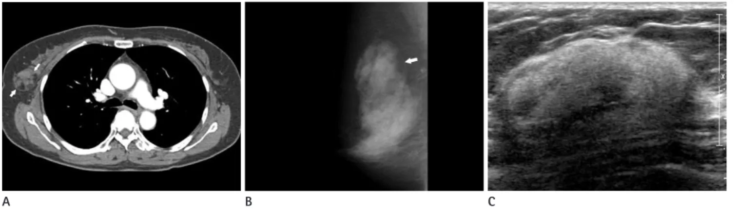
지방과 유선조직을 가지는 드문 종양으로 만져지는 경우가 많으며 “유방 내 유방”으로 칭해지기도 한다. CT에서는 잘 그 려지는 경계를 가지며 지방을 포함하는 혼합음영의 병변으로 나타난다. 유방촬영술에서 얇은 고음영의 가성피막으로 둘러 싸인 경계가 좋은 종괴로 보이며 초음파에서 경계가 잘 그려지 는 비균질한 혼합에코의 종괴로 보인다(Fig. 1)(1, 5).
섬유선종
섬유선종은 유방의 가장 흔한 종양으로 15세~40대 여성에 서 흔하며 다발성 혹은 양측성으로 나타날 수 있다. 유방 소엽 내에 결합 조직의 증식으로 발생하며 중년 이후의 여성에서 기 질 세포의 유리질화가 되면 석회화를 동반할 수 있다(1, 2).
J Korean Soc Radiol 2012;66(1):69-76
Received August 26, 2011; Accepted October 4, 2011 Corresponding author: Yun-Woo Chang, MD Department of Radiology, Soonchunhyang University Hospital, 22 Daesagwan-gil, Yongsan-gu,
Seoul 140-743, Korea.
Tel. 82-2-709-9396 Fax. 82-2-709-3928 E-mail: [email protected]
Copyrights © 2012 The Korean Society of Radiology
Chest CT often provides images of the breast when scanning is performed for pul- monary and cardiac disease or metastasis work up, and can allow for a confident diagnosis of breast lesions. This study is an exhibit with an introductory section for the usefulness of a chest CT and how to accurately describe breast lesions found on CT. In addition, a review of the benign and malignant lesions on chest CT with cor- relation of breast images was made.
Index terms
Multi-Detector Computed Tomography Chest
Breast Imaging
Breast Findings on Computed Tomography: Correlation with Mammography and Breast Ultrasonography
1흉부 전산화단층촬영에서 보이는 다양한 유방 질환의 영상소견:
유방촬영 및 유방 초음파의 비교
1Jae Hyun Kim, MD
1, Yun-Woo Chang, MD
1, Jung Hwa Hwang, MD
1, Yoo Na Kim, MD
1, Hye Shin Ahn, MD
1, Kui Hyang Kwon, MD
1, Hyung Hwan Kim, MD
21Department of Radiology, Soonchunhyang University Hospital, Seoul, Korea
2Department of Radiology, Soonchunhyang University Cheonan Hospital, Cheonan, Korea
CT에서 경계가 잘 그려지는 둥글거나 난원의 종양으로 보인 다(Fig. 2).
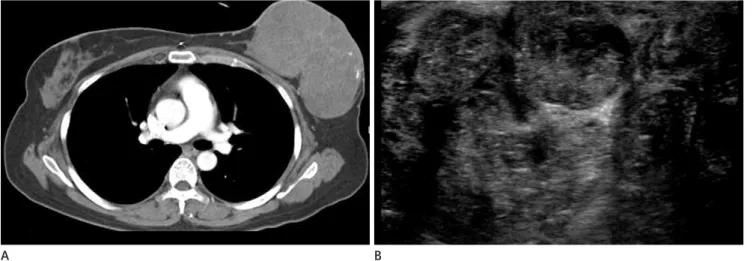
남성유방(Gynecomastia)
남성유방이 비종양성 증식을 보이는 것으로 단측 혹은 양측 성으로 올 수 있다. 청소년기와 50대, 두 번의 호발 연령을 가 지며 호르몬의 불안정한 상태를 유발할 수 있는 내적 혹은 외 적 호르몬의 불균형에서 생긴다. 내인성 요인으로 간경화, 호 르몬 분비 고환종양, 폐암이나 신장암의 종양부수증후군 혹은 갑상선기능항진증이 있으며 외인성 요인으로는 알코올이나 마 리화나, 단백동화 스테로이드(anabolic steroid)나 향정신성 약 물들이다(2, 6). CT에서 화염상(flame shape)의 연부 조직 감 쇠(soft tissue attenuation)로 나타난다(Fig. 3).
A B C
Fig. 1. 61-year-old female.
A. CT scan shows well-defined fat containing soft tissue density mass in right breast (arrows).
B. Mammography shows well-defined, fat containing, heterogeneous mass in right breast, representing hamartoma.
C. Sonography shows large circumscribed, ovoid mass with mixed internal echogenicity.
Fig. 2. 40-year-old woman.
A. Chest CT scan shows well-defined, round, homogeneous enhancing mass on right breast (arrow).
B. Sonography shows well-defined, round, hypoechogenic mass on right breast. This lesion was confirmed as fibroadenoma at biopsy.
Fig. 3. 14-year-old male with gynecomastia in both breast. Chest CT was performed due to evaluation of asthma. Chest CT scan shows soft tissue density in both subareloar regions (arrows), representing gyne- comastia.
A B
있는 악성종양과 감별해야 한다(2). 혈종은 시간이 지남에 따 라 점차 흡수되어 크기가 작아지므로 다른 종양과 구별하는 데 도움이 된다. CT에서 혈종은 장액종에 비해 상대적인 고음영 의 액체 저류로 보이며(Fig. 5) 혈액은 흡수되면서 장액종을 형성한다. 조영증강 후 주변부 조영증강이 보일 수 있어 농양과 도 유사하나 고열과 백혈구상승 등의 임상소견이 구별에 도움 이 된다(1).
악성 유방 병변
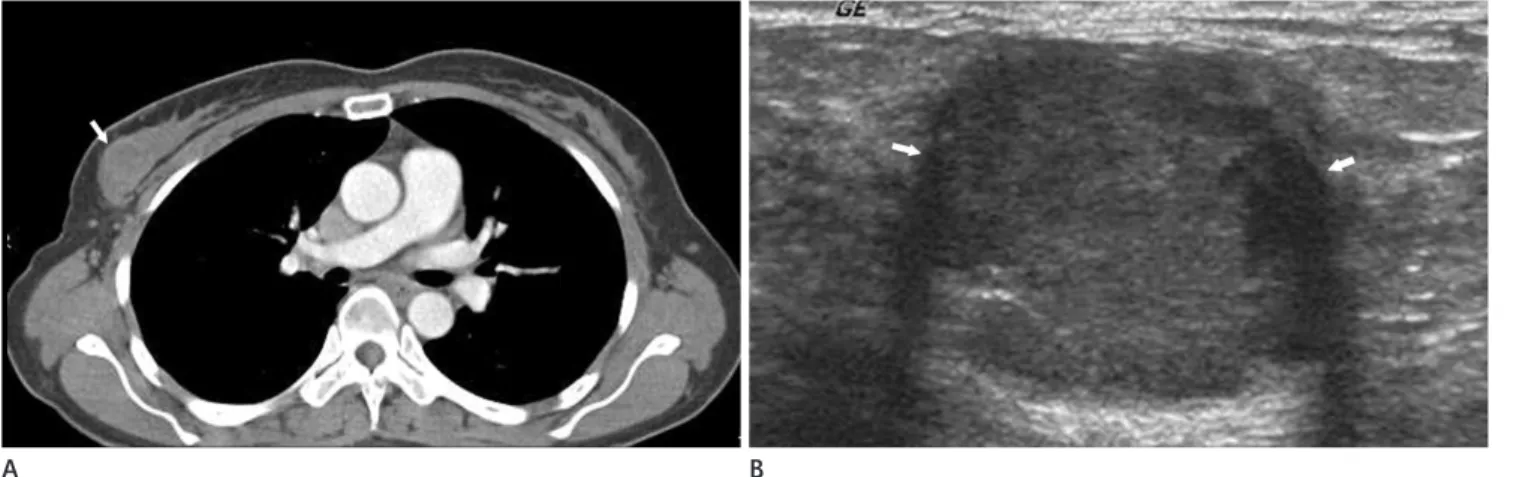
CT의 역동적 조영증강 기법을 사용하면 유방 악성종양은 불 규칙한 경계, 불규칙한 모양, 환형 조영증강을 보인다는 보고 이물질 주입에 따른 육아종(Granuloma due to Foreign
Body Injections)
실리콘이나 파라핀을 미용목적으로 유방 내에 주입한 경우 에 석회화나 육아종이 형성된다. CT에서 주입된 실리콘들은 다발성의 둥글고 밀도가 높은 결절로 보인다. 파라핀의 경우 높은 줄무늬 모양 음영의 기이한 구조적 왜곡으로 결절성의 연 부조직 음영보다는 거친 석회화로 나타난다(Fig. 4).
혈종과 장액종(Hematoma and Seroma)
외상, 수술 혹은 조직검사 후에 혈종이나 장액종이 생길 수 있으며 진단은 임상적인 병력과 연관하여 얻어진다. 수술 초기 의 혈종이나 장액종은 조직변성을 보이는 종양으로 보여 남아
Fig. 4. 58-year-old female with incidental breast lesions.
A. High resolution CT (HRCT) was performed to evaluation of fever and upper respiratory tract infection. HRCT scan shows multiple high density foreign bodies mainly in pectoralis muscle (arrows).
B. Mammography shows injection foreign bodies in right breast.
Fig. 5. 77-year-old female with contusion of breast due to traffic accident.
A. Chest CT scan shows non-enhancing, huge, soft tissue density mass in left breast.
B. Sonography was performed after 10 days, irregular, ill-defined, low echogenic cyst with septa and debris are seen in left breast, possible of liq- uefaction of hematoma correlation with history.
A
A
B
B
percellular stromal overgrowth), 현미경 소견에서 유사분열 수 가 10 high-power fields에 5개 이상, 기질 세포 다형태성 (stromal cellular pleomorphism)을 가지는 경우 악성 엽상종으 로 분류한다(10). 유방영상소견은 경계가 잘 그려지는 섬유선 종과 유사한 종양으로 낭성성분은 양성 엽상종보다 악성 엽상 종에서 더 잘 동반된다. CT에서 경계가 잘 그려지는 다양한 조 영증강의 종양으로 보인다(Fig. 6).
점액암종은 많은 양의 점액를 포함하는 종양으로 서서히 자 라는 비교적 예후가 좋은 종양으로 나이든 여성에서 호발 한다 (11). 유방 초음파 검사시 경계가 잘 그려지는 둥글고 난원의 종양으로 낭성과 고형성분이 혼합되어 보이므로 CT에서 양성 종양과 유사하게 보일 수 있으며(Fig. 7) 수질암종도 드문 유 방종양으로 35세 이하 젊은 여성에서 생기는 경우가 많고 유 가 있으며 침상 또는 불규칙한 경계가 악성진단에 정확한 소견
으로 알려져 있다(7). CT의 역동적 조영증강에서 배출(wash- out)되는 소견이나 소엽 분포를 따르는 부분적 조영증강은 악 성진단에 높은 양성 예측도를 보인다고 보고되었다(8).
경계가 잘 그려지는 악성종양
경계가 잘 그려지는 유방종양의 10~20%는 악성종양으로 악성 엽상종, 점액암종, 수질성 암종, 유두상 암종, 화생성 암 종 등이 있다(9). 이들 종양들은 양성종양처럼 보이는 악성종 양으로 유방 영상검사로 감별하기가 쉽지 않다.
악성 엽상종은 섬유선종과 유사하게 상피와 결합 조직 기질 을 가지는 종양으로 35~55세의 여성에서 호발하며 병리적으 로 침습적 종양 경계, 현저한 정도의 과세포 기질 과증식(hy-
Fig. 6. Malignant phyllodes tumor.
A. Multi-detector CT scan shows huge, relatively well-defined, mild enhancing mass with calcifications in left breast.
B. Sonography shows circumscribed mass with hyperechogenic internal septums.
Fig. 7. 36-year-old female with mucinous carcinoma.
A. CT scan shows well-defined, non/poorly enhancing mass in right inner breast (arrows).
B. Sonography shows well-defined, isoechoic mass in right breast (arrows).
A
A
B
B
(DCIS)이 동반되어 있음을 의미한다(1, 5). CT에서는 치밀한 침상형의 종괴로 조기 조영증강 그리고/또는 주변의 환형 조영 증강을 보인다. CT는 피부의 두꺼워짐, 임파절 침윤, 흉벽 침 윤 등을 확인하는 데 도움이 되며 양측성 병변을 발견할 수도 있다(Fig. 10). 유방촬영과는 달리 미세석회화는 0.5 mm 이 하의 크기를 가지므로 공간 분해능이 떨어지는 CT에서 발견하 기 어렵다(2).
염증유방암
염증유방암은 초기 진피 림프계 침윤을 보이는 예후가 좋지 않은 공격적인 종괴로 임상적으로 열감, 유방 피부의 발적, 오 렌지 껍질 같은 피부(erysipeloid edge, peau d’orange), 유두 뒤당김(nipple retraction)의 소견에 기초하여 진단한다. 유방염 이나 농양과 구분이 어려울 수 있다(1). CT에서 공격적인 현저 한 피부 두꺼워짐과 주변부 조영증강을 보이며 종종 명확히 구 분되는 종괴가 보이지 않을 수도 있다(Fig. 11). 병변의 범위를 방 초음파 검사에서 경계가 잘 그려지는 둥글거나 난원형의 종
양으로 내부에 괴사나 출혈을 유발하여 낭성성분을 가질수 있 다(12). CT 소견으로도 경계가 잘 그려지는 종괴로 보이며 균 질하지 않은 조영증강을 보일 수 있다(Fig. 8).
침습유두암종은 유방암의 2%를 차지하며 유관내형(intra- ductal type)과 낭내형(intracystic type)으로 나타날 수 있다 (13). CT에서도 유방촬영이나 초음파 소견과 같이 경계가 잘 그려지는 둥글거나 난원의 유방 종양으로 보이며 내부에 낭성 성분을 가지는 경우 내부에 조영증강이 되지 않을 수 있다 (Fig. 9).
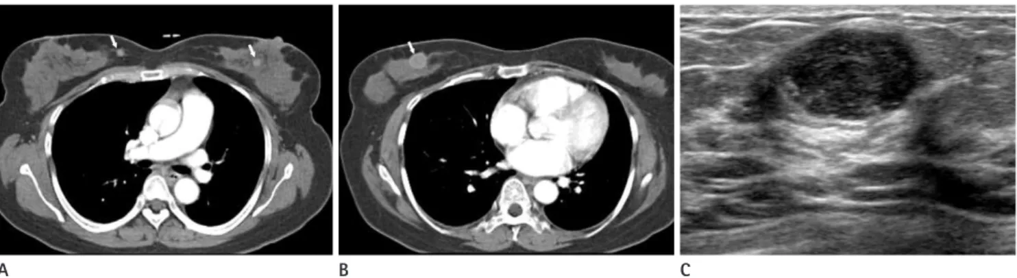
침윤유방암
침윤유방암의 80~90%를 차지하는 가장 흔한 악성종양으 로 유방촬영에서 불규칙, 침상 경계의 치밀한 종괴로 초음파에 서도 불규칙, 원형 또는 침상형의 종괴로 보인다. 유방촬영에 서 다형태성 미세석회화를 포함하는 경우는 유관 상피내암
Fig. 8. Medullary carcioma.
A. Multi-detector CT scan shows round, relatively well-defined, subtle enhancing mass in right breast (arrow).
B. Sonography shows a microlobulated, round, isoechogenic mass with posterior enhancement (arrows).
Fig. 9. Papillary carcinoma.
A. Multi-detector CT shows lobulated, round, cystic mass with solid enhancing component in right breast.
B. A lobulated, oval mass with well-circumscribed margins is seen on mammogramphy.
C. Sonography shows a well-circumscribed, oval, solid and cystic complex echoic mass.
A B
A B C
으나 유방외의 장기에서 유방으로의 전이는 드물며 유방암의 2%를 차지한다. 가장 흔한 유방 전이는 악성 흑생종이며 림프 종, 폐암과 유암종의 순으로 알려져 있다(15). 난소암, 신장암, 대장암이나 자궁경부암에서 유방으로 전이될 수도 있다. 전이 파악하는 데 있어 유방촬영술보다 도움이 된다(14).
유방 전이
유방 전이는 반대측 유방에서 다른 쪽으로의 전이는 볼 수 있 Fig. 10. Bilateral invasive ductal carcinoma.
A, B. Contrast-enhanced CT scan shows irregular, round, enhancing mass in right breast (arrow) and spiculated, irregular, enhancing mass in left breast (arrow).
C, D. These masses are well correlated with multi-detector CT, and ultrasonography. Pathology confirmed bilateral invasive ductal carcinoma.
Fig. 11. 59-year-old female with invasive ductal carcinoma and paget disease. Physical examination revealed erythema and flaking of nipple.
A. CT scan shows multiple irregular, ill-defined, heterogeneous enhancing masses with skin thickening in left breast and nipple.
B. Mammography shows irregular, ill-defined mass with thickening of nipple and periareolar region.
C
A A
D
B B
6. Kopans DB. The male breast. In Kopans DB. Breast Imaging.
Philadelphia: Lippincott-Raven, 1998:497-509
7. Inoue M, Sano T, Watai R, Ashikaga R, Ueda K, Watatani M, et al. Dynamic multidetector CT of breast tumors: diag- nostic features and comparison with conventional tech- niques. AJR Am J Roentgenol 2003;181:679-686
8. Miyake K, Hayakawa K, Nishino M, Nakamura Y, Morimoto T, Urata Y, et al. Benign or malignant?: differentiating breast lesions with computed tomography attenuation values on dynamic computed tomography mammography. J Comput Assist Tomogr 2005;29:772-779
9. Yoo JL, Woo OH, Kim YK, Cho KR, Yong HS, Seo BK, et al.
Can MR Imaging contribute in characterizing well-circum- scribed breast carcinomas? Radiographics 2010;30:1689- 1702
10. Liberman L, Bonaccio E, Hamele-Bena D, Abramson AF, Cohen MA, Dershaw DD. Benign and malignant phyllodes tumors: mammographic and sonographic findings. Radiol- ogy 1996;198:121-124
11. Memis A, Ozdemir N, Parildar M, Ustun EE, Erhan Y. Mucinous (colloid) breast cancer: mammographic and US features with histologic correlation. Eur J Radiol 2000;35:39-43
12. Meyer JE, Amin E, Lindfors KK, Lipman JC, Stomper PC, Genest D. Medullary carcinoma of the breast: mammo- graphic and US appearance. Radiology 1989;170:79-82 13. Lam WW, Tang AP, Tse G, Chu WC. Radiology-Pathology
conference: papillary carcinoma of the breast. Clin Imag- ing 2005;29:396-400
14. Kim SM, Park JM. Computed tomography of the breast.
암은 다발성으로 양측성의 표재성 위치를 보인다(Fig. 12).
결론
CT에서 발견되는 유방 질환들은 진단에 도움이 되는 올바른 정보들을 제공할 수 있다. CT는 액와부 전이를 평가하고 피부 나 흉곽의 병변을 평가하는 데 있어 유방촬영이나 초음파보다 유용한 정보를 제공할 수 있다. 그러나 CT에서 보이는 유방 질 환들은 종종 과대 평가 되거나 과소 평가 될 수 있으므로 유방 병변에 대한 정확한 임상정보하에 유방촬영이나 유방 초음파 를 통한 정확한 진단이 요구된다.
참고문헌
1. Harish MG, Konda SD, MacMahon H, Newstead GM. Breast lesions incidentally detected with CT: what the general ra- diologist needs to know. Radiographics 2007;27 Suppl 1:S37-S51
2. Yi JG, Kim SJ, Marom EM, Park JH, Jung SI, Lee MW. Chest CT of incidental breast lesions. J Thorac Imaging 2008;23:
148-155
3. Swensen SJ, Jett JR, Hartman TE, Midthun DE, Sloan JA, Sykes AM, et al. Lung cancer screening with CT: Mayo Clinic experience. Radiology 2003;226:756-761
4. Lin WC, Hsu HH, Li CS, Yu JC, Hsu GC, Yu CP, et al. Inciden- tally detected enhancing breast lesions on chest computed tomography. Korean J Radiol 2011;12:44-51
5. Marsteller LP, Shaw de Paredes E. Well defined masses in the breast. Radiographics 1989;9:13-37
A B C
Fig. 12. Melanoma metastasis.
A, B. Contrast enhanced CT scan shows multiple, well-defined, round masses with homogeneous or heterogeneous enhancement in both breasts (arrow).
C. Sonography shows well-defined heterogeneous low echoic masses. Melanoma is confirmed by core needle biopsy. This patient with a history of a malignant cutaneous melanoma of nasal cavity had been surgically excised.
correlation. In Kopans DB. Breast Imaging. Philadelphia:
Lippincott-Raven, 1998:511-615 Abnormal findings with mammographic and sonographic
correlation. J Comput Assist Tomogr 2003;27:761-770 15. Kopans DB. Pathologic, mammographic, and sonographic
흉부 전산화단층촬영에서 보이는 다양한 유방 질환의 영상소견:
유방촬영 및 유방 초음파의 비교
1김재현
1· 장윤우
1· 황정화
1· 김유나
1· 안혜신
1· 권귀향
1· 김형환
2흉부 전산화단층촬영은 폐나 심장 질환을 평가하거나 암의 전이를 찾기 위해 시행하나 예상치 않은 유방의 이상소견을 발견할 수 있다. 저자들은 흉부 전산화단층촬영에서 보이는 다양한 유방 질환의 영상 소견들을 양성 및 악성 병변으로 나 누어 유방촬영 및 유방 초음파 검사와 비교하여 보여주고자 한다.
1순천향대학교병원 영상의학과, 2순천향대학교 천안병원 영상의학과