Leiomyomas are benign tumors and they mainly oc- cur in the uterus. Primary leiomyomas in the central nervous system are extremely rare (1). Primary spinal leiomyomas have only rarely been reported in patients with acquired immune deficiency syndrome (AIDS), and this is irrespective of the concomitant presence of uterine leiomyoma (2, 3).
Although this histologically benign tumor is a rare finding, it can metastasize to many locations, which is a well known phenomenon that’s termed benign metasta- sizing leiomyomas (BMLs). The pathogenesis of metasta- sis has not been fully elucidated. However, women who have undergone hysterectomy for leiomyomas are most commonly affected (4-8), and the lung is the most com- mon site of involvement (4).
We report here on a case of a lumbosacral, extradural
leiomyoma in a woman who had a previous history of hysterectomy for treating her leiomyoma 20 years previ- ously.
Case Report
A 67-year-old woman presented in April 2004 to the neurosurgery department with a history of monoparesis of the left leg. Upon examination she exhibited a mild left lower extremity motor deficit (grade 4/5) without any sensory deficit.
She had undergone hysterectomy for leiomyoma 20 years prior to presentation. A contrast-enhanced com- puted tomography (CT) scan showed a lobulated, homo- geneously enhancing soft tissue mass in the right S1 par- avertebral region, the pelvic cavity and the spinal canal, and this was seen together with vertebral body erosion (Fig. 1A, B). The sagittal T1-weighted MR imaging demonstrated a soft tissue mass in the spinal canal from the L5 to S2 level and also in the prevertebral space (Fig.
2A). On the sagittal T2-weighted MR images, the lesions demonstrated nearly isosignal intensity to the adjacent muscles (Fig. 2B). The mass showed strong, homoge- neous contrast enhancement on a gadolinium-enhanced T1-weighted MR image. A contrast-enhanced axial T1 image showed the intraspinal mass exiting the spinal
J Korean Radiol Soc 2006;55:433-436
─ 433 ─
Metastatic Spinal Epidural Leiomyoma: A Case Report1
Yoo Na Seo, M.D., Seon Joo Lee, M.D.2, Yong Woo Kim, M.D., Yeong Mi Park, M.D., Seong Sook Cha, M.D., Jae Ik Bae, M.D., Choong Ki Eun, M.D., Gyung Kyu Lee, M.D.3
1Department of Radiology, College of Medicine, Inje University, Sanggye Paik Hospital
2Department of Radiology, College of Medicine, Inje University, Busan Paik Hospital
3Department of Radiology, College of Medicine, Hallym University, Hangang Sacred Heart Hospital
Received April 14, 2006 ; Accepted August 2, 2006
Address reprint requests to : Seon Joo Lee, M.D., Department of Radiology, Busan Paik Hospital, Inje University, 633-165 Kegum-dong, Busanjin-ku, Busan 614-735, Korea.
Tel. 82-51-890-6728 Fax. 82-51-896-1085 E-mail: [email protected]
We report here on a case of a spinal extradural leiomyoma in a 67-year-old woman, and this tumor was in a very unusual location for a leiomyoma. Because the patient underwent hysterectomy for a uterine leiomyoma 20 years ago, we can speculate that the spinal lesion was a metastatic leiomyoma.
Index words :Spinal cord Neoplasms Leiomyoma
canal through the bilateral neural foramina at the L5 lev- el. It formed a large bilobulated paraspinal component along the medial and lateral sides of the right psoas mus- cle, displacing it anterolaterally (Fig. 2C). Another large mass in the pelvic cavity (Fig. 1B) was connected with the intraspinal lesion through the left S2 neural fora- men.
Although the T2 signal intensity of the tumor was not as bright as a typical neurofibroma, this was our initial radiologic diagnosis due to the lesion’s characteristic imaging findings as described above. Total laminectomy at L5 and S1 with total excision of the tumor was per- formed. The lesion was intraspinal and extradural in lo- cation.
Yoo Na Seo, et al: Metastatic Spinal Epidural Leiomyoma
─ 434 ─
A B
Fig. 1. A, B. The contrast enhanced CT scan shows a lobulated, homogeneously enhancing, soft tissue mass in the right S1 paraver- tebral region, the pelvic cavity and within the spinal canal together with vertebral body erosion (arrows). This lesion is displacing the right psoas muscle (★) anterolaterally.
A B C
Fig. 2. A. The sagittal T1-weighted MR image demonstrates a soft tissue mass in the spinal canal from the L5 to S2 levels and in the prevertebral space (arrows).
B. The sagittal T2-weighted MR images shows the lesion has nearly isosignal intensity to the adjacent muscles.
C. The contrast-enhanced axial T1 image shows the extradural mass exiting the spinal canal through the bilateral neural foramina at the level of L5, and so creating a large bilobulated paraspinal component along the medial and lateral sides of the right psoas (★) muscle and displacing it anterolaterally (arrows).
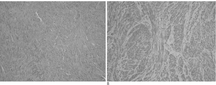
The final pathologic diagnosis was a leiomyoma with- out signs of malignant degeneration. Upon histologic ex- amination, the lesions showed intersecting short fasci- cles of acidophilic spindle cells without any significant cellular pleomorphism or mitotic activity. No malignant features were noted (Fig. 3A). Immunohistochemical evaluation revealed a positive reaction of the tumor cells to the smooth muscle marker (smooth muscle actin) (Fig. 3B).
Discussion
Leiomyomas are benign tumors that consist of well- differentiated smooth muscle tissue and vascular col- lagenous tissue, and they mainly occur in the uterus.
The other tumor locations include the gastrointestinal tract, mainly in the lower third part of the esophagus, the sinonasal tract or larynx, lung, trachea, bladder, liv- er and adrenal gland. The tunica media and smooth muscle of the skin are also known sites for leiomyomas (7). In addition, there have been reports of primary in- tracranial leiomyomas and primary spinal extradural leiomyomas in male pediatric patients and adult pa- tients with AIDS (1-3).
Uterine leiomyomas are on rare occasion associated with extrauterine benign smooth muscle tumors: this curious condition is referred to as “benign metastasizing leiomyoma”, which is a relatively well-known phenom- enon (4-8). Although most commonly seen in the lungs (4), other sites of metastatic involvement include the
lymph nodes, peritoneum, retroperitoneal structures, spine and the base of the skull (4-8).
Several hypotheses have been proposed to explain the pathogenesis of this poorly understood entity. Some in- vestigators have classified this tumor as a low grade leiomyosarcoma with malignant potential (9). Others have proposed a mechanism of implantation and prolif- eration of benign smooth muscle cells by an intravenous route or by mechanical means (9). Still others have pos- tulated that this is the result of a systemic leiomyomato- sis with multifocal, but independent smooth muscle proliferation (9).
We can speculate that there was tumor cell migration by an intravenous route: the most plausible explanation in this case is from the pelvic vein to the spinal epidural vein The mechanism of hematogenous spread is suben- dothelial vascular involvement of the uterine leiomy- oma, which possibly represents early vascular invasion via the formation of a tumor embolus (10).
As far as we could establish, our patient had no evi- dence of metastatic disease in other organs, including the lung. Moreover, a case of a benign metastasizing leiomyoma in a spinal extradural space without lung parenchymal involvement has been reported by Hekster et al (7).
The diagnosis upon imaging was initially mistaken as a neurofibroma because of its location and its character- istic shape. Yet after retrospectively reviewing the MR images, we found that the large mass exhibited lower T2 signal intensity than that of a neurofibroma, and it
J Korean Radiol Soc 2006;55:433-436
─ 435 ─
A B
Fig. 3. Photomicrograph from the high power magnification microscopy.
A. The tumor shows intersecting short fascicles of spindle cells. (hematoxylin-eosin stain, ×200)
B. The spindle cells reveal cytoplasmic positivity upon immunohistochemical staining for smooth muscle actin. (Labelled streptoa- vidin biotin, ×200)
showed rather strong homogeneous enhancement on the gadolinium enhanced T1 weighted images.
Complete resection of these lesions is usually possible and this can provide substantial improvement of the pa- tient’s functional status. Because of this potential for successful treatment, this disease entity should be kept in mind when diagnosing a paraspinal lesion that ex- hibits unusual imaging features.
References
1. Lai PH, Yang CF, Huang CH, Yeh LR, Lin SL, Pan HB. Primay in- tracranial leiomyoma: case report. Neuroradiology 1998;40:238-241 2. Steel TR, Pell MF, Turner JJ, Lim GH. Spinal epidural leiomyoma occurring in an HIV-infected man. case report. J Neurosurg 1993;
79:442-445
3. Choi S, Levy ML, Krieger MD, McComb JG. Spinal extradural leiomyoma in a pediatric patient with acquired immunodeficiency syndrome: case report. Neurosurgery 1997;40:1080-1082
4. Abramson S, Gilkeson RC, Goldstein JD, Woodard PK, Eisenberg R, Abramson N. Benign metastasizing leiomyoma: clinical, imag- ing, and pathologic correlation. AJR Am J Roentgenol 2001;176:
1409-1413
5. Alessi G, Lemmerling M, Vereecken L, De Waele L. Benign metas- tasizing leiomyoma to skull base and spine: a report of two cases.
Clin Neurol Neurosurg 2003;105:170-174
6. Joseph V, Chacko G, Raghuram L, Rajshekhar V. Benign metasta- sizing leiomyoma causing spinal cord compression. Surg Neurol 2003;60:575-577
7. Hekster RE, Lambooy N, van Hall EV, Kazzaz BA. van Rijssel EJ.
Hormone-dependent spinal leiomyoma. Surg Neurol 1994;41:330- 333
8. Gatti JM, Morvan G, Henin D, Aboulker J, Nahum H, Glowinski J. Leiomyomatosis metastasizing to the spine. A case report. J Bone Joint Surg Am 1983;65:1163-1165
9. Cho KR, Woodruff JD, Epstein JI. Leiomyoma of the uterus with multiple extrauterine smooth muscle tumors: a case report sug- gesting multifocal origin. Hum Pathol 1989;20:80-83
10. Patton KT, Cheng L, Papavero V, Blum MG, Yeldandi AV, Adley BP, et al. Benign metastasizing leiomyoma: clonality, telomere length and clinicopathologic analysis. Mod Pathol 2006;19:130-40
Yoo Na Seo, et al: Metastatic Spinal Epidural Leiomyoma
─ 436 ─
대한영상의학회지 2006;55:433-436
신경섬유종으로 오인된 척수에 생긴 전이성 양성 평활근종: 증례 보고1
1인제대학교 의과대학 상계백병원 영상의학과
2인제대학교 의과대학 부산백병원 영상의학과
3한림대학교 의과대학 한강성심병원 영상의학과
서유나・이선주2・김용우・박영미・차성숙・배재익・은충기・이경규3
평활근종은 자궁에 가장 흔히 생기는 양성 종양이다. 이 종양의 전이는 잘 알려진 사실이나 척수에 전이된 경우 는 아주 드문데, 저자들은 자궁근종으로 자궁적출술을 받았던 환자에서 척수에 전이된 1예를 경험하였기에 문헌고 찰과 함께 보고하고자 한다.