Korean J Gastroenterol Vol. 58 No. 4, 208-211 http://dx.doi.org/10.4166/kjg.2011.58.4.208
CASE REPORT
Korean J Gastroenterol, Vol. 58 No. 4, October 2011 www.kjg.or.kr
위 천공에 의한 복막염으로 나타난 위 림프상피종양 암종 1예
고평곤, 김의식, 김윤정, 이수윤, 문희석, 김석현, 이병석, 정현용
충남대학교 의학전문대학원 내과학교실
A Case of Gastric Lymphoepithelioma-like Carcinoma Presenting as Panperitonitis by Perforation of Stomach
Pyung Gohn Goh, Eui Sik Kim, Yun Jeung Kim, Soo Youn Lee, Hee Seok Moon, Seok Hyun Kim, Byung Seok Lee and Hyun Yong Jeong
Department of Internal Medicine, School of Medicine, Chungnam National University, Daejeon, Korea
Gastric lymphoepithelioma-like carcinoma is a rare carcinoma among gastric malignant tumor but has a good prognosis. The carcinoma has histologic feature characterized by small nest of cancer cells mixed with lymphoid stroma. We report a case with lymphoepithelioma-like carcinoma of stomach initially presenting as panperitonitis because of spontaneous tumor perforation.
A 56-year-old man visited our emergency room because of epigastric pain. A preoperative abdominal CT scan showed a massive pneumoperitoneum in the upper abdomen, and the presence of gastric cancer in the lesser curvature of the stomach. An emergent laparotomy was performed followed by radical subtotal gastrectomy. Pathologic examination revealed that the tumor was a lymphoepithelioma-like gastric carcinoma. (Korean J Gastroenterol 2011;58:208-211)
Key Words: Gastric lymphoepithelioma-like carcinoma; Spontaneous tumor perforation
Received August 10, 2010. Revised September 5, 2010. Accepted September 6, 2010.
CC This is an open access article distributed under the terms of the Creative Commons Attribution Non-Commercial License (http://creativecommons.org/licenses/
by-nc/3.0) which permits unrestricted non-commercial use, distribution, and reproduction in any medium, provided the original work is properly cited.
교신저자: 문희석, 301-721, 대전시 중구 문화로 33, 충남대학교 의학전문대학원 내과학교실
Correspondence to: Hee Seok Moon, Department of Internal Medicine, School of Medicine, Chungnam National University, 33, Munhwa-ro, Jung-gu, Daejeon 301-721, Korea. Tel: +82-42-280-7143, Fax: +82-42-257-5753, E-mail: [email protected]
Financial support: None. Conflict of interest: None.
서 론
림프상피종양 암종(lymphoepithelioma-like carcinoma) 은 비인두암과 조직학적으로 유사한 종양으로 림프구성 간질 에 악성종양 세포의 침윤을 특징으로 하고 있다.1 위에 발생하 는 림프상피종양 암종은 1921년 MacCarty와 Mahle에 의해 처음 보고되었고,2 전체 위암종의 1-4% 정도의 발생률을 보이 는 드문 암으로 알려져 있다. 위 림프상피종양 암종은 육안 형태면에서 조기 위암으로 발현하기도 하며, 진행성 위암으로 보이기도 한다. Watanabe 등에 의한 연구에서는 진행성 위 암의 경우에 Borrmann type 2형이 가장 많았으며, 약 70%에 서 궤양이 동반되었고,3 드물게 상피하종양으로 발현되는 경 우도 보고되었다.4 이번에 저자들은 위암종의 드문 합병증인 자발성 천공에 의한 복막염으로 내원하여, 수술 후 림프상피
종양 암종으로 진단된 증례를 경험하여 문헌고찰과 함께 보고 하는 바이다.
증 례
55세 남자가 내원 1주일 전부터 발생하여 점점 심해지는 심와부 통증을 주소로 응급실에 내원하였다. 고혈압으로 복약 중이었으며, 이 외에 다른 과거력은 없었다. 내원 당시 활력징 후는 혈압 100/80 mmHg, 맥박 96회/분, 호흡수 20회/분, 체 온 37.6oC이었다. 신체검사에서 복벽이 경직되어 있으면서 압 통과 반발통을 동반하고 있었다. 내원 시 시행한 말초 혈액 소견은 백혈구 10,900/mm3, 혈색소 8.5 g/dL, 혈소판 300,000/mm3이었으며, 혈청생화학검사는 AST/ALT 15/10 IU/L, ALP 72 IU/L, total bilirubin 0.7 mg/dL, BUN 19
Goh PG, et al. A Case of Gastric Lymphoepithelioma-like Carcinoma Presenting as Panperitonitis by Perforation of Stomach
209
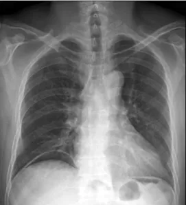
Vol. 58 No. 4, October 2011 Fig. 1. Chest X-ray finding. It showed free air below the right diaphragm.
Fig. 2. Abdominal CT finding. It showed massive pneumoperitoneum, especially in the upper abdomen. Cancer with occult perforation was suspected because of gastric wall thickening with small air bubble in the lesser curvature of the stomach.
Fig. 3. Gross finding of subtotally resected stomach. There was a large ulceroinfiltrative lesion in the antrum along the lesser curvature. The size of tumor was 6.9×6.6 cm sized. At the central portion of the tumor a tiny hole was noted, suggesting perforated site (yellow arrow).
mg/dL, 크레아티닌 0.7 mg/dL이었다. 흉부 단순 X-선 촬영 에서 오른쪽 횡격막 아래에 초승달 모양의 자유공기(free air) 가 관찰되었다(Fig. 1). 위장관 천공에 의한 복막염이 의심되 어, 응급으로 복부 CT를 시행하였다. 복부 CT에서 상복부 쪽 으로 다량의 복강내 공기가 관찰되었으며, 위의 소만측으로 위암이 의심되었다(Fig. 2). 위암에 의해 발생한 자발성 천공 과 이로 인한 복막염으로 진단을 하고 위암에 준하여 근치적 위아전절제술을 응급으로 시행하였다. 절제된 조직에서 전정 부 소만측에 6.9×6.6 cm 크기로 궤양침윤 형태의 병변이 있 었으며, 병변의 중심부위에 천공부위로 보이는 2-3 mm 정도
의 직경을 갖는 작은 구멍을 관찰할 수 있었다(Fig. 3). 조직병 리검사에서 종양세포들은 장막층까지 침윤하고 있었으며 (Fig. 4A), 종양세포들이 근육층을 밀고 들어가는 양상의 ‘push- ing border’를 형성하고 있었다(Fig. 4B). 고배율에서 림프구 성 기질에 불규칙한 모양이지만 경계는 비교적 명확한 모양을 띠는 세포 군집형태의 병변을 관찰할 수 있었다(Fig. 4C).
이상의 조직소견을 통해 위에 발생한 림프상피종양 암종으 로 진단할 수 있었다. 주변 림프절 4개의 침범이 확인되어, 수술 후 병기는 T4aN2M0로 stage IIIb에 해당되었다. 환자 는 수술 후 동시 항암화학 방사선치료를 시행받고, 현재는 경 구 항암제를 복용하면서 수술 후 1년째 재발의 소견 없이 외 래 추적관찰 중이다.
고 찰
위에 발생하는 림프상피종양 암종은 위암의 드문 형태로 림 프 모양의 간질에 작은 무리를 이루는 악성종양 세포의 침윤과 주변에 림프구가 침윤하는 조직학적 특징을 갖고 있다.1 이는 다른 위암종과는 구분되는 독특한 조직형태로, 주로 비인강에 서 가장 많이 발생하며, 이 외에도 구강, 타액선, 흉선, 폐, 자궁 경부, 요관, 식도 등 다양한 기관에서 발생할 수 있는 것으로 알려져 있다.5,6 우리나라에서는 위에 발생한 림프상피종양 암 종이 점막하 종양의 형태로 나타난 3예4,7,8와 내시경 점막하 박 리술로 치료된 2예9,10가 보고되었다.
림프상피종양 암종의 원인은 아직 정확히 밝혀지지는 않았 으나, 몇몇 연구에서 Epstein-Barr 바이러스(EBV)가 77.8- 100%에서 양성을 보인다고 보고하였고,11,12 Kume 등13은 Fas ligand, Hsu 등14은 Interleukin-10과의 연관성을 보고하 기도 하였다. 이번 증례에서 EBV의 검사는 이루어지지 않았
210
고평곤 등. 위 천공에 의한 복막염으로 나타난 위 림프상피종양 암종 1예The Korean Journal of Gastroenterology Fig. 4. Pathologic findings. (A) Tumor cells infiltrated into the serosa
(H&E, ×20). (B) Tumor cells consisted ‘pushing border’ around the muscle layer. It was characteristic of lymphoepithelioma-like carcinoma (H&E, ×20). (C) Nests of tumor cells were mixed with numerous lymphocytes. A lot of lymphocytes infiltrated into the surrounding muscle layers (H&E, ×200).
다.
이 종양은 아직 많은 연구가 이루어지지는 못하였지만, Watanabe 등이 위에서 발생한 42명의 림프상피종양 암종 환 자를 대상으로 발표한 연구에 따르면, 이들 중 조기위암이 17
명, 진행성 위암이 25명이었고, 평균 나이는 52.4세였다. 조기 위암의 경우 모두 전이는 없었으며, 77%에서 궤양이 동반되 었으며 IIa+IIc형이 가장 많았고, 진행성 위암에서는 48%에 서 국소 림프절 전이가 있었고 72%에서 궤양이 동반되었으 며, Borrmann 2형이 가장 많았다.3
또한 위 림프상피종양 암종은 위에 발생하는 선암에 비해 좋은 예후를 보이는 것으로 알려져 있는데, Watanabe 등은 5년 생존율을 점막하층으로 침범한 경우 100%, 고유근층을 침범한 경우 97.2%, 장막을 침범한 경우 77.5%로 보고하였 다.3
이처럼 림프구 침윤이 동반된 위 림프상피종양 암종과 같은 위암종의 예후가 다른 위암종에 비해 예후가 좋은 이유에 대 해, 림프구성 침윤이 암세포에 대해 숙주의 면역 반응을 통해 방어 작용에 좋은 역할을 하기 때문이라는 주장이 있었다.3 또 한, 간질에 림프구 침윤이 풍부한 종양의 경우 종양 기질내 결 체 조직이 거의 없기 때문에 종양 세포내로 항암제의 침투를 용이하게 하여 민감한 치료 반응을 보여 좋은 예후를 나타낸다 고 하였다.15
이번 증례는 위암의 합병증인 자발성 천공에 의한 복막염으 로 내원한 경우였다. 위암의 자발성 천공은 약 1-4%의 발생률 을 보이는 드문 합병증으로, 주로 장막의 침윤과 림프절 전이 를 동반한 진행성 암에서 발생하는 경우가 많다.16 위에 발생한 암종에 의해 위벽의 천공이 발생하는 이유는, 종양의 신생혈관 생성에 의해 조직이 허혈성 손상에 취약해지는 것과 감염성 인자에 의해 취약해지는 것 때문으로 여겨진다.17
이러한 자발성 천공으로 내원하여 진단된 위암종의 경우, 천공에 의한 복막 오염이나 암세포의 복막 전이의 가능성이 있고, 더군다나 복막염으로 인해 응급수술을 시행하게 되기 때문에 일반적으로 예후가 매우 좋지 않을 것으로 여겨진다.18 게다가 이번 증례 환자의 경우 수술 후 병기가 stage IIIb로 상당히 진행되어 있었기 때문에, 더욱 나쁜 예후가 예상되었 다. 하지만 환자는 수술 후 동시 항암화학 방사선치료 후 현재 까지 복부 CT 상 재발의 증거가 없는 상태이다. 이 환자에서 이처럼 좋은 예후를 보이는 이유로, 암종의 조직형태가 큰 영 향을 미쳤을 것으로 추정된다.
폐에 발생한 림프상피종양 암종의 경우 몇몇 연구에서 항 암치료 또는 동시 항암화학 방사선치료에 대한 보고가 있지
만,19,20 위 림프상피종양 암종의 경우 수술 후 치료에 대한
연구가 거의 되어있지 않은 상황이다. 하지만 위 림프상피종 양 암종과 같은 EBV와 연관된 암종의 경우 항암제에 좋은 반응을 보이는 것으로 알려져 있다.15
이번 증례는 위에 발생하는 드문 암인 림프상피종양 암종에 위 천공이라는 드문 합병증으로 내원하여 진단하게 된 경우이 다. 위 림프상피종양 암종에 대한 증례보고가 일본을 비롯한
Goh PG, et al. A Case of Gastric Lymphoepithelioma-like Carcinoma Presenting as Panperitonitis by Perforation of Stomach
211
Vol. 58 No. 4, October 2011
아시아에서 많은 점을 감안할 때, 우리나라에서도 더 많은 환 자들이 있을 수 있다고 생각된다. 이 암의 경우 진행성 암에서 도 이번 증례에서처럼 치료 후 좋은 예후를 보일 수 있으므로 기존의 위선암과는 다른 접근이 필요할 것으로 여겨지며, 이에 대한 더 많은 연구가 이루어져야 할 것이다.
REFERENCES
1. Wang HH, Wu MS, Shun CT, Wang HP, Lin CC, Lin JT.
Lymphoepithelioma-like carcinoma of the stomach: a subset of gastric carcinoma with distinct clinicopathological features and high prevalence of Epstein-Barr virus infection. Hepatogas- troenterology 1999;46:1214-1219.
2. MacCarty WC, Mahle AE. Relation of differentiation and lympho- cytic infiltration to postoperative longevity in gastric carcinoma.
J Lab Clin Med 1921;6:473-480.
3. Watanabe H, Enjoji M, Imai T. Gastric carcinoma with lymphoid stroma. Its morphologic characteristics and prognostic correlations. Cancer 1976;38:232-243.
4. Lee SH, Jang BI, Eun JR, Kim KO, Lee KH, Kim TN. A case of sub- mucosal gastric lymphoepithelioma-like carcinoma. Korean J Med 2009;76(Suppl 1):S35-S39.
5. Chan JK, Hui PK, Tsang WY, et al. Primary lymphoepithelio- ma-like carcinoma of the lung. A clinicopathologic study of 11 cases. Cancer 1995;76:413-422.
6. Chan JK, Yip TT, Tsang WY, Poon YF, Wong CS, Ma VW. Specific association of Epstein-Barr virus with lymphoepithelial carcino- ma among tumors and tumorlike lesions of the salivary gland.
Arch Pathol Lab Med 1994;118:994-997.
7. Hong CK, Chung WC, Choi HJ, et al. Two cases of advanced gastric carcinomas showing features of submucosal tumors. Korean J Gastrointest Endosc 2007;35:175-180.
8. Park WI, Kim HW, Park JH, et al. A case of gastric lymphoepithe- lioma-like carcinoma presenting as a submucosal tumor.
Korean J Gastrointest Endosc 2004;28:123-126.
9. Cho JH, Lee WS, Lee KR, et al. Gastric lymphoepithelioma-like
carcinoma diagnosed and treated by endoscopic submucosal dissection: review of the literature. Korean J Gastrointest Endosc 2010;40:256-260.
10. Moon HS, Kang SH, Seong JK, Jeong HY, Song KS.
Lymphoepithelioma-like gastric carcinoma resected by endo- scopic submucosal dissection (ESD). Endoscopy 2010;42 Suppl 2:E73-74.
11. Oda K, Tamaru J, Takenouchi T, et al. Association of Epstein-Barr virus with gastric carcinoma with lymphoid stroma. Am J Pathol 1993;143:1063-1071.
12. Nakamura S, Ueki T, Yao T, Ueyama T, Tsuneyoshi M.
Epstein-Barr virus in gastric carcinoma with lymphoid stroma.
Special reference to its detection by the polymerase chain re- action and in situ hybridization in 99 tumors, including a mor- phologic analysis. Cancer 1994;73:2239-2249.
13. Kume T, Oshima K, Yamashita Y, Shirakusa T, Kikuchi M.
Relationship between Fas-ligand expression on carcinoma cell and cytotoxic T-lymphocyte response in lymphoepithelio- ma-like cancer of the stomach. Int J Cancer 1999;84:339-343.
14. Hsu DH, de Waal Malefyt R, Fiorentino DF, et al. Expression of interleukin-10 activity by Epstein-Barr virus protein BCRF1.
Science 1990;250:830-832.
15. Matsunou H, Konishi F, Hori H, et al. Characteristics of Epstein-Barr virus-associated gastric carcinoma with lym- phoid stroma in Japan. Cancer 1996;77:1998-2004.
16. Jwo SC, Chien RN, Chao TC, Chen HY, Lin CY. Clinicopathologi- cal features, surgical management, and disease outcome of perforated gastric cancer. J Surg Oncol 2005;91:219-225.
17. Stechenberg L, Bunch RH, Anderson MC. The surgical therapy for perforated gastric cancer. Am Surg 1981;47:208-210.
18. Lee MS, Chae MK, Kim TY, et al. Surgical results for perforated gastric cancer. J Korean Gastric Cancer Assoc 2002;2:85-90.
19. Ho JC, Wong MP, Lam WK. Lymphoepithelioma-like carcinoma of the lung. Respirology 2006;11:539-545.
20. Ho JC, Lam WK, Ooi GC, Lam B, Tsang KW. Chemoradiotherapy for advanced lymphoepithelioma-like carcinoma of the lung.
Respir Med 2000;94:943-947.