CASE REPORT
독성 간염과 동반되어 나타난 간의 결절성 재생증식 1예
진선미, 송상희, 조양현, 신대규, 신선영, 김광일1, 박하나, 임규성
차의과학대학교 분당차병원 내과학교실, 병리학교실1
A Case of Nodular Regenerative Hyperplasia of the Liver Combined with Toxic Hepatitis
Sun Mi Jin, Sang Hee Song, Yang Hyun Cho, Dae Kyu Shin, Sun Young Shin, Gwang Il Kim1, Hana Park and Kyu Sung Rim Departments of Internal Medicine and Pathology1, CHA Bundang Medical Center, CHA University, Seongnam, Korea
Nodular regenerative hyperplasia (NRH) is an uncommon liver condition characterized by diffuse transformation of the hepatic parenchyma into regenerative nodules without fibrosis. Portal vasculopathy caused by abnormal hepatic venous flow may induce hepatocyte hyperplasia, which forms regenerative nodules. Underlying diseases or certain drugs may also be the cause of NRH. This condition is often underdiagnosed as the patients remain asymptomatic until development of portal hypertension, and histopathologic confirmation by liver biopsy is the only way of making a definite diagnosis. The management mainly involves prevention and treatment of the complications of portal hypertension. The frequency of diagnosis of NRH has increased rapidly in recent years, however, only a few cases have been reported in Korea. Here, we report on a case of NRH of the liver combined with toxic hepatitis. (Korean J Gastroenterol 2015;65:52-56)
Key Words: Nodular regenerative hyperplasia; Drug-induced liver injury; Liver diseases
Received July 7, 2014. Revised August 4, 2014. Accepted August 8, 2014.
CC This is an open access article distributed under the terms of the Creative Commons Attribution Non-Commercial License (http://creativecommons.org/licenses/
by-nc/3.0) which permits unrestricted non-commercial use, distribution, and reproduction in any medium, provided the original work is properly cited.
Copyright © 2015. Korean Society of Gastroenterology.
교신저자: 임규성, 463-712, 성남시 분당구 야탑로 59, 차의과학대학교 분당차병원 내과학교실
Correspondence to: Kyu Sung Rim, Department of Internal Medicine, CHA Bundang Medical Center, CHA University, 59 Yatap-ro, Bundang-gu, Seongnam 463-712, Korea. Tel: +82-31-780-5212, Fax: +82-31-780-5219, E-mail: [email protected]
Financial support: None. Conflict of interest: None.
INTRODUCTION
Nodular regenerative hyperplasia (NRH) is an uncommon liver condition characterized by diffuse transformation of the hepatic parenchyma into regenerative nodules without fibrosis. Nodules vary in size but are usually small.1 This dis- ease was initially described as “miliary hepatocellular ad- enomatosis” or “non-cirrhotic nodulation” before first being termed as NRH by Steiner in 1959.2 NRH can be diagnosed when nodulations evenly distributed across the whole liver are observed on an image study and no histological fibrosis between nodules in a patient with evidence of portal hypertension. NRH leads to non-cirrhotic portal hyper- tension, the complications of which are the cause of the main
symptoms of NRH. The disease progression is slow in most cases but may be accelerated due to other unexpected reasons.3 The frequency of diagnosis of NRH has increased rapidly in recent years, amounting to hundreds of cases worldwide in the last decade; however, not a single new case has been reported in Korea during the same period of time.
Here, we report on our experience with a patient of NRH com- bined with toxic hepatitis, together with a literature review.
CASE REPORT
A 54-year-old woman with no specific medical history vis- ited our hepatology clinic for jaundice and lower limb edema that had lasted for about 10 days. She had been suffering
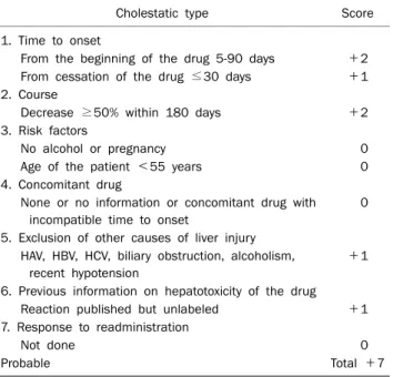
Fig. 2. Initial liver MRI shows large T2 low (A) and T1 high (B) nodular lesions in the liver.
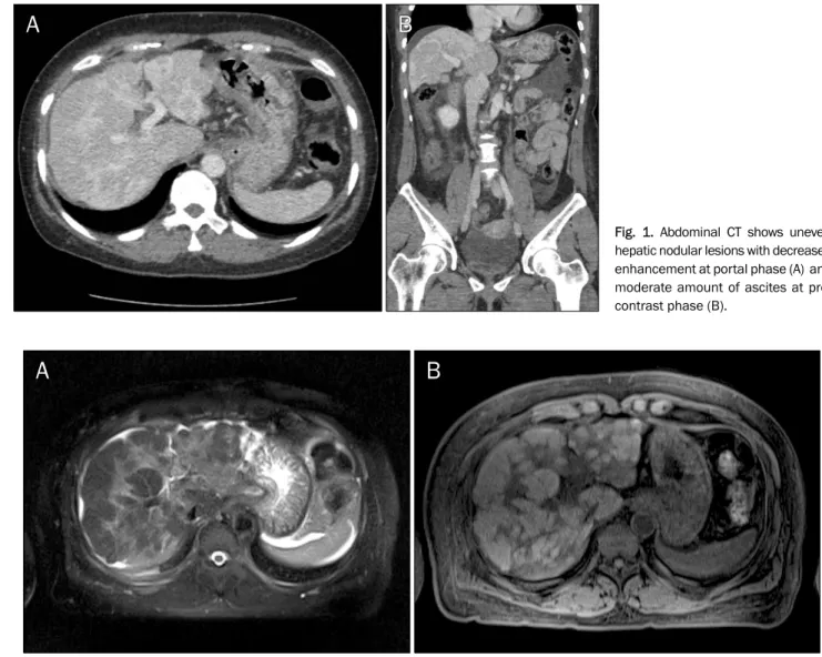
Fig. 1. Abdominal CT shows uneven hepatic nodular lesions with decreased enhancement at portal phase (A) and moderate amount of ascites at pre- contrast phase (B).
from nausea and abdominal discomfort for almost a month, for which she had taken herbal medicines for one week.
During this time she developed jaundice and lower limb edema. Her primary care physician had detected abnormal- ities in her liver function tests and referred her to a specialist.
Eventually, she was admitted to our hospital. She had under- gone esophagogastroduodenoscopy at the primary care for her upper abdominal discomfort and was taking medication for duodenal ulcer, which was detected at that time. Herbal medicines were discontinued. She had no history of smoking or drinking alcohol.
At the time of admission, the patient’s vital signs were as follows: blood pressure 130/80 mmHg, pulse rate 80 beats/min, respiratory rate 16 times/min, and body temper- ature 36.7oC. She was alert and well oriented. Physical exami-
nation revealed pitting edema of the lower limbs and icteric sclera. Hepatomegaly or splenomegaly was not detected.
Laboratory tests showed hemoglobin level 11.7 g/dL; plate- let count 71,000/L; white blood cell count 4,000/L (neutrophils 40% and lymphocyte 46%); blood urea nitrogen 7.0 mg/dL; creatinine 0.8 mg/dL; prothrombin time 18.9 sec;
international normalized ratio 1.67; activated partial throm- boplastin time 33.6 sec; ferritin 1,879 ng/mL; albumin 2.4 g/dL; AST 229 IU/L; ALT 224 IU/L; ALP 3,590 IU/L; and total bilirubin 6.69 mg/dL. Diagnostic markers of viral hepatitis, including IgM anti-HAV, HBsAg, anti-HCV, HCV-RNA, and IgM anti-HEV were all negative. Autoimmune markers, including anti-nuclear, anti-smooth muscle, anti-mitochondrial, anti- dsDNA, and anti-cardiolipin antibodies, were also negative.
Ceruloplasmin, serum copper, and serum iron were all within
Fig. 3. (A) Most hepatocytes are arranged in two-to-three cell thick plates, and small cell or large cell changes (arrowheads) are seen (H&E, ×100).
(B) Masson trichrome stain (×40) shows absence of fibrosis and presence of portal tracts (arrows).
Table 1. Roussel Uclaf Causality Assessment Method Scale Score of Our Patient
Cholestatic type Score
1. Time to onset
From the beginning of the drug 5-90 days +2 From cessation of the drug ≤30 days +1 2. Course
Decrease ≥50% within 180 days +2
3. Risk factors
No alcohol or pregnancy 0
Age of the patient <55 years 0
4. Concomitant drug
None or no information or concomitant drug with incompatible time to onset
0
5. Exclusion of other causes of liver injury HAV, HBV, HCV, biliary obstruction, alcoholism,
recent hypotension
+1
6. Previous information on hepatotoxicity of the drug
Reaction published but unlabeled +1
7. Response to readministration
Not done 0
Probable Total +7
The type of liver injury was considered as ‘cholestatic’ since R ratio (serum activity of ALT/serum activity of ALP) was 0.4.
the normal range.
Abdominal ultrasonography at the primary care showed coarse echotexture of the liver parenchyma; however, no nod- ules were observed. Abdominal CT scan at our hospital showed high-attenuation nodules distributed across the liver on the precontrast phase and decreased enhancement com- pared to the adjacent liver parenchyma (Fig. 1A). A moderate amount of ascites was observed but portal vein thrombosis was not present (Fig. 1B). Liver MRI demonstrated numerous large nodular lesions in both T2 low signal and T1 high signal images (Fig. 2). Repeat esophagogastroduodenoscopy showed a healing duodenal ulcer but no esophageal or gas- tric varix. Colonoscopy findings were unremarkable.
Liver biopsy was performed and microscopic examination revealed that the lesion was composed of hepatocytes ar- ranged in two-to three cell-thick plates and diffuse small cell or large cell changes were seen (Fig. 3A). Masson trichrome stain protocol for collagen fibers showed no fibrosis and there was no evidence of dysplastic or carcinomatous changes of hepatocytes (Fig. 3B). Conclusively, the investigations showed marked regenerative hepatocytic changes without fibrosis, which conformed to the definition of NRH.
Based on the above mentioned findings, the patient was finally diagnosed with NRH associated with toxic hepatitis.
Conservative treatment including hepatotonics, diuretics, and albumin was administered. The patient was discharged two weeks after admission, because her general condition showed significant improvement, along with slight improve- ment in her liver function tests; showing AST/ ALT 157/32
IU/L, ALP 1,102 IU/L, and total bilirubin 6.4 mg/dL. Three months after discharge, the laboratory findings were almost normalized; AST/ALT 40/14 IU/L, ALP 319 IU/L, total bilirubin 1.2 mg/dL, ferritin 384 ng/mL, and albumin 3.1 g/dL. In addi- tion, she had recovered completely from the jaundice and lower limb edema. According to the Roussel Uclaf Causality Assessment Method scale for determining drug-induced liver injury, this patient was scored as ‘probable’ (Table 1). Five months after discharge, MRI showed mild enlargement of in-
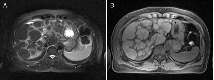
Fig. 4. Follow-up liver MRI shows progression of nodular regenerative hyperplasia in the liver. These nodular lesions show T2 low (A) and T1 high (B) signal intensity.
numerable nodular lesions in the liver and increase of num- bers compared to the previous MRI. This finding suggested some progression of NRH in the liver (Fig. 4). However the lab- oratory findings were still within normal limit. She has been on regular follow-up on an outpatient basis.
DISCUSSION
NRH is a rare disease; the current knowledge regarding NRH has been obtained mainly from case reports and only a few large studies. NRH is underdiagnosed and under- reported because of the difficulty of the diagnostic approach.
Only a few cases of NRH have been reported in Korea.4-6 An autopsy study of 2,500 patients found the presence of NRH in 64 patients, 2.6% of the overall incidence.1 Many of these patients had accompanying systemic disorders, and the incidence of NRH among patients aged 80 years and old- er was seven times higher than that among patients aged 60 years and younger, thereby suggesting that older patients with an associated systemic disease are more susceptible to NRH.1
The pathogenesis of NRH is not yet well established, how- ever, adaptive hyperproliferative response of hepatocytes to alteration in portal hepatic venous perfusion is assumed to be the main mechanism.7 NRH is also highly associated with underlying diseases, including autoimmune, inflammatory, and neoplastic disorders.8 In addition, NRH has often been reported in patients after chemotherapy with certain drugs9 and use of immunosuppressant such as thiopurine, which
may cause damage to hepatic venous endothelial cells.10,11 However, our patient had no medical history or disorder that might be considered as a cause of NRH.
NRH is not a common disease; however, it should be sus- pected in patients with symptoms of portal hypertension, such as ascites, esophageal varix, and splenomegaly, with normal liver enzymes and no other findings suggestive of liver cirrhosis. Eliminating other common causes of non-cirrhotic portal hypertension, such as virus, alcohol, drugs, and auto- immune disorder, is also essential for the diagnosis. NRH is asymptomatic most of the time until the development of por- tal hypertension,12 in which case it is often detected by in- cidental findings on imaging studies. In our patient, AST and ALT increased to levels higher than 200 IU/L, and she had tak- en herbal medicines before the symptoms appeared. It is suspected that she had been asymptomatic with an under- lying NRH for a long period of time and that her current symp- toms were aggravated because of acute hepatitis. We ruled out other common causes of acute hepatitis and presumed that the herbal product could be the highly possible cause of hepatitis. Diagnostic images, such as ultrasound, CT, and MRI have low sensitivity and specificity for diagnosis of NRH and are known to show only non-specific, diffuse liver disease or several millimeters of tiny regenerative nodules evenly dis- tributed across the liver.13,14 However, reports indicate that, although rare, macronodules may be detected by imag- ing,15,16 which was applicable in our case. In the end, histo- pathologic confirmation by liver biopsy is the gold standard for a definite diagnosis.
In management of patients with NRH, the priority is treat- ment of causal factors and control of symptoms arising from portal hypertension. Ascites and esophageal varix may be managed by conventional standard treatments such as diu- retics, endoscopic variceal ligation, and portosystemic shunt.
Liver transplantation is rarely attempted.
Disease progression is slow in most cases. The expected prognosis is presumably better than in cirrhosis or other chronic liver diseases; however, long-term prognosis is still unknown. NRH was associated with hepatocellular carcinoma in several reported cases; however, there is no evidence sug- gesting that these NRH cases were pre-malignant lesions.3,17 Further studies are needed in order to obtain long-term da- ta on the natural history of NRH and to determine the etiology of NRH, particularly the molecular pathogenesis associated with portal vasculopathy and tumorigenesis.
In conclusion, NRH is an uncommon liver condition, which probably has a benign progressive course. We presented a patient who showed a macronodular hepatic lesion on radio- logic finding and was finally diagnosed with NRH based on histology results. We believe that the herbal medicine that the patient had been taking before the symptoms appeared, might have led to the development of toxic hepatitis in addi- tion to the underlying NRH.
REFERENCES
1. Wanless IR. Micronodular transformation (nodular regenerative hyperplasia) of the liver: a report of 64 cases among 2,500 au- topsies and a new classification of benign hepatocellular nodules. Hepatology 1990;11:787-797.
2. Steiner PE. Nodular regenerative hyperplasia of the liver. Am J Pathol 1959;35:943-953.
3. Nzeako UC, Goodman ZD, Ishak KG. Hepatocellular carcinoma and nodular regenerative hyperplasia: possible pathogenetic relationship. Am J Gastroenterol 1996;91:879-884.
4. Lee DH, Lee JI, Ko YT, Kim YW. Nodular regenerative hyperplasia of the liver: radiologic findings. J Korean Radiol Soc 1997;37:
119-122.
5. Kim HK, Lee YH, Chung DS, Kim OD, Whang JB, Park JB. Nodular
regenerative hyperplasia of the liver in an infant: case report. J Korean Radiol Soc 2002;47:689-692.
6. Shim SG, Sohn JH, Lee JW, et al. A case of nodular regenerative hyperplasia of liver that mimicked primary biliary cirrhosis.
Korean J Hepatol 2004;10:313-318.
7. Wanless IR, Godwin TA, Allen F, Feder A. Nodular regenerative hy- perplasia of the liver in hematologic disorders: a possible re- sponse to obliterative portal venopathy. A morphometric study of nine cases with an hypothesis on the pathogenesis. Medicine (Baltimore) 1980;59:367-379.
8. Morris JM, Oien KA, McMahon M, et al. Nodular regenerative hy- perplasia of the liver: survival and associated features in a UK case series. Eur J Gastroenterol Hepatol 2010;22:1001-1005.
9. Rubbia-Brandt L, Lauwers GY, Wang H, et al. Sinusoidal ob- struction syndrome and nodular regenerative hyperplasia are frequent oxaliplatin-associated liver lesions and partially pre- vented by bevacizumab in patients with hepatic colorectal metastasis. Histopathology 2010;56:430-439.
10. Buster EH, van Vuuren HJ, Zondervan PE, Metselaar HJ, Tilanus HW, de Man RA. Thiopurine-methyltransferase and inosine tri- phosphate pyrophosphatase polymorphism in a liver transplant recipient developing nodular regenerative hyperplasia on low-dose azathioprine. Eur J Gastroenterol Hepatol 2008;20:
68-72.
11. Daniel F, Cadranel JF, Seksik P, et al. Azathioprine induced nod- ular regenerative hyperplasia in IBD patients. Gastroenterol Clin Biol 2005;29:600-603.
12. Ueno S, Tanabe G, Sueyoshi K, et al. Hepatic hemodynamics in a patient with nodular regenerative hyperplasia. Am J Gastroen- terol 1996;91:1012-1015.
13. Clouet M, Boulay I, Boudiaf M, et al. Imaging features of nodular regenerative hyperplasia of the liver mimicking hepatic metastases. Abdom Imaging 1999;24:258-261.
14. Casillas C, Martí-Bonmatí L, Galant J. Pseudotumoral pre- sentation of nodular regenerative hyperplasia of the liver: imag- ing in five patients including MR imaging. Eur Radiol 1997;7:
654-658.
15. Reshamwala PA, Kleiner DE, Heller T. Nodular regenerative hy- perplasia: not all nodules are created equal. Hepatology 2006;44:7-14.
16. Doğan E, Ozgür R, Ercan V, Tekin A, Senkal O, Cevikbaş U.
Nodular regenerative hyperplasia of the liver: a case report. Turk J Gastroenterol 2003;14:64-67.
17. Kobayashi S, Saito K, Nakanuma Y. Nodular regenerative hyper- plasia of the liver in hepatocellular carcinoma. An autopsy study.
J Clin Gastroenterol 1993;16:155-159.