J Lung Cancer 2011;10(2):102-104 http://dx.doi.org/10.6058/jlc.2011.10.2.102
102
Fatal Broncho-Mediastinal Fistula in a Patient with Non- Small Cell Lung Cancer after Photodynamic Therapy
Photodynamic therapy (PDT) can be used as palliative therapy to reduce obstructive symptoms in patients with advanced lung cancer. Herein, we report on the case of a patient with fatal broncho-mediastinal fistula after PDT. A 57-year-old woman was diagnosed as non-small cell lung cancer (squamous cell carcinoma, cT4N3). She received PDT on the endobronchial mass ob- structing her right main bronchus twice in 48 hours interval. Two weeks later, concurrent chemoradiation therapy (CCRT) with weekly Paclitaxel/Carboplatin was started. During maintenance chemotherapy, a new nodule in her scalp developed and turned out to be a metastatic nodule. A broncho-mediastinal fistula was suspicious on follow-up chest computed tomography and a broncoscopy revealed an extensively damaged medial right main bronchial wall.
On the day following bronchoscopy, the patient died of sudden massive hemoptysis. (J Lung Cancer 2011;10(2):102 104)
Key Words: Lung cancer, Photodynamic therapy
Eun Young Heo, M.D.1 Yu Jung Kim, M.D.2 and Seok-Chul Yang, M.D., Ph.D.3
1Department of Internal Medicine, SMG- SNU Boramae Medical Center, Seoul,
2Division of Hematology and Medical Oncology, Department of Internal Medi- cine, Seoul National University of Bun- dang Hospital, Seongnam, 3Division of Pulmonary and Critical Care Medicine, Department of Internal Medicine and Lung Institute, Seoul National Univer- sity College of Medicine, Seoul, Korea Received: November 19, 2011 Revised: December 5, 2011 Accepted: December 7, 2011 Address for correspondence Seok-Chul Yang, M.D., Ph.D.
Division of Pulmonary and Critical Care Medicine, Department of Internal Medi- cine, Seoul National University College of Medicine, 28, Yeongeon-dong, Jong- no-gu, Seoul 110-799, Korea Tel: 82-2-2072-0354 Fax: 82-2-762-9662 E-mail: [email protected]
Photodynamic therapy (PDT) with radiotherapy can be used as a palliative therapy in late stage non-small cell lung cancer patients with endobronchial obstructive lesions to reduce symptoms. Physicians must consider the potential side effects of PDT. We report a case with fatal broncho-mediastinal fistula after PDT.
CASE REPORT
A 57-year old woman was admitted to our hospital for evaluation of a lung mass. She was a current 30 pack-year smoker and had no other past medical history. Four months prior to hospital admission, the patient began to experience cough, dyspnea, and occasional blood tinged sputum. Therefore,
she visited a local clinic and was treated for bronchial asthma.
Despite asthma treatment for 2 months, there was no improve- ment in symptoms and, she visited another hospital and had a chest computed tomography (CT) scan on which her lung mass was detected. Initial chest CT showed a left mediastinal mass adjacent to the left innominate vein and aorta, the right main bronchus encircling the mass, multiple mediastinal and hilar lymphadenopathies, and a left supraclavicular lymphaden- opathy. She was diagnosed with non-small cell lung cancer (squamous cell carcinoma, cT4N3) after needle aspiration biopsy of the left supraclavicular lymph node. A bronchoscopy revealed nearly total obstruction of the right main bronchus due to an endobronchial mass, and tumor infiltration was observed from the carina to the left main bronchus 2 cm from the carina.
Broncho-mediastinal Fistula after PDT Therapy 103
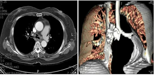
Fig. 1. Chest CT finding of bron- cho-mediastinal fistula after photo- dynamic therapy and concurrent chemoradiation therapy.
Fig. 2. Bronchoscopic finding of extensively damaged carina and the medial wall of right main bronchus (A, carina; B, right main bronchus).
Forty-eight hours after intravenous Photogem (TimTec Corp., Newark, DE, USA) (3 mg/kg) as a photosensitizer, she received PDT on the endobronchial mass twice in 48 hours interval. The diode laser (Ceralas, 630 nm wavelength, 200 J/cm2) with a 2 cm length frontal type optic fiber was used for light irradiation.
On clean-up bronchoscopy after PDT, the patency of the right main bronchus was observed as 70∼80%. Two weeks later, concurrent chemoradiation therapy (CCRT) with weekly Pacli- taxel/Carboplatin (Paclitaxel: 45 mg/m2, Carboplatin AUC: 2) was started. Radiotherapy (60 Gy) was administered to the tumors and mediastinal lymph nodes. Five cycles of chemo- therapy with weekly Paclitaxel/Carboplatin were administered together with radiation. After the completion of chemoradiation, the disease was partially responsive. Therefore, a maintenance dose of Paclitaxel/Carboplatin (80% of the original dose) was administered for 2 more months. During maintenance chemo- therapy, a small scalp nodule developed, and it was determined to be a metastatic lesion by skin biopsy. The patient had been
off chemotherapy due to disease progression and the status of disease was reevaluated. The main lung mass remained in a decreased status, but the broncho-mediastinal fistula and left femur shaft metastasis was found to be suspicious (Fig. 1). The patient was referred to an emergency room for further evaluation and management. On the following day, a bron- choscopy was performed to identify the broncho-mediastinal fistula on previously treated area. On bronchoscopy, the medial right main bronchial wall was extensively damaged and connected to the left main bronchus, and was filled with yellowish necrotic material (Fig. 2). The right upper lobar bronchus wall looked intact. On the day following broncho- scopy, the patient died of sudden massive hemoptysis.
DISCUSSION
PDT was established as an effective local therapy for the palliative care of obstructing endobronchial lung cancer (1-3).
104 J Lung Cancer 2011;10(2):102-104
PDT is a two-step process that involves intravenous admini- stration of a photosensitizer, followed by illumination with a suitable wavelength of light after 2 to 4 days. The cytotoxic effects of PDT cause tissue necrosis by the production of singlet oxygen, which is produced by the interaction of the photosensitizer and laser light in the presence of oxygen (1).
Tissue necrosis by PDT is superficial, up to a maximal depth of 10 mm. The greatest advantage of PDT is protection of adjacent normal tissues. In addition, PDT can be repeated in cases of recurrence or new primary tumors in previously treated areas (4). Therefore, PDT has been used previously as a treatment for endobronchial lesions in advanced lung cancer to reduce respiratory symptoms and improve the quality of life.
A few randomized studies and several non-controlled pro- spective studies reported the efficacy and toxicity of PDT in advanced lung cancer patients. Moghissi et al. (1) reported that 8 out of 100 patients died within 30 days of the first treatment.
Four of these patients died of pulmonary hemorrhage and all complained of hemoptysis initially. LoCicero et al. (5) reported 10 cases who had received PDT for the palliation of late stage obstructing lung cancer. Six out of 10 patients subsequently received external beam radiation therapy. Three of these patients developed significant problems following radiation therapy. In a paper describing PDT as an induction modality in NSCLC, bronchopleural fistula occurred in 4 patients who had received chemotherapy, radiotherapy, PDT, and pneumo- nectomy simultaneously (6).
In our case, massive hemoptysis and broncho -nodal fistula after PDT and combined chemotherapy and radiation therapy were observed in obstructed non-small cell lung cancer female patient. The pathogeneses of these complications cannot be explained clearly. However, there have been several experi- mental studies regarding vascular toxicities related to PDT.
Aggregating platelets that cause vasoconstriction and obstruc- tion of tumor blood flow have been observed, which leads to the closure of the tumor vasculature that limits the oxygen
supply to the tumor (7). Kostron et al. showed that ionizing radiation could activate hematoporphyrin derivatives without light, thus inhibiting tumor growth in a rat model (8).
We assume that more severe tissue necrosis could be caused by immediate vascular damage related to PDT itself as well as the synergistic effects of the photosensitizer and ionizing radiation by Paclitaxel based CCRT performed immediately after PDT.
In conclusion, we must carefully consider whether PDT is suitable since severe toxicity is possible when administered together with other therapeutic modalities in patients with advanced lung cancer.
REFERENCES
1. Moghissi K, Dixon K, Stringer M, Freeman T, Thorpe A, Brown S. The place of bronchoscopic photodynamic therapy in advanced unresectable lung cancer: experience of 100 cases.
Eur J Cardiothorac Surg 1999;15:1-6.
2. Imamura S, Kusunoki Y, Takifuji N, et al. Photodynamic therapy and/or external beam radiation therapy for roentgen- ologically occult lung cancer. Cancer 1994;73:1608-1614.
3. Jones BU, Helmy M, Brenner M, et al. Photodynamic therapy for patients with advanced non-small-cell carcinoma of the lung. Clin Lung Cancer 2001;3:37-41.
4. Triesscheijn M, Baas P, Schellens JH, Stewart FA. Photody- namic therapy in oncology. Oncologist 2006;11:1034-1044.
5. LoCicero J 3rd, Metzdorff M, Almgren C. Photodynamic therapy in the palliation of late stage obstructing non-small cell lung cancer. Chest 1990;98:97-100.
6. Ross P Jr, Grecula J, Bekaii-Saab T, Villalona-Calero M, Otterson G, Magro C. Incorporation of photodynamic therapy as an induction modality in non-small cell lung cancer. Lasers Surg Med 2006;38:881-889.
7. Stewart F, Baas P, Star W. What does photodynamic therapy have to offer radiation oncologists (or their cancer patients)?
Radiother Oncol 1998;48:233-248.
8. Kostron H, Swartz MR, Miller DC, Martuza RL. The inter- action of hematoporphyrin derivative, light, and ionizing radiation in a rat glioma model. Cancer 1986;57:964-970.