INTRODUCTION
Ovarian cancer is one of the most severe gynecological ma- lignancies known and is the fourth leading cause of cancer- related deaths in the United States [1], and has become the most lethal of all gynecological malignancies in Singapore women. Early stage disease has excellent prognosis but late stage disease has contributed to the high mortality rate of between 80% and 90% compared to early stage disease of between 10% and 30% [2]. Similar trends have been seen in
Combined panel of serum human tissue kallikreins and CA-125 for the detection of epithelial ovarian cancer
Stephen Chee Liang Koh1, Chan Yiong Huak2, Delfi Lutan3, Johny Marpuang3, Suwiyoga Ketut4, Nyoma Gede Budiana4, Agustria Zainu Saleh5, Mohamad Farid Aziz6, Hariyono Winarto6, Heru Pradjatmo7, Nguyen Khac Han Hoan8, Pham Viet Thanh8, Mahesh Choolani1
1Department of Obstetrics and Gynaecology, 2Biostatistics Unit, National University Health System (NUHS), Yong Loo Lin School of Medicine, National University of Singapore, Singapore; 3Department of Obstetrics and Gynaecology, Adam Malik Hospital, University of North Sumatera, Medan; 4Department of Obstetrics & Gynaecology, Sanglah Denpasar Hospital, University of Udayana, Denpasar;
5Department of Obstetrics & Gynaecology, Dr Mohammad Hoesin General Hospital, Uiiversity of Sriwijaya, Palembang; 6Department of Obstetrics & Gynaecology, Division of Oncology, University of Indonesia, Jakarta; 7Department of Obstetrics & Gynaecology, Dr Sardjito Hospital, University Gadja Mada, Yogyakarta, Indonesia; 8Tu Du Hospital, Ho Chi Minh, Vietnam
Received Nov 28, 2011, Revised Apr 13, 2012, Accepted Apr 16, 2012 Correspondence to Stephen Chee Liang Koh
Department of Obstetrics and Gynaecology, National University Health System (NUHS), Yong Loo Lin School of Medicine, National University of Singapore, NUHS Tower Block, Level 12, 1E Kent Ridge Road, Singapore 119228. Tel: 65-6772-2670, Fax: 65-6775-1160, E-mail: [email protected]
Copyright © 2012. Asian Society of Gynecologic Oncology, Korean Society of Gynecologic Oncology
Objective: To determine the predictive accuracy of the combined panels of serum human tissue kallikreins (hKs) and CA-125 for the detection of epithelial ovarian cancer.
Methods: Serum specimens collected from 5 Indonesian centers and 1 Vietnamese center were analyzed for CA-125, hK6, and hK10 levels. A total of 375 specimens from patients presenting with ovarian tumors, which include 156 benign cysts, 172 epithelial ovarian cancers (stage I/II, n=72; stage III/IV, n=100), 36 germ cell tumors and 11 borderline tumors, were included in the study analysis. Receiver operating characteristic analysis were performed to determine the cutoffs for age, CA-125, hK6, and hK10. Sensitivity, specificity, negative, and positive predictive values were determined for various combinations of the biomarkers.
Results: The levels of hK6 and hK10 were significantly elevated in ovarian cancer cases compared to benign cysts. Combination of 3 markers, age/CA-125/hk6 or CA-125/hk6/hk10, showed improved specificity (100%) and positive predictive value (100%) for prediction of ovarian cancer, when compared to the performance of single markers having 80-92% specificity and 74-87%
positive predictive value. Four-marker combination, age/CA-125/hK6/hK10 also showed 100% specificity and 100% positive predictive value, although it demonstrated low sensitivity (11.9%) and negative predictive value (52.8%).
Conclusion: The combination of human tissue kallikreins and CA-125 showed potential for improving prediction of epithelial ovarian cancer in patients presenting with ovarian tumors.
Keywords: Age, Epithelial ovarian cancer, Tumor markers
Singapore women showing mortality of advanced stage at 77.1% and 20% in the early stage disease over a 5-year pe- riod [3]. The overall 5-year survival rate has not changed over the past thirty years despite the availability of new cytotoxic treatments [4] which showed dismal outcome, especially in advanced stage disease. Early detection remains the most important approach to improve long-term survival for ovar- ian cancer. Currently, CA-125 discovered nearly 30 years ago [5] remained the well-accepted serum biomarker for ovarian cancer but is neither specific nor sensitive enough for diag- nosis [6-8]. It has clinical value for disease monitoring and has been used as an aid for early detection of relapse and for evaluation of response to treatment [9-11]. Prognostic indica- tors or biomarkers improve the accuracy of predicting patient outcomes, and until reliable screening or diagnostic strategies become available, they may contribute to the optimal tailored individual management of patients with ovarian cancer.
The current trend to focus on combined multiparametric analysis of different biomarkers to improve clinical outcomes and diagnosis will offer several advantages [12-15]. The group of serine proteases known as human tissue kallikreins (hK) has a rich source of cancer biomarkers and suited for multi- parametric analysis. Human tissue kallikreins (KLK for gene, hK for protein) is a family of 15 members encoded by a group of genes tandemly localized on chromosome 19q13.3-4 [16].
It is highly expressed in sex organs such as the breast, ovary, prostate and testis. Emerging diagnostic markers notably hK6, hK10, and hK11 are highly expressed in the majority of ovarian cancer cases and especially in the advanced stage diseases [17-20]. The conventional CA-125 marker has shown negative values in 40% to 50% of early stage ovarian cancer and upreg- ulated in benign tumors and other cancers [8,21]. However, tissue kallikrein proteins hK6 and hK10 together with osteo- pontin levels have been reported to be promising potential markers that might complement CA-125 in ovarian cancer [22].
It was suggested that the expression of hK6 may be an early event during ovarian cancer development and have potential use as biomarker for early detection of ovarian cancer [23] and prediction of progression-free survival [24]. In a small ovarian cancer cohort study, elevated levels of CA-125, hK6 and hK10 were seen. However, elevated serum hK6 levels was associ- ated with mortality outcome within 12 months of diagnosis compared to those who survived over five years from epithe- lial ovarian cancer [3].
The objective of this collaborative study in South-East Asia from five Indonesian centers and one Vietnamese center in Ho Chi Minh City was to determine the value of ovarian cancer biomarkers hK6/hK10 and their association with CA-125 levels and age in patients with ovarian tumors for the prediction of
epithelial ovarian cancer.
MATERIALS AND METHODS 1. Subjects
The study received approval from the National University of Singapore Institutional Review Board (NUS-IRB reference codes 05-025 and 06-067) and the respective ethics commit- tee of various institutions involved in the study. Inclusion crite- ria were women with a pelvic mass detected by ultrasound at the hospital clinic and scheduled for operation. This was a pro- spective study and patients diagnosed with ovarian tumors were recruited to the study. Informed Consent was sought be- fore they were allowed to take part in the study. Recruitment of subjects began in May 2005 in Medan, Indonesia and there- after in various Indonesian centers; Denpasar (February 2006), Palembang (October 2006), Yogyakarta (December 2006), Ja- karta (September 2007), and Ho Chi Minh City, Vietnam (April 2008) until August 2010. A total of 375 samples with con- firmed patient information data were received; benign cysts 156, epithelial ovarian cancer 172 (stage I/II, n=72; stage III/
IV n=100), germ cell cancer 36 and borderline 11. Administra- tion of chemotherapy consisting of either cyclophosphamide, taxol and platinum drugs was recorded only for 26.2% (45/172) epithelial ovarian cancer patients. Most of the cancer patients were lost to follow-up and survival outcome analysis was not possible as most of them returned to their hometown soon af- ter their diagnosis and surgery or after chemotherapy and did not returned for follow-up visits. Benign cyst cohorts were sig- nificantly younger with mean age of 38.3±1.2 years (95% CI, 35.9 to 40.8) compared to ovarian cancer patients with mean age of 45.5±1.0 years (95% CI, 43.5 to 47.5).
2. Blood collection
Blood sampling was performed in the morning before the scheduled operation. A clean venepuncture was performed together for hemostatic analysis, about 5 mL of plain blood was transferred in to clean plastic tubes. The blood tubes were then left at room temperature to clot for about 2 hours before they are centrifuged at 2,000 g for 15 minutes. The available sera was stored in aliquots and immediately kept at either -40oC or -70oC until dispatched to Singapore for storage at -80oC and analysis.
3. Laboratory assays
CA-125 assay was performed by IMMULITE 1000 systems (Siemens, Los Angeles, CA, USA). Elisa assay for serum hK6 and hK10 was performed at the Pathology & Laboratory Medicine,
Mount Sinai Hospital, Toronto, Canada, after consultation. It is a two-step sandwich immunoassay described earlier by Dia- mandis et al. [25].
4. Statistical analysis
All statistical analysis was performed using SPSS ver. 18.0 (SPSS Inc., Chicago, DE, USA). Group means were tested and compared by t-test. A p-value of less than 0.05 was considered statistically significant. Receiver operating characteristics (ROC) analysis were performed to determine the optimal cutoffs for age, CA-125, hK6 and hK10 levels. The sensitivity, specificity, positive predictive value (ppv) and negative predictive value (npv) for single and combination of markers were determined for the detection of ovarian cancer, early and advanced stage of disease.
RESULTS
The types of benign cysts and epithelial ovarian cancer in
the cohorts studied are shown in Table 1. The comparison of mean values of age, CA-125, hK6, and hK10 between be- nign cysts and epithelial ovarian cancer are shown in Table 2.
Benign cyst cohorts from the Vietnamese center (n=44) and the Indonesian centers (n=112) showed no significant differ- ences in the parameters studied. Hence, they were grouped together in the final analysis. Ovarian cancer cohorts were significantly older with a mean age of 45.5 years compared to 38.3 years in benign cyst cohorts and showed significant upregulation (p<0.001) of CA-125, hK6, and hK10 levels com- pared to benign cyst cohorts. In early stage ovarian cancer cases, significantly elevated levels of CA-125 (p=0.04), hK6 (p=0.03) in addition to older age (p=0.005) were observed compared to benign cyst cohorts. In addition, significant upregulation of CA-125 (p<0.001), hK6 (p=0.001), and hK10 (p=0.001) levels together with older age (p=0.002) were also seen in advanced stage diseases compared to benign cyst cohorts. When comparing early and advanced stage diseases, CA-125 (p=0.03) and hK6 (p=0.04) were significantly elevated in advanced stage disease with no significant differences in
Table 1. Types of benign cyst and epithelial ovarian cancer of cohorts in the study
Dermoid Mucinous Serous Endometroid Clear cell Others
Benign cysts (n=156) 34 37 30 26 - 29
Ovarian cancer (n=172)
Stage - I/II III/IV I/II III/IV I/II III/IV I/II III/IV I/II III/IV
No. - 22 28 21 18 8 9 3 4 18 41
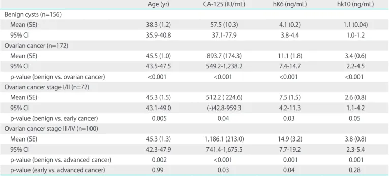
Table 2. Descriptive statistics and group mean test for age, CA-125, human tissue kallikrein 6 (hK6), and hK10 in benign cyst and epithelial ovarian cancer (early and advanced stage disease)
Age (yr) CA-125 (IU/mL) hK6 (ng/mL) hk10 (ng/mL)
Benign cysts (n=156)
Mean (SE) 38.3 (1.2) 57.5 (10.3) 4.1 (0.2) 1.1 (0.04)
95% CI 35.9-40.8 37.1-77.9 3.8-4.4 1.0-1.2
Ovarian cancer (n=172)
Mean (SE) 45.5 (1.0) 893.7 (174.3) 11.1 (1.8) 3.4 (0.6)
95% CI 43.5-47.5 549.2-1,238.2 7.4-14.7 2.2-4.5
p-value (benign vs. ovarian cancer) <0.001 <0.001 <0.001 <0.001
Ovarian cancer stage I/II (n=72)
Mean (SE) 45.3 (1.5) 512.2 ( 224.6) 7.5 (1.5) 2.6 (0.8)
95% CI 43.1-49.0 (-)42.8-959.3 4.2-11.3 1.1-4.2
p-value (benign vs. early cancer) 0.005 0.04 0.03 0.05
Ovarian cancer stage III/IV (n=100)
Mean (SE) 45.3 (1.3) 1,186.1 (213.0) 14.9 (3.2) 3.8 (0.8)
95% CI 42.3-47.9 741.4-1,675.5 7.7-19.2 2.3-5.4
p-value (benign vs. advanced cancer) 0.002 <0.001 0.001 0.001
p-value (early vs. advanced cancer) 0.99 0.03 0.04 0.28
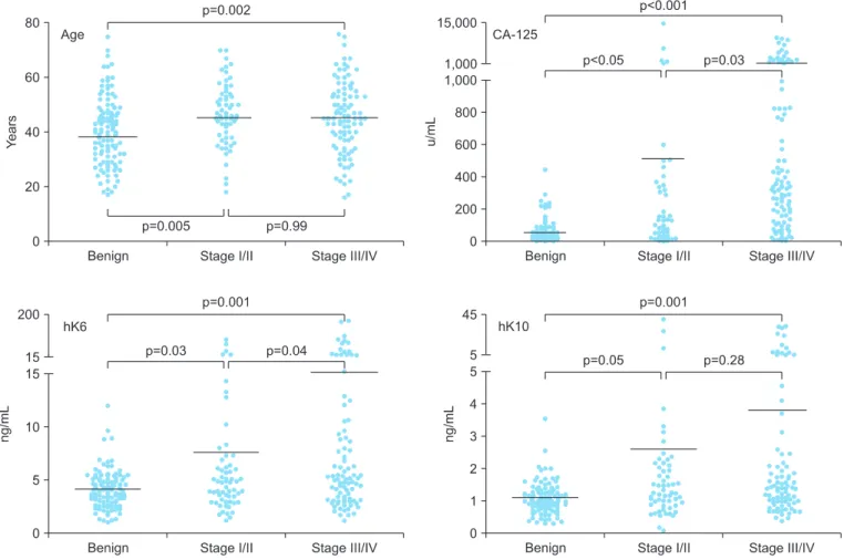
hK10 level and age. The variable scatter plots for the mark- ers studied in benign cyst, early and advanced stage cancer are shown in Fig. 1. No significant differences in age, hK6 and hK10 levels between germ cell cancer (n=36) and benign cyst cohorts except CA-125 level which was significantly elevated (p=0.04) with mean value of 377.3±224.2 IU/mL (data not shown). The highest hK6 and hK10 levels were expressed by mucinous cancer (mean, 10.0±2.6 ng/mL and 3.3±1.2 ng/mL respectively), whilst in benign cyst cohorts, the endometroid type showed relatively elevated levels of hK6 (mean, 4.6±0.6 ng/mL) with hK10 mean levels of between 1.0 to 1.2 ng/mL seen for different benign types including endometroid cysts.
Optimal cutoffs determined by ROC analysis for age, CA-125, hK6, and hK10 were 42 years, 93.0 IU/mL, 6.0 ng/mL, and 1.4 ng/mL, respectively and they were all above the mean levels of benign cyst cohorts (Table 2). The combination of either hK6 or hK10 with CA-125 showed potential as ovarian cancer prediction. The 3-combined markers of either age/CA-125/
hK6 or CA-125/hK6/hK10 had 100% specificity and ppv with
npv 54.1%/55.6% and sensitivity 18.8%/19.2% respectively for ovarian cancer prediction. The 4-combined markers age/
CA-125/hK6/hK10 also showed 100% specificity and ppv with sensitivity 11.9%. When all 4 combined markers are negative, no advanced cancer is predicted with 100% specificity and ppv. The predictive values of various combinations of epithe- lial ovarian cancer markers are shown in Table 3.
DISCUSSION
Promising predictive tumor markers have been evaluated in patients with ovarian cancer. These combinations of markers showed promise to improve sensitivity and specificity, and in one study of four-marker panel which included CA-125, apo- lipoprotein A-1, transthyretin and transferrin, it was reported to improve sensitivity (96%), specificity (98%) for early stage ovarian cancer but has yet to be validated in clinical trials [26].
An earlier study using four-analyte test (leptin, prolactin, os-
Fig. 1. Scatter plots with mean levels indicated comparing the variable distribution of age, CA-125, human tissue kallikrein 6 (hK6), hK10 between benign cyst and ovarian cancer stage I/II and stage III/IV.
teopontin, and insulin-like growth factor II) exhibited sensitiv- ity 95%, ppv 95%, and npv 94% [27]. Proteases have emerged as important prognosticators in ovarian cancer [28] and the human tissue kallikrein family showed promise as biomark- ers for ovarian cancer diagnosis, prognosis and monitoring [17,18,20]. Elevated serum or tissue levels of hKs have been individually implicated as diagnostic and prognostic factors in ovarian cancer [18,29-32]. In advanced stage disease, serous histological type and large residual tumor are known indica- tors for aggressiveness and poor outcome in ovarian cancer [30]. Proteases are widely believed to be involved in carcino- genesis and the concentration of proteases released by the primary tumor may reflect the ability of a tumor to spread [33,34]. Combined serum hK6 and hK10 can increase the di- agnostic sensitivity of CA-125 in patients with early stage (I/II) ovarian cancer [29]. In the current study, the combination of hK6 and/ or hK10 with CA-125 improved the specificity and ppv for the detection of ovarian cancer, despite the lower sensitivity.
In this study from South-East Asia, the ovarian cancer cohorts was significantly older with upregulated levels of CA-125, hK6, and hK10 than those observed in benign cyst cohorts. Early stage cancer showed older cohorts with significant upregula- tion of CA-125 and hK6 levels when compared with benign cyst cohorts. Only CA-125 and hK6 levels were further raised
in advanced stage disease compared to early stage. The ovar- ian cancer cohorts from this study were younger (mean, 45.5 years) than reported for the incidence in postmenopausal women in the United States (mean, 60 years) and Scandinavia (mean, 61 years) [35,36]. Expression of the highest levels of hK6/hK10 in ovarian cancer were seen in mucinous cancer, contrary to the serous type reported [32]. In benign tumors, endometroid type expresses the highest hK6 levels whilst hK10 was not appreciable. In germ cell tumors, neither hK6 nor hK10 were upregulated except for raised CA-125 levels.
The proposed combination of biomarkers, including hK6, hK10, CA-125, and age, which cutoffs were determined by ROC analysis showed potential in improving the prediction of epithelial ovarian cancer in patients presenting with ovarian tumors. We analyzed various combination of the 4 markers for their ability to predict ovarian cancer, in early and late stage disease. Using this approach, the combination of 3 markers including age/CA-125/hK6 and hk6/hK10/CA-125 showed 100% specificity and ppv despite lower sensitivity, similar to the 4-marker combination of age/CA-125/hK6/hK10. No advanced cancer is predicted if the 4 combined markers are negative. The template proposed for predicting ovarian can- cer may not meet the WHO criteria for screening tests which defines that high sensitivity and specificity are required during development of screening tests to detect early stage disease Table 3. Analysis of single and combination of markers for the prediction of ovarian cancer
Ovarian cancer Early cancer (stage I/II) Advanced cancer (stage III/IV) ROC Sen Spec ppv npv ROC Sen Spec ppv npv ROC Sen Spec ppv npv Single marker Age 0.634 66.1 54.6 61.7 59.3 0.607 67.6 47.0 26.7 83.6 0.567 64.9 47.5 35 75.7 CA-125 0.763 64.9 89.0 86.7 69.7 0.505 43.1 61.8 24.2 79.3 0.799 80.8 78.9 62.5 90.4 hK10 0.650 49.0 80.3 74.2 57.6 0.585 47.6 68.2 30.9 81.4 0.603 50.0 71.1 43.3 76.3 hK6 0.620 35.2 92.1 83.6 55.6 0.525 28.6 79.5 29.9 78.4 0.618 40.4 85.2 53.7 77.2 2 Markers Age + CA-125 0.687 42.3 95.5 91.0 60.6 0.544 31.0 77.9 28.2 80.1 0.683 50.5 87.2 62.8 80.5 Age + hK10 0.605 28.1 91.8 79.6 52.8 0.549 25.4 83.2 31.5 78.5 0.583 30.2 86.1 48.1 74.2 Age + hK6 0.580 23.2 95.2 84.4 52.5 0.519 19.4 86.9 31.1 77.9 0.578 25.6 90.3 53.3 74.5 CA-125 + hK10 0.660 34.0 96.8 91.4 59.1 0.531 23.5 82.6 27.6 79.4 0.661 42.0 90.5 63.8 79.8 CA-125 + hK6 0.631 28.4 99.4 97.9 57.5 0.509 18.6 86.4 27.7 79.1 0.646 35.9 93.9 70.2 78.4 hK6 + hK10 0.602 21.3 98.6 94.1 53.7 0.524 15.2 89.2 29.4 78.0 0.601 26.2 94.1 64.7 75.7 3 Markers Age + CA-125 + hK10 0.603 21.4 98.1 91.9 54.9 0.516 14.3 88.9 27 78.3 0.611 27.0 94.2 64.9 76.5 Age + CA-125 + hK6 0.583 18.8 100 100 54.1 0.503 11.3 90.9 25.8 78.4 0.599 24.5 96.5 74.2 75.7 Age + hK10 + hK6 0.560 12.8 98.6 90.9 51.1 0.500 7.2 92.6 22.7 77.0 0.572 17.2 96.7 68.2 74.1 hK10 + CA-125 + hK6 0.600 19.2 100 100 55.6 0.522 13.2 91.5 30.0 79.2 0.603 23.9 96.0 70.0 76.4 4 Markers Age + hk10 + CA-125 + hK6 0.563 11.9 100 100 52.8 0.507 7.1 94.4 26.3 78.3 0.571 15.6 97.8 73.7 74.6
All markers negative 0.658 35.2 96.4 89.54 63.4 - 0.633 26.5 100 100 38.5
Cut offs: age, 42 years; CA-125, 93.0 IU/mL; hK10, 1.4 ng/mL; hK6, 6.0 ng/mL.
hK, human tissue kallikrein; npv, negative predictive value; ppv, positive predictive value; ROC, receiver operating characteristics (area under the curve); Sen, sensitivity; Spec, specificity.
[37]. Screening tests must achieve at least 75% sensitivity and specificity of greater than 99.6% to achieve a positive predic- tive value of 10% for the detection of all stages of ovarian can- cer [38]. Moreover, there may be limitations to this approach as full ultrasonography details are lacking but further study with the inclusion of ultrasonography can better enhance the potential value of these markers.
The analysis of combined markers may be a better choice than the use of CA-125 alone to predict ovarian cancer. Whilst the markers may be combined with CA-125 in improving the overall specificity of ovarian cancer detection, further investi- gation is needed to refine and evaluate the marker panels as a screening and detection modality [39]. The combination of multiple biomarkers and early screening modalities may be the key to obtain the most accurate forms of ovarian cancer detection. It is also important to identify screening techniques with low false positive rates and high positive predictive value so that negative surgical interventions can be minimized [39].
In conclusion, ovarian cancer cohorts from the region were older with upregulated levels of CA-125, hK6, and hK10. The combination of hK6 and hK10 with CA-125 and age dem- onstrated the potential for improved prediction of epithelial ovarian cancer, early and late stage disease in patients pre- senting with ovarian tumor.
CONFLICT OF INTEREST
No potential conflict of interest relevant to this article was reported.
ACKNOWLEDGMENTS
The study was made possible through the financial support from the Lee Foundation. The collaboration study involving Indonesian centers was initiated through Professor Djaffar Siddik, former Head of the University Department of Obstet- rics and Gynaecology, Adam Malik Hospital, Medan, Sumatra, Indonesia and the author with assistance from Professor Her- man Hariman of the University of North Sumatera Faculty of Medicine. The expert technical assistance from Chua SE, Yuen WK and Ng BL is gratefully acknowledged.
REFERENCES
1. Jemal A, Tiwari RC, Murray T, Ghafoor A, Samuels A, Ward E, et al. Cancer statistics, 2004. CA Cancer J Clin 2004;54:8-
29.
2. Schink JC. Current initial therapy of stage III and IV ovarian cancer: challenges for managed care. Semin Oncol 1999;26:2-7.
3. Koh SC, Razvi K, Chan YH, Narasimhan K, Ilancheran A, Low JJ, et al. The association with age, human tissue kallikreins 6 and 10 and hemostatic markers for survival outcome from epithelial ovarian cancer. Arch Gynecol Obstet 2011;284:183-90.
4. Cannistra SA. Cancer of the ovary. N Engl J Med 2004;351:
2519-29.
5. Bast RC Jr, Feeney M, Lazarus H, Nadler LM, Colvin RB, Knapp RC. Reactivity of a monoclonal antibody with human ovarian carcinoma. J Clin Invest 1981;68:1331-7.
6. Rosenthal AN, Jacobs IJ. The role of CA 125 in screening for ovarian cancer. Int J Biol Markers 1998;13:216-20.
7. Maggino T, Gadducci A. Serum markers as prognostic factors in epithelial ovarian cancer: an overview. Eur J Gynaecol Oncol 2000;21:64-9.
8. Bast RC Jr, Xu FJ, Yu YH, Barnhill S, Zhang Z, Mills GB. CA 125: the past and the future. Int J Biol Markers 1998;13:
179-87.
9. Rustin GJ, Nelstrop A, Stilwell J, Lambert HE. Savings obtained by CA-125 measurements during therapy for ovarian carcinoma: the North Thames Ovary Group. Eur J Cancer 1992;28:79-82.
10. Rustin GJ, Nelstrop AE, McClean P, Brady MF, McGuire WP, Hoskins WJ, et al. Defining response of ovarian carcinoma to initial chemotherapy according to serum CA 125. J Clin Oncol 1996;14:1545-51.
11. Eisenhauer EA, Vermorken JB, van Glabbeke M. Predictors of response to subsequent chemotherapy in platinum pretreated ovarian cancer: a multivariate analysis of 704 patients. Ann Oncol 1997;8:963-8.
12. Spentzos D, Levine DA, Ramoni MF, Joseph M, Gu X, Boyd J, et al. Gene expression signature with independent prognostic significance in epithelial ovarian cancer. J Clin Oncol 2004;22:4700-10.
13. Kaern J, Aghmesheh M, Nesland JM, Danielsen HE, Sandstad B, Friedlander M, et al. Prognostic factors in ovarian carcinoma stage III patients: can biomarkers improve the prediction of short- and long-term survivors? Int J Gynecol Cancer 2005;15:1014-22.
14. Bast RC Jr, Badgwell D, Lu Z, Marquez R, Rosen D, Liu J, et al. New tumor markers: CA125 and beyond. Int J Gynecol Cancer 2005;15 Suppl 3:274-81.
15. Badgwell D, Bast RC Jr. Early detection of ovarian cancer.
Dis Markers 2007;23:397-410.
16. Yousef GM, Diamandis EP. The new human tissue
kallikrein gene family: structure, function, and association to disease. Endocr Rev 2001;22:184-204.
17. Diamandis EP, Yousef GM. Human tissue kallikreins: a family of new cancer biomarkers. Clin Chem 2002;48:
1198-205.
18. Diamandis EP, Okui A, Mitsui S, Luo LY, Soosaipillai A, Grass L, et al. Human kallikrein 11: a new biomarker of pro state and ovarian carcinoma. Cancer Res 2002;62:295-300.
19. Diamandis EP, Scorilas A, Fracchioli S, Van Gramberen M, De Bruijn H, Henrik A, et al. Human kallikrein 6 (hK6): a new potential serum biomarker for diagnosis and prognosis of ovarian carcinoma. J Clin Oncol 2003;21:1035-43.
20. Obiezu CV, Diamandis EP. Human tissue kallikrein gene family: applications in cancer. Cancer Lett 2005;224:1-22.
21. Mills GB, Bast RC Jr, Srivastava S. Future for ovarian cancer screening: novel markers from emerging technologies of transcriptional profiling and proteomics. J Natl Cancer Inst 2001;93:1437-9.
22. Rosen DG, Wang L, Atkinson JN, Yu Y, Lu KH, Diamandis EP, et al. Potential markers that complement expression of CA125 in epithelial ovarian cancer. Gynecol Oncol 2005;99:267-77.
23. Ni X, Zhang W, Huang KC, Wang Y, Ng SK, Mok SC, et al. Characterisation of human kallikrein 6/protease M expression in ovarian cancer. Br J Cancer 2004;91:725-31.
24. Zheng Y, Katsaros D, Shan SJ, de la Longrais IR, Porpiglia M, Scorilas A, et al. A multiparametric panel for ovarian cancer diagnosis, prognosis, and response to chemotherapy. Clin Cancer Res 2007;13:6984-92.
25. Diamandis EP, Yousef GM, Soosaipillai AR, Grass L, Porter A, Little S, et al. Immunofluorometric assay of human kallikrein 6 (zyme/protease M/neurosin) and preliminary clinical applications. Clin Biochem 2000;33:369-75.
26. Nosov V, Su F, Amneus M, Birrer M, Robins T, Kotlerman J, et al. Validation of serum biomarkers for detection of early-stage ovarian cancer. Am J Obstet Gynecol 2009;200:639.e1-5.
27. Mor G, Visintin I, Lai Y, Zhao H, Schwartz P, Rutherford T,
et al. Serum protein markers for early detection of ovarian cancer. Proc Natl Acad Sci U S A 2005;102:7677-82.
28. Duffy MJ. Proteases as prognostic markers in cancer. Clin Cancer Res 1996;2:613-8.
29. Borgono CA, Diamandis EP. The emerging roles of human tissue kallikreins in cancer. Nat Rev Cancer 2004;4:876-90.
30. Diamandis EP, Yousef GM, Soosaipillai AR, Bunting P.
Human kallikrein 6 (zyme/protease M/neurosin): a new serum biomarker of ovarian carcinoma. Clin Biochem 2000;33:579-83.
31. Dorn J, Schmitt M, Kates R, Schmalfeldt B, Kiechle M, Scorilas A, et al. Primary tumor levels of human tissue kallikreins affect surgical success and survival in ovarian cancer patients. Clin Cancer Res 2007;13:1742-8.
32. Trope C. Prognostic factors in ovarian cancer. Cancer Treat Res 1998;95:287-352.
33. Aznavoorian S, Murphy AN, Stetler-Stevenson WG, Liotta LA. Molecular aspects of tumor cell invasion and metastasis. Cancer 1993;71:1368-83.
34. Duffy MJ. The role of proteolytic enzymes in cancer invasion and metastasis. Clin Exp Metastasis 1992;10:145- 55.
35. Kramer BS, Gohagan J, Prorok PC, Smart C. A National Cancer Institute sponsored screening trial for prostatic, lung, colorectal, and ovarian cancers. Cancer 1993;71:589- 93.
36. Bjorge T, Engeland A, Sundfor K, Trope CG. Prognosis of 2,800 patients with epithelial ovarian cancer diagnosed during 1975-94 and treated at the Norwegian Radium Hospital. Acta Obstet Gynecol Scand 1998;77:777-81.
37. Wilson JM, Jungner G. Principles and practice of screening for disease. Geneva: World Health Organization; 1968.
38. Chu CS, Rubin SC. Screening for ovarian cancer in the general population. Best Pract Res Clin Obstet Gynaecol 2006;20:307-20.
39. Rein BJ, Gupta S, Dada R, Safi J, Michener C, Agarwal A.
Potential markers for detection and monitoring of ovarian cancer. J Oncol 2011;2011:475983.