© 2014 Korean Breast Cancer Society. All rights reserved. http://ejbc.kr | pISSN 1738-6756
INTRODUCTION
Breast cancer is a common malignancy and the second lead- ing cause of death in women [1]. Although there are several ef-
fective options for treatment, including surgery, radiation, che- motherapy, and endocrine therapy, the rate of mortality of breast cancer remains high. While the definite pathogenesis of breast cancer remains unclear, it is postulated that genetic al- teration of normal cells may cause breast cancer.
Synucleins are a family of small proteins composed of three members, Synuclein α (SNCA), Synuclein β (SNCB), and Synuclein γ (SNCG) [2]. While Synucleins are highly expressed in neuronal cells, and SNCA and SNCB have been linked to neurodegenerative diseases [3,4]; SNCG is primarily involved in neoplastic diseases, with no involvement in neurodegenera- tive disease pathogenesis [5-10]. SNCG, which was initially identified as breast cancer specific gene 1 (BCSG1), is highly expressed in advanced breast cancers, but not in normal or be- nign breast tissue [5]. When overexpressed, SNCG stimulates hormone-dependent growth of breast cancer cells [11]. Expres- sion of SNCG in breast cancer cells also leads to a significant increase in invasiveness and metastasis [6]. Downregulation of
siRNA-Mediated Suppression of Synuclein γ Inhibits MDA-MB-231 Cell
Migration and Proliferation by Downregulating the Phosphorylation of AKT and ERK
Jingsong He*, Ni Xie1,*, Jianbo Yang2, Hong Guan3, Weicai Chen, Huisheng Wu, Zishan Yuan, Kun Wang, Guojin Li, Jie Sun, Limin Yu
Department of Breast Surgery and 1Biobank, The First Affiliated Hospital of Shenzhen University, Second People’s Hospital of Shen Zhen, Shen Zhen, China;
2Department of Laboratory Medicine and Pathology, Masonic Cancer Center, University of Minnesota, Minneapolis, MN, USA;3Department of Pathology, The First Affiliated Hospital of Shenzhen University, Second People’s Hospital of Shen Zhen, Shen Zhen, China
ORIGINAL ARTICLE
Purpose: Synuclein-γ (SNCG), which was initially identified as breast cancer specific gene 1, is highly expressed in advanced breast cancers, but not in normal or benign breast tissue. This study aimed to evaluate the effects of SNCG siRNA-treatment on breast cancer cells and elucidate the associated mech- anisms. Methods: Vectors containing SNCG and negative control (NC) siRNAs were transfected into MDA-MB-231 cells; mRNA levels were determined by real-time polymerase chain reaction.
Cell proliferation was evaluated using the MTT assay, cell migra- tion was assessed by the Transwell assay, apoptosis and cell cycle analyses were conducted with the flow cytometer, and Western blot analysis was performed to determine the relative levels of AKT, ERK, p-AKT, and p-ERK expression. Results:
SNCG mRNA levels were significantly reduced in MDA-MB-231 cells transfected with SNCG siRNA. Our results indicate that in SNCG siRNA-treated cells, cell migration and proliferation de- creased significantly, apoptosis was induced, and the cell cycle was arrested. Western blot analysis indicated that the protein levels of p-AKT and p-ERK were much lower in the SNCG siR- NA-treated groups, than in the control and NC groups. Conclu- sion: SNCG siRNA could decrease the migration and prolifera- tion of breast cancer cells by downregulating the phosphoryla- tion of AKT and ERK.
Key Words: Breast neoplasms, Extracellular signal-regulated MAP kinases, Human SNCG protein, Proto-oncogene proteins c-akt, Small interfering RNA
Correspondence to: Jinsong He
Department of Breast Surgery, The First Affiliated Hospital of Shenzhen University, Second People’s Hospital of Shen Zhen, Shen Zhen 518035, China Tel: +86-18011520187, Fax: +86-28-85503201
E-mail: [email protected]
*These authors contributed equally to this work as first authors.
This study was supported by the Basic Research Program for Scientific and Technological Innovation of Shenzhen City (Grant No. JC201105170637, JCYJ20130329110955809, JCYJ20120613171430264), the Key Program for Science and Technology of Shenzhen City (Grant No. 201101005) and the International Joint Program for Scientific and Technological Innovation of Shenzhen City (Grant No. GJHZ20120614154716498,
GJHZ20130412153906740).
Received: July 25, 2014 Accepted: September 4, 2014
Cancer
SNCG expression sensitizes breast cancer cells to antimicrotu- bule agent-induced cytotoxicity [12-14]. These studies indicate that SNCG is a tumor marker and downregulation of SNCG may be an effective strategy in breast cancer treatment. In the present study, MDA-MB-231 breast cancer cells were trans- fected with SNCG siRNA constructs to investigate the effects of downregulated SNCG expression and its associated mech- anisms.
METHODS
Cell line
The human breast cancer cell line MDA-MB-231 (Beijing Dingguo Changsheng Biotech Co., Ltd., Beijing, China) was cultured in Dulbecco’s modified Eagle’s medium (DMEM;
Gibco, Carlsbad, USA) supplemented with 10% heat-inacti- vated fetal bovine serum, 100 U/mL penicillin, and 100 mg/L streptomycin (Gibco) under 5% CO2, at 37°C in a humidified incubator.
siRNA
SNCG siRNA-1 (sequence: 5'-GGGCTTCTCCATCGC- CAAG-3'), SNCG siRNA-2 (sequence: 5'-GCCAAGAC- CAAGGAGAATG-3') and negative control (NC) siRNA (se- quence: 5'-GCAGATAGGTAGGCGTTAT-3') were synthe- sized by Shanghai Genechem Biotech Co., Ltd. (Shanghai, China).
Transfection
Human breast cancer MDA-MB-231 cells were transfected with no vectors or with the different SNCG siRNA and NC siRNA constructs, using Lipofectamine RNAiMAX (Invitro- gen, Carlsbad, USA) according to the manufacturer’s instruc- tions. Seventy-two hours after transfection, cells were harvest- ed for quantitative polymerase chain reaction (PCR) or West- ern blotting.
Reverse transcription-PCR
Total cellular RNA was extracted from MDA-MB-231 cells using Trigol reagent (Beijing Dingguo Changsheng Biotech Co., Ltd.) and then reverse-transcribed into cDNA using a re- verse transcription kit (Beijing Dingguo Changsheng Biotech Co., Ltd.). The cDNA was then amplified in a mixture contain- ing 1 µL cDNA, 0.5 µL primer each, 12.5 µL of 2× Mix (Bei- jing Dingguo Changsheng Biotech Co., Ltd.), 1 µL of 10×
Sybr Green I (Genview, El Monte, USA), 9.5 µL double dis- tilled water (ddH2O) under the following conditions: initial denaturation at 94°C for 2 minutes; 35 cycles of (94°C for 30 seconds, 56°C for 30 seconds, and 72°C for 30 seconds); and a
final extension at 72°C for 10 minutes. The PCR products were resolved on a 2% agarose gel by electrophoresis and visualized under ultraviolet light.
The primer sequences were as follows: SNCG forward prim- er, 5'-TGTGGTGAGCAGCGTCAA-3'; SNCG reverse primer, 5'-ATGGTGTCCAAGGCAGAGG-3'; β-actin forward primer, 5'-ATCATGTTTGAGACCTTCAACA-3'; β-actin reverse primer, 5'-CATCTCTTGCTCGAAGTCCA-3'.
A standard curve was generated by determining the mean cycle threshold values on 10-fold serial dilutions of the ampli- fied products. Melting curve analysis was performed by heat- ing to 62°C for 20 seconds, followed by incremental tempera- ture increase to 95°C (spread over 20 minutes, and held for 15 seconds), with continuous measurement of fluorescence.
Transwell assay
Human breast cancer MDA-MB-231 cells were transfected with no vectors, SNCG siRNA 1 or 2 constructs, or with NC siRNA constructs; after incubating for 72 hours, the cells were seeded in the transwell upper chamber. The transwell assay was performed according to the manufacturer’s instructions.
Transwell chambers were then incubated for 48 hours at 37°C in a humidified incubator with 5% CO2, following which, the lower chamber was stained with hematoxylin and photo- graphed.
MTT assay
Seventy-two hours after transfection, MDA-MB-231 cells were seeded in sextuplicate in 96 well plates, at a density of 3,000 cells/well and incubated for 0, 24, 48, and 72 hours. At the end of incubation, 20 µL of 5 mg/mL MTT (Sigma, St.
Louis, USA) were added to each well. The plates were incubat- ed in a humidified incubator at 37°C, under 5% CO2 for 4 hours, following which 150 µL dimethyl sulfoxide was added.
The plates were gently agitated until the formazan was com- pletely dissolved, and the absorbance was measured at 490 nm wavelength.
Flow cytometry analyses
Cell cycle analysis was performed on the flow cytometer, af- ter propidium iodide (PI) staining. MDA-MB-231 cells, in the logarithmic phase of growth, were seeded in 6-well plates, and incubated overnight. The cells, except in control group, were transfected with SNCG siRNA-1, SNCG siRNA-2, or NC siR- NA. After 72 hours, the cells were collected, washed with Dul- becco’s phosphate buffered saline (DPBS; Genview, El Monte, USA), fixed in 70% ethanol, and incubated overnight at 4°C;
ethanol was removed by centrifugation. The cell pellets were washed with DPBS followed by incubation with 300 μL PI so-
lution, for 30 minutes in dark at 37°C; cells were then analyzed by flow cytometry (Beckman Coulter, Brea, USA).
Flow cytometeric analysis for apoptosis was carried out with an Annexin V-FITC/PI Apoptosis Detection Kit (Shanghai Genechem Biotech Co., Ltd.). The cells were divided into six groups, as in the cell cycle analysis. Cells were harvested 72 hours after transfection. According to the manufacturer’s in- structions, the binding buffer, Annexin V/FITC, and PI were added individually, followed by incubation in the dark at room temperature for 15 minutes. Apoptosis was then detect- ed by flow cytometry.
Western blotting
Whole cell lysates were harvested and samples (50 μg pro- tein/lane) were fractionated by sodium dodecyl sulfate poly- acrylamide gel electrophoresis (SDS-PAGE) and transferred to polyvinylidene difluoride (PVDF) membranes. The mem- branes were incubated in 5% skimmed milk for 1 hour at room temperature and overnight at 4°C with primary anti- bodies; β-actin was used as a loading control. Bands were vi- sualized using an ECL chemiluminescent kit (Genview) and quantitated by Quantity One (Bio-Rad, Hercules, USA).
Statistics analysis
SPSS version 17.0 (SPSS Inc., Chicago, USA) was used for statistical analysis. All data were expressed as mean±SD [(x)
±s], and the statistical differences among different groups were assessed by one-way analysis of variance. The two groups were compared using independent simple t-test; p<0.05 indi- cated a significant difference, and p<0.01 indicated that there was a very significant difference.
Relative mRNA expression of SNCG
Control NC SNCG siRNA-1 SNCG siRNA-2 0.0020
0.0015
0.0010
0.0005
0
*, †
*, †
Figure 1. The relative concentration of breast cancer specific gene 1 mRNA in each group. Synuclein-γ (SNCG) and β-actin mRNA levels were determined by fluorescent quantitative polymerase chain reaction.
Levels of SNCG mRNA were found to be much lower in SNCG siR- NA-1 and SNCG siRNA-2 groups than negative control (NC) (p<0.01, respectively) and control groups (p<0.01, respectively).
*p<0.01 vs. control group; †p<0.01 vs. NC group.
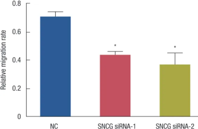
Relative migration rate
NC SNCG siRNA-1 SNCG siRNA-2 0.8
0.6
0.4
0.2
0
* *
Figure 2. The relative migration rate of each group. The relative migra- tion rate of Synuclein-γ (SNCG) siRNA-1 and SNCG siRNA-2 groups decreased significantly (p<0.05, respectively).
NC=negative control.
*p<0.05 vs. NC group.
RESULTS
SNCG mRNA level in MDA-MB-231
SNCG and β-actin mRNA levels were determined using flu- orescent quantitative PCR; the relative concentrations of SNCG mRNA were calculated (SNCG mRNA relative concentra- tion=SNCG mRNA concentration/β-actin mRNA concentra- tion). As shown in Figure 1, levels of SNCG mRNA were found to be much lower in groups that received SNCG siRNA-1 and SNCG siRNA-2, than in the NC (p<0.01, p< 0.01) and control groups (p<0.01, p<0.01). These results suggest that SNCG siRNA can significantly downregulate the SNCG mRNA levels in breast cancer cells.
Migration of MDA-MB-231
Cells that migrated through the membrane were counted in five random fields for each group, and the relative migration rate was calculated (relative migration rate=number of mi- grated cells/number of migrated cells in the control group).
Figure 2 shows that the relative migration rates of SNCG siR- NA-1 and SNCG siRNA-2 groups were decreased significantly (p<0.05, p<0.05). Our results indicate that SNCG siRNAs can inhibit breast cancer cell migration.
Proliferation of MDA-MB-231 cells
The results obtained from the MTT assay show that cell pro- liferation decreased significantly in the groups treated with SNCG siRNA-1 and -2 (p<0.01, p<0.01), while there were no differences between the NC and control group (p>0.05) (Fig- ure 3). The MTT assay demonstrated that SNCG siRNAs can inhibit the proliferation of breast cancer cells.
E
PI
103
102
101
100
100 101 102 103
Annexin V-FITC F
PI
103
102
101
100
100 101 102 103 Annexin V-FITC
D
PI
103
102
101
100
100 101 102 103 Annexin V-FITC
A
PI
103
102
101
100
100 101 102 103
Annexin V-FITC B
PI
103
102
101
100
100 101 102 103
Annexin V-FITC C
PI
103
102
101
100
100 101 102 103 Annexin V-FITC
Figure 4. Apoptosis of MDA-MB-231 in each group. Synuclein-γ (SNCG) siRNA induced apoptosis in MDA-MB-231 cells. (A) Control-1 group; (B) control-2 group; (C) negative control (NC)-1 group; (D) NC-2 group; (E) SNCG siRNA-1 group; (F) SNCG siRNA-2 group.
PI=propidium iodide.
Coulter Inc., Fullerton, USA). As shown in Figure 4, 26.9%
cells in the SNCG siRNA-1 group and 33.2% cells in the SNCG siRNA-2 group were annexin V/FITC-positive, much higher than in the NC1/2 and control 1/2 groups. These results sug- gest that the SNCG siRNA decreased the proliferation of breast cancer cells through the induction of apoptosis.
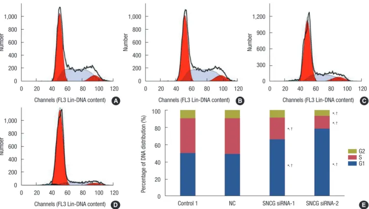
Cell cycle of MDA-MB-231 cells
The effect of SNCG siRNA on cell cycle was analyzed by FACS (Beckmann Coulter Inc., Fullerton, USA). As shown in Figure 5, a higher percentage of cells in SNCG siRNA-1 (65.89%±2.45%), and SNCG siRNA-2 groups (78.76%±
2.24%) were in the G0/G1 phase, than in the NC and control groups (48.67%±0.49%, p<0.05; 49.37%±2.06%, p<0.05, re- spectively). A lower percentage of cells of SNCG siRNA-1 and -2 groups (25.69%±1.57%, 15.00%±1.17%, respectively) were in the S phase, than in the NC and control groups (42.14%±
0.26%, p<0.05; 41.08%±2.45%, p<0.05, respectively). Cells from the SNCG siRNA-1 group (7.52%±1.13%) in the G2/M phase were significantly lower than in the NC and control groups (9.51%±0.47%, p<0.05; 9.92%±0.46%, p<0.05);
while a lower percentage of cells from the SNCG siRNA-2 group were in the G2/M phase (8.17%±0.87%), but the differ- Apoptosis of MDA-MB-231 cells
The rate of apoptosis for MDA-MB-231 cells was analyzed by fluorescence-activated cell sorting (FACS) (Beckmann
Figure 3. MTT assay at 0, 24, 48, and 72 hours after transfection. To determine the amount of cells alive, absorbance was measured on 490 nm wavelength. The cell proliferation decreased significantly in Synuclein-γ (SNCG) siRNA-1 and SNCG siRNA-2 groups (p<0.01, p<0.01) while there was no difference between negative control (NC) group and control group (p>0.05).
OD=optical density.
*p<0.01 vs. control group; †p<0.01 vs. NC group.
OD 490 nm of MDA-MB-231 cells
1.5
1.0
0.5
0
0 24 48 72
Control NCSNCG siRNA-1 SNCG siRNA-2
*, †*, †
*, †
*, †
*, †
*, †
Time (hr)
Figure 5. Cell cycle of MDA-MB-231 in each group. The cell cycle of MDA-MB-231 cells was arrested significantly. (A) Control group; (B) negative control (NC) group; (C) Synuclein-γ (SNCG) siRNA-1 group; (D) SNCG siRNA-2 group; (E) the histogram for the cell percentage of each mitotic phase in each group.
*p<0.05 vs. control group; †p<0.05 vs NC group.
Number
Channels (FL3 Lin-DNA content) 1,000
800 600 400 200 0
0 20 40 60 80 100 120 A
Number
Channels (FL3 Lin-DNA content) 1,000
800 600 400 200 0
0 20 40 60 80 100 120
Number
Channels (FL3 Lin-DNA content) 1,000
800 600 400 200 0
0 20 40 60 80 100 120 B
Number
Channels (FL3 Lin-DNA content) 1,200
900 600 300 0
0 20 40 60 80 100 120 C
D
Percentage of DNA distribution (%)
100 80 60 40 20 0
Control 1 NC SNCG siRNA-1 SNCG siRNA-2
G2S G1
*, †
*, † *, †
*, †
*, †
E
ence was not statistically significant. These results indicate that transfection with SNCG siRNA was able to arrest the cell cycle of breast cancer cells.
Protein expression in MDA-MB-231 cells
Human breast cancer MDA-MB-231 cells transfected SNCG siRNA constructs showed a relative downregulation in
A B
Figure 6. Western blotting. The levels of p-AKT and p-ERK were less in Synuclein-γ (SNCG) siRNA-1 and SNCG siRNA-2 groups than negative con- trol (NC) and control groups while there were not significant differences in the expression of AKT and ERK among the four groups. (A) Western blot- ting of AKT, p-AKT, ERK, p-ERK and their corresponding internal reference (β-Actin); (B) the histogram for the content of proteins in each group.
Control NC SNCG siRAN-1 SNCG siRAN-2
p-AKT
β-Actin
ERK
p-ERK
β-Actin
Relative protein content
AKT p-AKT ERK p-ERK
2.0
1.5
1.0
0.5
0
Control NC
SNCG siRNA-1 SNCG siRNA-2 AKT
p-AKT and p-ERK levels (Figure 6), while there were no sig- nificant differences in the expression levels of AKT and ERK among the four groups. These data suggest that the SNCG siRNA downregulated the p-AKT and p-ERK levels, which in turn, may have been involved in the process of cell apoptosis induced by SNCG siRNA.
DISCUSSION
SNCG, also known as BCSG1, is a kind of γ-Synuclein en- coded by the human γ-Synuclein gene; it is expressed in a high percentage of advanced and metastatic breast tumors, but not in normal or benign breast tissues [5]. Abnormal ex- pression of SNCG is also implicated in various cancer types, including glial tumors, ovarian, liver, esophagus, colon, gas- tric, lung, prostate, pancreas, bladder, and cervical cancers [7,9,15-21]. In breast cancer, SNCG expression was closely correlated with the disease stage, lymph node involvement, metastasis, tumor size, and human epidermal growth factor receptor 2 status; however, SNCG expression was indepen- dent of the expression of the estrogen (ER) and progesterone receptors [10]. Overexpression of SNCG in breast cancer cells can facilitate cell proliferation [11], increase migration, and enhance metastasis in nude mice [6]. Moreover, SNCG is also associated with ERα overexpression [22], antimicrotubule drug resistance [23], and an accelerated rate of chromosomal instability [24]. All these studies suggest that knockdown of SNCG expression may be an effective therapy in breast cancer treatment. In our study, SNCG mRNA expression was effec- tively suppressed by siRNA in human breast cancer MDA- MB-231 cells, and the migration and proliferation of cells de- creased significantly.
According to the results of cell cycle analysis, in the SNCG siRNA-treated groups, the number of cells in the G0/G1 phase increased, while those in the S phase decreased, when com- pared with the control and NC groups. These results suggest that the downregulation of SNCG expression in breast cancer cells was able to inhibit mitosis by blocking the cells in the G0/
G1 phase. It has been reported that SNCG physically interacts with, and inhibits the function of the mitotic checkpoint pro- tein BubR1 in breast cancer cells [23]. The G1 checkpoint plays an important role in cell damage repair since the cells with DNA damage will be blocked in the G1 phase; the dam- aged cells that cannot be repaired may directly undergo apop- tosis [25], which is consistent with the results of the apoptosis analysis. Our results revealed that the cell cycle was arrested and apoptosis was induced by the downregulation of SNCG expression in breast cancer cells.
Western blot analysis showed that p-AKT and p-ERK levels
were lower in the SNCG siRNA-treated cells, than in the con- trol and NC cells. These results suggest that the SNCG siRNA decreased the migration and proliferation of breast cancer cells by downregulating the levels of p-AKT and p-ERK. Thus, the overexpression of SNCG may enhance the migration and viability of breast cancer cells through regulation of AKT and ERK pathways.
In conclusion, the migration and proliferation of breast cancer cells were inhibited by suppressing SNCG expression through downregulation of AKT and ERK phosphorylation.
CONFLICT OF INTEREST
The authors declare that they have no competing interests.
REFERENCES
1. Smolarek AK, Suh N. Chemopreventive activity of vitamin E in breast cancer: a focus on γ- and δ-tocopherol. Nutrients 2011;3:962-86.
2. Clayton DF, George JM. The synucleins: a family of proteins involved in synaptic function, plasticity, neurodegeneration and disease. Trends Neurosci 1998;21:249-54.
3. Polymeropoulos MH, Lavedan C, Leroy E, Ide SE, Dehejia A, Dutra A, et al. Mutation in the alpha-synuclein gene identified in families with Parkinson’s disease. Science 1997;276:2045-7.
4. Spillantini MG, Schmidt ML, Lee VM, Trojanowski JQ, Jakes R, Goed- ert M. Alpha-synuclein in Lewy bodies. Nature 1997;388:839-40.
5. Ji H, Liu YE, Jia T, Wang M, Liu J, Xiao G, et al. Identification of a breast cancer-specific gene, BCSG1, by direct differential cDNA sequencing.
Cancer Res 1997;57:759-64.
6. Jia T, Liu YE, Liu J, Shi YE. Stimulation of breast cancer invasion and me- tastasis by synuclein gamma. Cancer Res 1999;59:742-7.
7. Bruening W, Giasson BI, Klein-Szanto AJ, Lee VM, Trojanowski JQ, Godwin AK. Synucleins are expressed in the majority of breast and ovar- ian carcinomas and in preneoplastic lesions of the ovary. Cancer 2000;
88:2154-63.
8. Wu K, Weng Z, Tao Q, Lin G, Wu X, Qian H, et al. Stage-specific expres- sion of breast cancer-specific gene gamma-synuclein. Cancer Epidemi- ol Biomarkers Prev 2003;12:920-5.
9. Liu H, Liu W, Wu Y, Zhou Y, Xue R, Luo C, et al. Loss of epigenetic con- trol of synuclein-gamma gene as a molecular indicator of metastasis in a wide range of human cancers. Cancer Res 2005;65:7635-43.
10. Guo J, Shou C, Meng L, Jiang B, Dong B, Yao L, et al. Neuronal protein synuclein gamma predicts poor clinical outcome in breast cancer. Int J Cancer 2007;121:1296-305.
11. Jiang Y, Liu YE, Goldberg ID, Shi YE. Gamma synuclein, a novel heat- shock protein-associated chaperone, stimulates ligand-dependent es- trogen receptor alpha signaling and mammary tumorigenesis. Cancer Res 2004;64:4539-46.
12. Zhou Y, Inaba S, Liu J. Inhibition of synuclein-gamma expression in- creases the sensitivity of breast cancer cells to paclitaxel treatment. Int J Oncol 2006;29:289-95.
13. Pan ZZ, Bruening W, Giasson BI, Lee VM, Godwin AK. Gamma-synu-
clein promotes cancer cell survival and inhibits stress- and chemothera- py drug-induced apoptosis by modulating MAPK pathways. J Biol Chem 2002;277:35050-60.
14. Singh VK, Zhou Y, Marsh JA, Uversky VN, Forman-Kay JD, Liu J, et al.
Synuclein-gamma targeting peptide inhibitor that enhances sensitivity of breast cancer cells to antimicrotubule drugs. Cancer Res 2007;
67:626-33.
15. Lavedan C, Leroy E, Dehejia A, Buchholtz S, Dutra A, Nussbaum RL, et al. Identification, localization and characterization of the human gam- ma-synuclein gene. Hum Genet 1998;103:106-12.
16. Zhao W, Liu H, Liu W, Wu Y, Chen W, Jiang B, et al. Abnormal activa- tion of the synuclein-gamma gene in hepatocellular carcinomas by epi- genetic alteration. Int J Oncol 2006;28:1081-8.
17. Zhou CQ, Liu S, Xue LY, Wang YH, Zhu HX, Lu N, et al. Down-regula- tion of gamma-synuclein in human esophageal squamous cell carcino- ma. World J Gastroenterol 2003;9:1900-3.
18. Liu C, Guo J, Qu L, Bing D, Meng L, Wu J, et al. Applications of novel monoclonal antibodies specific for synuclein-gamma in evaluating its levels in sera and cancer tissues from colorectal cancer patients. Cancer Lett 2008;269:148-58.
19. Li Z, Sclabas GM, Peng B, Hess KR, Abbruzzese JL, Evans DB, et al.
Overexpression of synuclein-gamma in pancreatic adenocarcinoma.
Cancer 2004;101:58-65.
20. Iwaki H, Kageyama S, Isono T, Wakabayashi Y, Okada Y, Yoshimura K, et al. Diagnostic potential in bladder cancer of a panel of tumor markers (calreticulin, gamma -synuclein, and catechol-o-methyltransferase) identified by proteomic analysis. Cancer Sci 2004;95:955-61.
21. Fung KM, Rorke LB, Giasson B, Lee VM, Trojanowski JQ. Expression of alpha-, beta-, and gamma-synuclein in glial tumors and medulloblasto- mas. Acta Neuropathol 2003;106:167-75.
22. Jiang Y, Liu YE, Lu A, Gupta A, Goldberg ID, Liu J, et al. Stimulation of estrogen receptor signaling by gamma synuclein. Cancer Res 2003;
63:3899-903.
23. Gupta A, Inaba S, Wong OK, Fang G, Liu J. Breast cancer-specific gene 1 interacts with the mitotic checkpoint kinase BubR1. Oncogene 2003;
22:7593-9.
24. Inaba S, Li C, Shi YE, Song DQ, Jiang JD, Liu J. Synuclein gamma inhib- its the mitotic checkpoint function and promotes chromosomal insta- bility of breast cancer cells. Breast Cancer Res Treat 2005;94:25-35.
25. Zhang W, Ohnishi K, Shigeno K, Fujisawa S, Naito K, Nakamura S, et al.
The induction of apoptosis and cell cycle arrest by arsenic trioxide in lymphoid neoplasms. Leukemia 1998;12:1383-91.