INTRODUCTION
Drug-induced neutropenia (DIN) is a rare, sporadic, and transient disorder. The patients with DIN experience a high rate of infectious complications and have a mortality rate of approximately 10% (1, 2). The most common drugs associ- ated with DIN are antithyroidal drugs, anticonvulsants, and antibiotics such as cephalosporins, penicillins, sulfonamides, and chloramphenicol, although the pathogenesis of DIN is poorly understood (3).
Here, we describe the brief history of a child with pneu- mococcal pneumonia, who experienced severe neutropenia during antibiotic treatment and in whom we detected 4 kinds of antibiotic-dependent antineutrophil antibodies.
CASE REPORT
A 22-month-old boy presented to another hospital with fever and cough for 3 days. On admission to our institution, he was irritable and dyspneic. Physical examinations revealed markedly decreased breathing sounds over the left lung fields.
The radiologic findings showed diffuse infiltration in the left
lower lung fields without shifting of pleural fluid. Laboratory studies on admission disclosed the following values: hemo- globin 11.2 gm/dL, white blood cell (WBC) 9,800/μL (dif- ferential count; segmented neutrophil 82.6%, lymphocyte 14.8%, monocyte 2.6%), platelet 281,000/μL, erythrocyte sedimentation rate (ESR) 55 mm/hr, C-reactive protein (CRP) 32.2 mg/dL. We started antibiotics with augmentin (100 mg/
kg/day, intravenously) and tobramycin (5 mg/kg/day, intra- venously) as well as intravenous immunoglobulin (IVIG, 1 gm/kg) after septic workup including blood cultures. Three days later, we switched antibiotics to vancomycin (50 mg/kg/
day, intravenously), cefotaxim (200 mg/kg/day, intravenous- ly), and roxithromycin (8 mg/kg/day, orally) because the blood culture revealed Streptococcus pneumoniae being sensitive to van- comycin and the antibody titer for Mycoplasma pneumoniae was 1:160 even with a negative cold agglutinin test. His clinical condition became well without fever after 8 days of antibi- otic therapy.
On hospital day 12, before which it was not evident, fluid shifting on a chest radiography was detected and also con- firmed by chest computed tomograohy (CT) scans. A chest tube was inserted and pleural fluid analysis showed compat- ible findings with transudate, possibly due to previous antibi-
975
Young-Ho Lee1,*, Ha-Baik Lee1,*, Jung-Yun Kim1, Yeon-Jung Lim1, Su-A Shin1, and Tae-Hee Han2
Department of Pediatrics1, Hanyang University College of Medicine, Seoul; Department of Laboratory Medicine2, Inje University Sanggye Paik Hospital, Seoul, Korea
*These 2 authors are equally contributed to this paper as first author.
Address for correspondence Young-Ho Lee, M.D.
Department of Pediatrics, Hanyang University College of Medicine, 17 Haengdang-dong, Seongdong-gu, Seoul 133-792, Korea
Tel : +82.2-2290-8383, Fax : +82.2-2297-2380 E-mail : cord@hanyang.ac.kr
This work was supported by the research fund of Hanyang University (HY-2006-000-0000-3455).
J Korean Med Sci 2009; 24: 975-8 ISSN 1011-8934
DOI: 10.3346/jkms.2009.24.5.975
Copyright � The Korean Academy of Medical Sciences
Antibiotic-induced Severe Neutropenia with Multidrug-Dependent Antineutrophil Antibodies Developed in A Child with Streptococcus pneumoniae Infection
Drug-induced neutropenia (DIN), particularly that in which antibiotic-dependent an- tineutrophil antibodies have been detected, is a rare disorder. We report the case of a child with pneumococcal pneumonia, who experienced severe neutropenia during various antibiotic treatments. We detected 4 kinds (cefotaxim, augmentin, vancomycin, and tobramycin) of antibiotic-dependent antineutrophil antibodies by using the mixed passive hemagglutination assay (MPHA) technique with this child.
Key Words : Neutropenia; Antineutrophil Antibody; Anti-Bacterial Agents
Received : 13 October 2007 Accepted : 2 May 2008
otic therapy. Furthermore, polymerase chain reactions for Mycobacterium tuberculosis and Mycoplasma pneumoniae in pleu- ral fluid were also negative. The serologic test for Mycoplas- ma pneumoniae antibody was 1:160 and cold agglutinin test was negative. The blood tests disclosed hemoglobin 6.2 g/dL, WBC 18,500/μL (differential count; segmented neutrophil 62%, lymphocyte 35%, monocyte 3%), platelet 979,000/μL, reticulocyte 2.3%, ESR 68 mm/hr, CRP 4.32 mg/dL. He did not receive packed red blood cells (RBC) because of a pos- itive direct Coombs test and warm antibody on the screen- ing test. He received aspirin because of a high platelet count.
On hospital day 15, fever developed again and persisted until hospital day 18, when progressive, generalized, erythe- matous maculopapular rashes also appeared and a complete blood count (CBC) revealed hemoglobin 7.8 g/dL, WBC 4,200/μL (differential count; segmented neutrophil 29%, lymphocyte 59%, monocyte 9%, eosinophil 1%, atypical lymphocyte 1%, myelocyte 1%), platelet 224,000/μL, and reticulocyte 7.3%. Generalized skin rashes and edema were aggravated until hospital day 22, when IVIG and solumedrol was administered with replacement of vancomycin by same doses of augmentin and tobramycin after removal of the chest tube. Thereafter, the fever and skin rashes disappeared, even the CBC findings on hospital day 26 showed severe neutrope- nia (absolute neutrophil count: 492/μL).
On hospital day 32, the fever developed again without any significant symptoms or signs. CBC findings on next day dis- closed severe neutropenia with WBC 3,700/μL (differential count; segmented neutrophil 1%, lymphocyte 91%, mono- cyte 8%). Severe neutropenia persisted until withdrawal of augmentin and tobramycin as well as administration of gran-
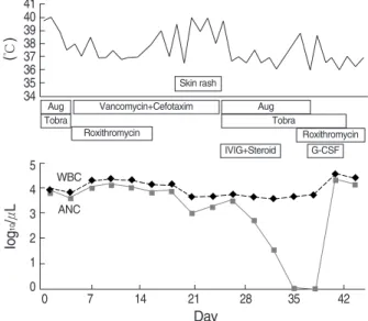
ulocyte colony-stimulating factor (G-CSF, 5 mcg/kg/day, sub- cutaneously) for 3 days from hospital day 36 when the blood samples for antineutrophil antibodies were collected. The brief clinical course as well as the changing pattern of absolute neu- trophil counts is presented in Fig. 1.
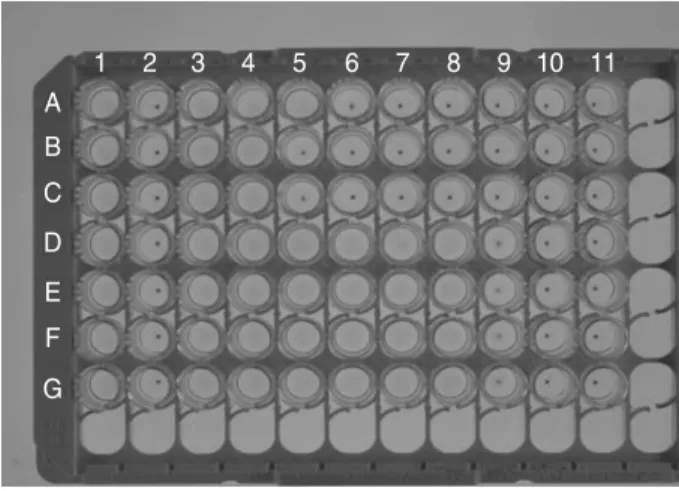
To detect neutrophil antibody, we used the mixed passive hemagglutination assay (MPHA), using extracted neutrophil antigens coated onto microplates (4-6). Neutrophil antibody IgG was detected in the patient’s serum. The serum was reac- tive with the patient’s neutrophil but not with donors’ neu- trophil (Fig. 2). Furthermore, positive control sera were reac- tive with all donors’ neutrophil because human neutrophil antigen (NA 1 and NA 2) are present in the all donors’ neu- trophil including patient’s neutrophil, but patient’s own anti- bodies did not have specificity for NA 1 and NA 2. The se- rum was 1:8 positive with the patient's neutrophil. The serum incubated with cefotaxim, augmentin, vancomycin, and to- bramycin was positive (all 1:64) with the patient’s neutrophil, but the serum incubated with aspirin and roxithromycin was less reactive (1:4) with the patient’s neutrophil than the intact serum (Fig. 3). The serum coincubated with drugs was not reactive with any of the donor’s neutrophils (Fig. 4). In con- clusion, there was an anti-neutrophil autoantibody that had specificity for antibiotics (cefotaxim, augmentin, vancomycin, and tobramycin).
DISCUSSION
DIN is an idiosyncratic reaction that is mediated by im- mune or allergic and toxic mechanisms, and results in pro- found neutropenia (7). Although antineutrophil antibodies also appear in a range of diseases associated with neutrope-
976 Y.-H. Lee, H.-B. Lee, J.-Y. Kim, et al.
Fig. 1. The clinical course and changing pattern of absolute neu- trophil counts according to antibiotic treatment.
Aug, Augmentin; Tobra, Tobramycin; IVIG, intravenous immuno- globulin; G-CSF, granulocyte-colony stimulating factor; WBC, white blood cell count; ANC, absolute neutrophil count.
(℃)
41 40 39 38 37 36 35 34
Iog10/μL
5 4 3 2 1 0
0 7 14 21 28 35 42
Day
WBC ANC
Skin rash
Aug Tobra
Vancomycin+Cefotaxim
Roxithromycin
Aug Tobra
Roxithromycin G-CSF IVIG+Steroid
Fig. 2. The detection of granulocyte antibody in the patient’s serum.
The serum is reactive with the patient’s granulocyte but not with all of the donors’ granulocytes. Row A, patient’s granulocyte coated well; Row B, donor 1 granulocyte; Row C, donor 2 granulocyte;
Row D, donor 3 granulocyte; Column 1, positive control; Column 2, negative control; Column 3, patient’s serum.
1 2 3
A
B
C
D
nia, such as pure white blood cell aplasia, immune neutrope- nia, Felty’s syndrome, and systemic lupus erythematosus, antineutrophil antibodies have been detected in most of the cases of antibodies to autologous or normal granulocytes in immune-mediated DIN (8). In DIN, antibody binding to a target cell usually requires the presence of the causative drug and the complement is often consumed (9). Other mecha- nisms, involving cytotoxic T cells, haptens, autoimmunity and oxidative modifications of drugs as well as direct dam- age to myeloid precursors have been reported (10-13).
The causes of neutropenia include infectious diseases (espe- cially viral), exposure to chemicals, nutritional deficiencies, immune disorders, and congenital or chronic neutropenia (7).
However, most, but not all, instances of neutropenia result from exposure to drugs, and either the drug itself or a metabo- lite may be causative (3). The annual incidence of symptomat- ic nonchemotherapy DIN is between 2.4 and 15.4 cases per million people per year (7). This incidence increases with age, as only 10% of cases are reported in children and young adults and more than half of these episodes occur in people aged over 60 yr (14).
The drugs most commonly associated with neutropenia are antibiotics (particularly beta-lactam and trimethoprim- sulfamethoxasole) as well as antithyroid drugs, antiplatelet agents, nonsteroidal anti-inflammatory agents, and norami- dopyrin (3). We experienced a case of DIN in a child in whom was detected antineutrophil antibodes by MPHA technique
to 4 kinds of antibiotics, but not to roxithromycin and acetyl- salicylic acid. Even if it is not usual to detect antibodies simul- taneously for 4 kinds of antibiotics, we could not try to con- firm the basic mechanism with further studies. In this child, fever and allergic skin reactions developed respectively at 17 days, 10 days prior to neutropenia, which suggested a van- comycin induced drug reaction. Our patient also showed low hemoglobin level at 20 days prior to detection of severe neu- tropenia, which could be considered to be immune mediat- ed hemolytic anemia. However, we could not find laboratory evidence of hemolysis except from the positive direct Coombs test, which was considered to be the effect of previous admin- istration of intravenous immunoglobulin. We believed that the decreased hemoglobin level was caused by the suppression of bone marrow function due to severe infection, although we did not perform bone marrow examinations, and even- tually the decreased hemoglobin level was normalized with- out transfusion in accordance with the control of infection.
In conclusion, we could suggest that neutropenia occurred in this patient was mediated by autoantibodies to antibiotics, based on the immune mediated clinical findings which show- ed prior to detection of antineutrophil antibodies in patient’s serum. Thus we report this patient with DIN presenting an- tineutrophil antibodies to antibiotics.
REFERENCES
1. Andres E, Kurtz JE, Maloisel F. Nonchemotherapy drug-induced agranulocytosis: experience of the Strasbourg teaching hospital (1985- 2000) and review of the literature. Clin Lab Haematol 2002; 24:
Antibiotic-induced Neutropenia with Antineutrophil Antibodies 977
Fig. 3. The granulocyte antibody has specificity for antibiotics. The patient’s granulocyte coated wells are used. The patient’s serum is serially diluted to 1:256 and 5 μL of each drug (1 mg/mL) is added.
The serum is 1:8 reactive without drugs and 1:64 reactive with cefo- taxim, augmentin, vancomycin, and tobramycin, but 1:4 reactive with roxithromycin and aspirin. Row A, no drug; Row B, aspirin; Row C, roxithromycin; Row D, cefotaxim; Row E, augmentin; Row F, va- ncomycin; Row G tobramycin; Column 1, positive control; Column 2, negative control; Column 3, patient’s serum; Column 4 patient’s serum (1:2); Column 5 patient’s serum (1:4); Column 6 patient’s serum (1:8); Column 7 patient’s serum (1:16); Column 8 patient’s serum (1:32); Column 9 patient’s serum (1:64); Column 10 patient’s serum (1:128); Column 11 patient’s serum (1:256).
1 2 3 4 5 6 7 8 9 10 11 A
B C D E F G
Fig. 4. The patient’s serum coincubated with the drug is reactive with none of the donor’s granulocytes. The donors’ granulocyte- coated wells are used. Row A, donor1; Row B, donor 2; Row C, donor 3; Row D, donor 4; Column 1, positive control; Column 2, negative control; Column 3, patient’s serum only; Column 4, pa- tient’s serum and aspirin; Column 5 patient’s serum and roxith- romycin; Column 6, patient’s serum and cefotaxim; Column 7, patient’s serum and augmentin; Column 8, patient’s serum and vancomycin; Column 9, patient’s serum and tobramycin.
1 2 3 4 5 6 7 8 9
A B C D
99-106.
2. van der Klauw MM, Goudsmit R, Halie MR, van’t Veer MB, Her- ings RM, Wilson JH, Stricker BH. A population-based case-cohort study of drug-associated agranulocytosis. Arch Intern Med 1999;
159: 369-74.
3. Young NS, Alter BP, eds. Aplastic Anemia, Acquired and Inherited.
Philadelphia, PA: W.B. Saunders Co., 1994; 229-67.
4. Araki N, Nose Y, Kohsaki M, Mito H, Ito K. Anti-granulocyte anti- body screening with extracted granulocyte antigens by a micro-mixed passive hemagglutination method. Vox Sang 1999; 77: 44-51.
5. Han TH, Chey MJ, Han KS. Granulocyte antibodies in Korean ne- onates with neutropenia. J Korean Med Sci 2006; 21: 627-32.
6. Han TH, Chey MJ, Han KS. A case of neonatal alloimmune neu- tropenia associated with anti-human neutrophil antigen-1a (HNA- 1a) antibody. J Korean Med Sci 2006; 21: 351-4.
7. Kaufman DW, Kelly JP, Levy M, Shapiro S, eds. The Drug Etiology of Agranulocytosis and Aplastic Anaemia. New York: Oxford Uni- versity Press, 1991; 148-58.
8. Salama A, Schu_tz B, Kiefel V, Breithaupt H, Mueller-Eckhardt C.
Immune-mediated agranulocytosis related to drugs and their metabo- lites: mode of sensitization and heterogeneity of antibodies. Br J Ha- ematol 1989; 72: 127-32.
9. Claas FH. Immune mechanisms leading to drug-induced blood dys- crasias. Eur J Haematol Suppl 1996; 60: 64-8.
10. Gerson WT, Fine DG, Spielberg SP, Sensenbrenner LL. Anticon- vulsant-induced aplastic anemia: increased susceptibility to toxic drug metabolites in vitro. Blood 1983; 61: 889-93.
11. Weinshilboum RM. Pharmacogenetics of methyl conjugation. Prog Clin Biol Res 1983; 135: 167-81.
12. Young NS. Drug-related blood dyscrasias. Introduction. Eur J Ha- ematol Suppl 1996; 60: 6-8.
13. Nyfeler B, Pichler WJ. The lymphocyte transformation test for the diagnosis of drug allergy: sensitivity and specificity. Clin Exp Aller- gy 1997; 27: 175-81.
14. Kurtz JE, Andre$s E, Maloisel F, Kurtz-Illig V, Heitz D, Sibilia J, Imler M, Dufour P. Drug-induced agranulocytosis in older people.
A case series of 25 patients. Age Ageing 1999; 28: 325-6.
978 Y.-H. Lee, H.-B. Lee, J.-Y. Kim, et al.