소아 급성림프모구백혈병에서 CDH1 , p16 , DAPK 유전자의 DNA 메틸화
한승민ㆍ권승연ㆍ김효선ㆍ한정우ㆍ유철주
연세대학교 의과대학 소아과학교실, 연세암연구소
Aberrant DNA Methylation of CDH1 , p16 and DAPK in Childhood Acute Lymphoblastic Leukemia
Seung Min Hahn, M.D., Seung Yeon Kwon, M.D., Hyo Sun Kim, M.D., Jung Woo Han, M.D.
and Chuhl Joo Lyu, M.D., Ph.D.
Department of Pediatrics, Yonsei Cancer Research Center, Yonsei University College of Medicine, Seoul, Korea
Background: Hypermethylation of tumor suppressor gene has been reported in various types of leukemia with potential involvement in the inactivation of regulatory cell cycle and apoptosis genes.
Methods: To evaluate the methylation status at initial diagnosis and morphologic com- plete remission (CR) period in childhood acute lymphoblastic leukemia (ALL), we ana- lyzed the methylation status of three key genes (CDH1, p16 and DAPK) in 43 childhood ALL patients and 7 healthy bone marrow (BM) donors.
Results: CDH1 was methylated in 26 (60.4%) patients, p16 in two (4.6%) patients and DAPK in six (13.9%) patients at the time of diagnosis. Twenty nine (67.4%) patients had methylation of at least one gene. None of the healthy BM donors showed methyl- ation of the above genes. Age was the only factor which showed significant association with the presence of DNA methylation (P=0.03). None of the other clinicopathological factors showed association with initial methylation status. At the time of morphologic CR, all patients who had aberrant DNA methylation at the time of diagnosis had no detectable residual methylation.
Conclusion: Since hypermethylation was found in around two thirds of pretreatment ALL patients and none in healthy BM donor, we suggest hypermethylation of some important genes is a biologic marker of childhood ALL. We recommend that further studies with a large number of patients should be conducted.
pISSN 2233-5250 / eISSN 2233-4580 http://dx.doi.org/10.15264/cpho.2015.22.1.60 Clin Pediatr Hematol Oncol 2015;22:60∼66
Received on April 1, 2015 Revised on April 17, 2015 Accepted on April 23, 2015
Corresponding Author: Chuhl Joo Lyu Department of Pediatrics, Yonsei University College of Medicine, Yonsei-ro 50, Seodaemun-gu, Seoul 120-752, Korea
Tel: +82-2-2228-2060 Fax: +82-2-393-9118 E-mail: [email protected]
Key Words: DNA methylation, Pediatric acute lymphoblastic leukemia
Introduction
Hypermethylation of DNA, which involves the addition of a methyl group to the carbon 5 position of the cytosine ring in a cytosine-guanine (CpG) pair, is known to be the
most common and well-described epigenetic mechanism.
CpG pairs are usually underrepresented in most of the
mammalian genome. However, short regions rich in CpG
content known as CpG islands are found in the proximal
promoter regions of almost half of the genes in the human
genome, and are generally unmethylated in normal cells
[1-3]. Methylation of these promoter-associated areas has been associated with gene silencing which serves as an al- ternative mechanism by inactivating functionally relevant genes related to diverse human pathologies including can- cer and aging [4,5]. In cancer, the aberrant hypermethylation of CpG islands surrounding gene promoter regions is the most well-investigated epigenetic change to occur in tumors.
CpG island hypermethylation has been described in various types of human malignancies, and is associated with in- appropriate transcriptional silencing of functionally important cancer related genes, including tumor-suppressor genes [6,7].
Hypermethylation of promoter-associated CpG islands al- so has been found in acute lymphoblastic leukemia (ALL).
Initial studies in ALL were targeted on the analysis of single genes such as calcitonin [8-10], p73 [11,12], p15 [13-15], E-cadherin (CDH1) [16], and so on. Further studies focusing upon hypermethylation of multiple genes have proved that concurrent methylation of multiple genes is a common fea- ture in both adult and childhood ALL [17,18]. The studies in Philadelphia chromosome (Ph)-negative adult ALL and acute myeloid leukemia (AML) have shown that residual methylation at remission could predict poor prognosis, sug- gesting that DNA methylation could be a potential bio- marker in ALL [19,20]. Furthermore, many studies implicated that methylation pattern in childhood ALL patients at diag- nosis were different from that of adults [21-24]. To study these issues, we examined methylation status of three key genes in leukemia, CDH1, p16 and DAPK, at the time of initial diagnosis and at the time of morphologic complete remission (CR). CDH1 is a member of membrane glycoprotein, its function as a tumor suppressor and its loss due to meth- ylation enables disaggregation of cancer cells from one an- other and increases their metastatic potential. p16 is a im- portant member of cyclin dependent kinase inhibitor, regu- lated by a feedback loop with Rb. DAPK is a Ca
+2/Calmod- ulin regulated serine/threonine kinase, associated with the cytoskeleton, and participates in a wide range of apoptotic pathways.
We also aimed to investigate the correlation of methyl- ation status with clinicopathological features.
Materials and Methods
1) Patients and samples
We studied 43 patients (24 males, 19 females) who were diagnosed as de novo childhood ALL between January 2006 and February 2010. Diagnosis was established according to the standard morphologic, cytochemical, and immunophe- notypic criteria. All patients were risk stratified and treated with protocols based on recognized prognostic factors, in- cluding cytogenetic analysis. Criteria for CR were defined as restoration of normal hematopoiesis with a blast cell fraction of less than 5% by light microscopic examination of the bone marrow (BM) [25] after 4 weeks of induction chemotherapy. The median age at the time of diagnosis was 4.8 years (range, 0.9-17 years). Patients were grouped ac- cording to the National Cancer Institute (NCI) risk classi- fication criteria [26] for statistical analyses. Other clinical characteristics of the patients are described in Table 1. The median follow up period of the patients was 5.4 years (range, 4.1-7.8 years). Seven healthy individuals were in- cluded in this study for comparison. All of them were BM donors for their siblings. The median age of the donors was 8.2 years (range, 3.5-22 years).
Study samples were obtained from BM aspirates from the above patients at the time of diagnosis (prior to chemo- therapy) and at the time of morphologic CR (after 4 weeks of induction chemotherapy). Patients with sample from one of the time points were not included in this study. All 43 patients included in this study had achieved CR after in- duction chemotherapy. BM aspiration samples were also obtained from 7 healthy individuals who were BM donors for their siblings. Consent for sample collection was ob- tained from all patients and healthy individuals following the institutional guidelines. This study was reviewed and approved by the Institutional Review Board.
2) Methylation specific PCR (MSP)
To study methylation patterns at the time of initial diag-
nosis and CR in childhood ALL, we analyzed the methyl-
ation status of three key genes related to its pathogenesis,
CDH1, p16 and DAPK, in 43 patients. Gene selection was
based on the previous methylation studies in ALL [17,21,27, 28]; we selected the three genes that are known to be fre- quently methylated in childhood ALL [18,29].
Mononuclear cells were isolated using Ficoll-Hypaque density gradient centrifugation, and genomic DNA was ex- tracted with QIAmp DNA blood mini kit (QIAGEN, CA, USA) following procedures according to the manufacturer’s instructions. Genomic DNA was modified using the sodium bisulfate as previously described by Herman et al. [30]. One microgram of genomic DNA was treated with sodium bi- sulfate to convert all unmethylated cytosine residues to ur- acil using EZ DNA Methylation kit (Zymo Research, CA, USA) following the manufacturer’s instructions. Modified DNA was stored at −70
oC until used. PCR was performed using specific primers for CDH1, p16 and DAPK genes in 43 pairs of samples from the patients and 7 samples from healthy donor. Two hundred nanograms of sodium bi- sulfate modified DNA was used as a template in PCR re- action containing 25 pmoles of each primer, 200 M each dNTP, 1 U AmpliTaq Gold DNA polymerase with 10X PCR buffer and 25 mM MgCl
2solution (Applied Biosystems, CA, USA) in a final volume of 25 L. PCR conditions and pri- mers specific for methylated and unmethylated genes were the same as those used in the literature by Gutierrez et al.
[18]. After amplification, 10 L of the PCR product was sep- arated on a 2% agarose gel containing ethidium bromide.
For each MSP reaction, a CpGenone universal methylated DNA (Millipore, MA, USA) and commercial normal human genomic DNA [Human Genomic DNA: Female (Promega, MI, USA)] was used as controls for methylation and unme- thylation. Distilled water was used as a negative control for PCR in each set of reaction. Specificity of each reaction was demonstrated when methylated gene consistently yielded a band from universally methylated DNA.
3) Statistical analysis
For statistical purposes, patients were classified into two different groups: unmethylated (no methylated genes) and methylated (at least one methylated gene). Association of methylation status with patient characteristics were ana- lyzed using
test or Fisher exact test. Overall survival (OS) was measured from the day of diagnosis until death from
any cause and was censored only for patients known to be alive at last contact. Disease-free survival (DFS) was measured from the day that CR was established until either relapse or death without relapse, and it was censored only for patients who were alive without evidence of relapse at the last follow-up. Comparisons among the groups were performed by analysis of variance with SPSS ver. 12.0 for Windows (SPSS Inc., Chicago, IL, USA). Estimated 5-year disease free survival and overall survival were based on the Kaplan-Meier method, and differences were tested using the log-rank test. An effect was considered statistically sig- nificant if the P-value was 0.05 or less.
Results
1) Methylation pattern at initial diagnosis and CR
Among 43 patients, methylation frequency of each gene at the time of initial presentation was as follows: 60.4%
(n=26) for CDH1, 4.6% (n=2) for p16, and 13.9% (n=6) for DAPK. 67.4% (n=29) of all patients had methylation of at least one gene, whereas 32.6% (n=14) of the patients had no methylation at all. Five (11.6%) patients had methylation of two genes. Methylation of CDH1 showed considerably high frequency of methylation as previously reported [18].
However, p16 and DAPK were rarely methylated in this study.
To evaluate the dynamics of DNA methylation changes as a result of therapy, we also analyzed the methylation status of the above three genes at the time of morphologic CR with the paired samples. All patients who showed meth- ylation at the time of initial presentation (n=29) revealed clear absence of methylation of all three genes at the time of CR. One patient who did not have methylation at initial presentation revealed methylation of CDH1 gene at the time of CR. The patient was an eight year-old girl who ex- perienced bone marrow and CNS relapse after six months, and finally died of disease progression. Except this one pa- tient, neither residual methylation nor new methylation was detected at the time of CR in childhood ALL patients.
2) Clinical characteristics and methylation profile
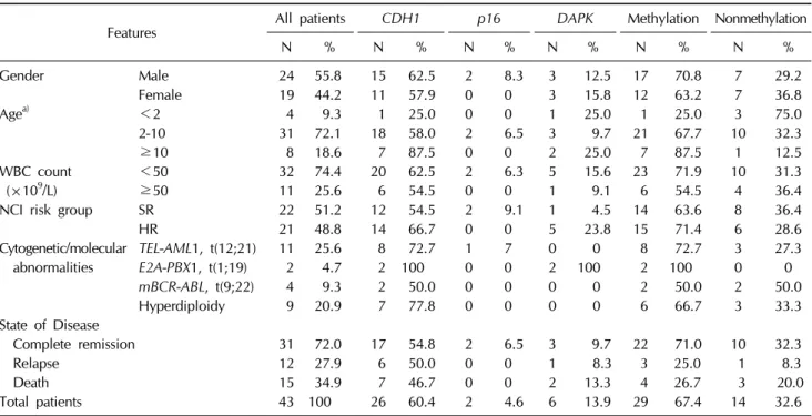
We studied the association between DNA methylation
Table 1. Clinical characteristics and methylation profile of the patients
Features All patients
CDH1 p16 DAPK
Methylation NonmethylationN % N % N % N % N % N %
Gender Male 24 55.8 15 62.5 2 8.3 3 12.5 17 70.8 7 29.2
Female 19 44.2 11 57.9 0 0 3 15.8 12 63.2 7 36.8
Agea) <2 4 9.3 1 25.0 0 0 1 25.0 1 25.0 3 75.0
2-10 31 72.1 18 58.0 2 6.5 3 9.7 21 67.7 10 32.3
≥10 8 18.6 7 87.5 0 0 2 25.0 7 87.5 1 12.5
WBC count <50 32 74.4 20 62.5 2 6.3 5 15.6 23 71.9 10 31.3
(×109/L) ≥50 11 25.6 6 54.5 0 0 1 9.1 6 54.5 4 36.4
NCI risk group SR 22 51.2 12 54.5 2 9.1 1 4.5 14 63.6 8 36.4
HR 21 48.8 14 66.7 0 0 5 23.8 15 71.4 6 28.6
Cytogenetic/molecular TEL-AML1, t(12;21) 11 25.6 8 72.7 1 7 0 0 8 72.7 3 27.3 abnormalities
E2A-PBX1, t(1;19)
2 4.7 2 100 0 0 2 100 2 100 0 0mBCR-ABL, t(9;22)
4 9.3 2 50.0 0 0 0 0 2 50.0 2 50.0Hyperdiploidy 9 20.9 7 77.8 0 0 0 0 6 66.7 3 33.3 State of Disease
Complete remission 31 72.0 17 54.8 2 6.5 3 9.7 22 71.0 10 32.3
Relapse 12 27.9 6 50.0 0 0 1 8.3 3 25.0 1 8.3
Death 15 34.9 7 46.7 0 0 2 13.3 4 26.7 3 20.0
Total patients 43 100 26 60.4 2 4.6 6 13.9 29 67.4 14 32.6
Methylation, patients with DNA methylation of at least one gene; nonmethylation, patients with no methylation at all; WBC, white blood cell; NCI, National Cancer Institute; SR, standard risk; HR, high risk.
a)