Acknowledgments
This work was supported by the year 2016 clinical re- search grant from Pusan National University Hospital.
AuthorsÊ Disclosures of Potential Conflicts of Interest No potential conflicts of interest relevant to this article were reported.
REFERENCES
1. Zagaria A, Anelli L, Coccaro N, et al. 5'RUNX1-3'USP42 chimeric gene in acute myeloid leukemia can occur through an insertion mechanism rather than translocation and may be mediated by ge- nomic segmental duplications. Mol Cytogenet 2014;7:66.
2. Masetti R, Togni M, Astolfi A, et al. Whole transcriptome se- quencing of a paediatric case of de novo acute myeloid leukaemia with del(5q) reveals RUNX1-USP42 and PRDM16-SKI fusion transcripts. Br J Haematol 2014;166:449-52.
3. Ji J, Loo E, Pullarkat S, Yang L, Tirado CA. Acute myeloid leuke- mia with t(7;21)(p22;q22) and 5q deletion: a case report and liter- ature review. Exp Hematol Oncol 2014;3:8.
4. Panagopoulos I, Gorunova L, Brandal P, Garnes M, Tierens A, Heim S. Myeloid leukemia with t(7;21)(p22;q22) and 5q deletion.
Oncol Rep 2013;30:1549-52.
5. Jeandidier E, Gervais C, Radford-Weiss I, et al. A cytogenetic study of 397 consecutive acute myeloid leukemia cases identified three with a t(7;21) associated with 5q abnormalities and exhibit- ing similar clinical and biological features, suggesting a new, rare acute myeloid leukemia entity. Cancer Genet 2012;205:365-72.
6. Giguère A, Hébert J. Microhomologies and topoisomerase II con- sensus sequences identified near the breakpoint junctions of the recurrent t(7;21)(p22;q22) translocation in acute myeloid leukemia. Genes Chromosomes Cancer 2011;50:228-38.
7. Foster N, Paulsson K, Sales M, et al. Molecular characterisation of a recurrent, semi-cryptic RUNX1 translocation t(7;21) in mye- lodysplastic syndrome and acute myeloid leukaemia. Br J Haematol 2010;148:938-43.
8. Chessells JM, Harrison CJ, Kempski H, et al. Clinical features, cy- togenetics and outcome in acute lymphoblastic and myeloid leu- kaemia of infancy: report from the MRC Childhood Leukaemia working party. Leukemia 2002;16:776-84.
9. Park KU, Lee DS, Lee HS, Kim CJ, Cho HI. Granulocytic sarcoma in MLL-positive infant acute myelogenous leukemia: fluo- rescence in situ hybridization study of childhood acute myeloge- nous leukemia for detecting MLL rearrangement. Am J Pathol 2001;159:2011-6.
10. Preiss BS, Kerndrup GB, Pedersen RK, Hasle H, Pallisgaard N;
Lymphoma-Leukemia Study Group of the Region of Southern Denmark. Contribution of multiparameter genetic analysis to the detection of genetic alterations in hematologic neoplasia. An evaluation of combining G-band analysis, spectral karyotyping, and multiplex reverse-transcription polymerase chain reaction (multiplex RT-PCR). Cancer Genet Cytogenet 2006;165:1-8.
11. Paulsson K, Békássy AN, Olofsson T, Mitelman F, Johansson B, Panagopoulos I. A novel and cytogenetically cryptic t(7;21) (p22;q22) in acute myeloid leukemia results in fusion of RUNX1 with the ubiquitin-specific protease gene USP42. Leukemia
2006;20:224-9.
12. Kim YK, Kim YS, Yoo KJ, et al. The expression of Usp42 during embryogenesis and spermatogenesis in mouse. Gene Expr Patterns 2007;7:143-8.
13. Hock AK, Vigneron AM, Carter S, Ludwig RL, Vousden KH.
Regulation of p53 stability and function by the deubiquitinating enzyme USP42. EMBO J 2011;30:4921-30.
Toxic megacolon and interstitial pneumonia caused by cytomegalovirus infection in a pediatric patient with acute lymphoblastic leukemia
receiving chemotherapy
TO THE EDITOR: Cytomegalovirus (CMV) is a major cause of morbidity and mortality after transplantation. However, the risk of CMV infection increases with more intense treat- ment outside transplantation settings. We describe a case of fatal CMV disease in a pediatric patient with standard risk acute lymphoblastic leukemia (ALL). She developed life-threatening toxic megacolon and interstitial pneumonia, complicated with bacterial sepsis, during induction chemo- therapy. With a diagnosis of ganciclovir (GCV)-resistant CMV disease, she was successfully treated with foscarnet and has remained in complete remission for >2 years. We discuss the association between CMV and immune suppression, the clinical utility of diagnostic tools, and CMV antiviral resistance.
Cytomegalovirus is a commonly encountered viral in- fection following hematopoietic stem cell transplantation (HSCT). While CMV infection is occasionally characterized as asymptomatic or as a mononucleosis-like syndrome in immunocompetent hosts, the disease can be fatal in im- munocompromised patients including transplant recipients.
The disease results from a combination of altered cellular immunity, uncontrolled viral replication with multiorgan involvement, and end-organ disease secondary to direct viral cytopathic effects. While it rarely occurs in pediatric patients receiving only the chemotherapy, critically ill patients with a transient depression in immunity are predisposed to CMV reactivation [1-3]. Herein, we describe a pediatric patient with ALL who developed toxic megacolon and antiviral medication-resistant interstitial pneumonia caused by CMV reactivation.
CASE
A 5-year-old girl with standard risk B-precursor ALL received induction chemotherapy according to established protocol [4]. She was considered as a rapid early responder with M1 marrow on day 8 of induction chemotherapy con- sisting vincristine, dexamethasone, L-asparaginase, and in-
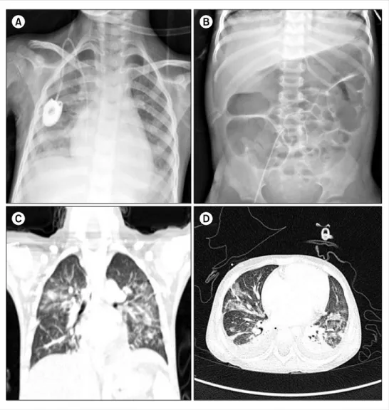
Fig. 1. The patient developed fever, dyspnea, and abdominal disten- sion on day 28 of induction. Chest X-ray (A) showed a newly deve- loped pneumonic consolidation.
Abdominal X-ray (B) showed a gas-filled colonic dilatation resem- bling toxic megacolon. A high- resolution computed tomography (HRCT; C, D) demonstrated the development of consolidations and ground-glass opacities in both lung fields.
and abdominal pain for a week, symptoms suggestive of vincristine-induced ileus. Physical examination revealed hypotension, shortness of breath, chest retraction, and ab- dominal distension. Her hemoglobin level was 8.9 g/dL, WBC count was 100 cells/μL, and platelet count was 51,000 cells/μL. Chest X-ray and computed tomography (CT) scans showed pneumonic consolidations suggestive of fungal pneumonia, and abdominal X-rays showed a gas-filled co- lonic dilation resembling toxic megacolon (Fig. 1). With the exception of blood cultures positive for Aeromonas sor- bia infection, all serologic tests, and a C. difficile toxin assay showed nonspecific result. A bronchoalveolar lavage (BAL) could not confirm the cause of pneumonia. Despite the administration of broad-spectrum antibiotics, trimetho- prim/sulfamethoxazole and voriconazole, and supportive treatment, the patient deteriorated rapidly and developed hypotensive shock associated with a large volume of hematochezia. After resuscitation, angiography detected ac- tive bleeding from the superior mesenteric artery, and de-
and hemorrhagic colitis involving entire sigmoid colon.
Biopsies showed chronic inflammatory ulcers, and im- munohistochemical staining for the CMV antigen was pos- itive (Fig. 2). The presence of CMV antigenemia in three leukocytes and a reverse transcription-polymerase chain re- action (RT-PCR) based blood viral load of 4,260 copies/mL confirmed active CMV infection. Intravenous GCV dramati- cally improved the toxic megacolon and hematochezia.
Despite becoming negative for CMV infection by anti- genemia and RT-PCR analysis after 7 days of GCV treatment, and with negative test results for other pathogens, her pneu- monia continued to worsen. Chest CT demonstrated in- creased multifocal patchy consolidations and ground-glass opacities in both lung fields (Fig. 3). Repeated bronchoscopy on day 55 detected CMV in the BAL fluid based on a qual- itative PCR analysis. We suspected a GCV-resistant CMV pneumonia and changed her treatment to foscarnet, to which intravenous immunoglobulin was added every other day for 2 weeks. Her pneumonia gradually improved, and
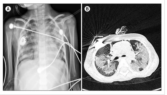
Fig. 3. Serial chest X-rays on day 54 showed progressive haziness in both lungs despite 2 weeks of ganciclovir treatment (A). Chest CT (B) on the same day (day 54) showed increased multifocal patchy consolidations and ground-glass opacities in both lung fields compared with previous chest CT images.
Fig. 2. Sigmoidoscopy performed on day 40 revealed active bleeding with multiple hemorrhagic ulcers throughout the colon (A). Endoscopic biopsy revealed a chronic inflammatory ulcer (B). Histopathologic staining did not show any inclusion-bearing cells typical of CMV infection (owl's eye inclusions); however, immunohistochemical staining identified immunoreactivity for CMV antigen (C).
she was weaned from the ventilator on day 70. She sub- sequently made a full recovery and continued receiving chemotherapy, and has remained in remission for the last 24 months.
DISCUSSION
CMV seropositivity, corticosteroid use, hypoalbuminem- ia, prolonged lymphocytopenia, bacterial pneumonia, and sepsis are known risk factors for CMV disease in critically ill patients [2, 3]. Intact cellular immune responses are essen- tial in controlling acute CMV infection, and preventing recrudescence and end-organ disease. In patients with ma- lignancies, lymphocytopenia is significantly associated with serious CMV pneumonia and gastrointestinal (GI) CMV dis- ease, suggesting that chemotherapy-associated cellular im-
mune dysfunction affects CMV reactivation [3, 5, 6].
Torres et al. [6] reported that the site of CMV involvement in the GI varies with the type of cancer; particularly, color- ectal involvement is more common in solid tumors, whereas upper GI tract involvement is more frequent in hemato- logical malignancies. However, our patient exhibited upper GI tract-sparing severe CMV colitis. CMV disease also occurs more commonly at sites susceptible to tissue inflammation;
GI disease occurs in patients with mucosal toxicity from chemotherapy, transplantation, graft-versus-host disease, or inflammatory bowel disease, whereas pneumonitis occurs more commonly in patients with pre-existing/coexistent pulmonary diseases [1]. Our patient was treated with dex- amethasone (6 mg/m2/day) for 28 days and showed persistent lymphocytopenia (>500 cells/μL) and neutropenia. She
effects. We could not identify the cause of her respiratory failure, and could not find any evidence of CMV pneumonia.
However, we eventually diagnosed GCV-resistant CMV pneumonia, a suspected late reactivation of latent CMV infection following acute lung injury [7].
The diagnoses of CMV colitis and CMV pneumonia in this patient were somewhat delayed. The toxic megacolon was too severe to perform colonoscopy, and the earlier gas- trofibroscopic findings were normal. She had severe ulcer- ative colitis accompanying a life-threatening amount of hematochezia; although CMV antigenemia assay was pos- itive in only three leukocytes, and the CMV viral load was 4,260 copies/mL. Following GCV treatment, the results of CMV antigenemia and PCR analyses continued to be neg- ative throughout the course of disease; therefore, it was difficult to implicate CMV for the gradually worsening inter- stitial pneumonia. Consequently, repeated bronchoscopy with BAL was needed, and CMV pneumonia was eventually diagnosed. CMV end-organ disease diagnosis relies on the detection of CMV by one or more appropriate diagnostic tests, including culture, antigen detection by immunohisto- chemistry, histopathological examination, or in situ DNA hybridization, accompanied by signs and symptoms of the affected organs. The CMV antigenemia assay and quantita- tive PCR analysis have been increasingly used in recent years, as they are highly sensitive and non-invasive proce- dures that provide important prognostic information for patients at risk of developing CMV disease. Some reports have described a high risk of CMV end-organ disease in patients with high viral loads and/or persistent viremia [8], although in general, the detection of CMV by antigenemia or PCR analysis has not been a reliable predictor of CMV end-organ disease in settings outside transplantation. [9, 10]. While CMV antigenemia and blood CMV PCR show high levels of specificity for the diagnosis of CMV colitis (82% and 88%, respectively), the sensitivity values are low (47% and 44%, respectively) [11]. Our case also showed that blood CMV antigenemia and PCR analyses are of limited clinical value in diagnosing CMV GI disease and pneumonia.
When patients present symptoms of an unexplained end-or- gan disease, more aggressive investigations including gastro- fibroscopy; colonoscopy; and bronchoscopy with biopsies should be performed [9, 10].
GCV is the first-line antiviral therapy for CMV infection in immunocompromised patients. Because of her early neg- ative CMV-antigenemia and improvement in CMV colitis following GCV treatment, we suspected antiviral resistance at a much later stage due to her worsening pneumonia.
In practice, CMV antiviral resistance is diagnosed based on increasing/high-plateaued viral load despite admin- istration of adequate antiviral therapy for >2 weeks and is confirmed based on genotypic and/or phenotypic testing.
However, clinical and virological resistance are not always
previous viral load, long-term GCV use (usually >6 weeks), and delayed immune recovery, although resistance may also occur in patients with asymptomatic viremia [12, 13]. The most common (>95%) antiviral-resistant CMV strains con- tain mutations of the phosphotransferase UL97, conferring resistance to GCV, followed by mutations of the viral DNA polymerase UL54 associated with more severe resistance [14]. Our patient experienced progressive pneumonia, and CMV PCR analysis of her BAL fluid remained positive after 2 weeks of GCV treatment. Although we did not perform genotypic testing, this was suggestive of clinical resistance.
We consequently switched GCV to foscarnet empirically, and her pneumonia improved.
The unusual occurrence of CMV infection in this patient emphasizes the importance of closely monitoring im- munosuppressed patients receiving chemotherapy, includ- ing dexamethasone. The immunosuppressive effects of dex- amethasone and bacterial sepsis may have reactivated a la- tent CMV infection and may have predisposed the patient to secondary infections and delayed immune recovery, thereby increasing the risk of a fatal outcome. Additionally, emerging drug-resistant viral mutations present new chal- lenges to successful treatment. GCV is the drug of choice for CMV treatment; however, an extensive diagnostic work- up is imperative when no clinical improvements are ob- served within 2 weeks. Furthermore, in suspected antiviral resistance cases, an antiviral therapy change should be con- sidered early on.
Hyunseop Kwon1, Hyun Hee Lee1, Chung Ryul Paik1, Yun-Jeong Lim2, Jeong A. Park1
Departments of 1Pediatrics, 2Radiology, Inje University Haeundae Paik Hospital, Busan, Korea
Correspondence to: Jeong A. Park Department of Pediatrics, Inje University Haeundae-Paik Hospital, 875, Haeundae-ro, Haeundae-gu, Busan 48108, Korea E-mail: [email protected]
Received on Jan. 15, 2016; Revised on Feb. 28, 2016; Accepted on Mar. 16, 2016 https://doi.org/10.5045/br.2016.51.4.281
Acknowlegments
This work was supported by Grant from Inje University, 2010.
AuthorsÊ Disclosures of Potential Conflicts of Interest No potential conflicts of interest relevant to this article were reported.
REFERENCES
1. Ng AP, Worth L, Chen L, et al. Cytomegalovirus DNAemia and
disease: incidence, natural history and management in settings other than allogeneic stem cell transplantation. Haematologica 2005;90:1672-9.
2. Osawa R, Singh N. Cytomegalovirus infection in critically ill pa- tients: a systematic review. Crit Care 2009;13:R68.
3. Torres HA, Aguilera E, Safdar A, et al. Fatal cytomegalovirus pneu- monia in patients with haematological malignancies: an autop- sy-based case-control study. Clin Microbiol Infect 2008;14:1160-6.
4. Mitchell HR, Lu X, Myers RM, et al. Prospective, longitudinal as- sessment of quality of life in children from diagnosis to 3 months off treatment for standard risk acute lymphoblastic leukemia:
Results of Children's Oncology Group study AALL0331. Int J Cancer 2016;138:332-9.
5. Jaber S, Chanques G, Borry J, et al. Cytomegalovirus infection in critically ill patients: associated factors and consequences. Chest 2005;127:233-41.
6. Torres HA, Kontoyiannis DP, Bodey GP, et al. Gastrointestinal cytomegalovirus disease in patients with cancer: a two decade ex- perience in a tertiary care cancer center. Eur J Cancer 2005;41:
2268-79.
7. Cook CH, Zhang Y, McGuinness BJ, Lahm MC, Sedmak DD, Ferguson RM. Intra-abdominal bacterial infection reactivates la- tent pulmonary cytomegalovirus in immunocompetent mice. J Infect Dis 2002;185:1395-400.
8. Gondo H, Minematsu T, Harada M, et al. Cytomegalovirus (CMV) antigenaemia for rapid diagnosis and monitoring of CMV-associated disease after bone marrow transplantation. Br J Haematol 1994;86:130-7.
9. Green ML, Leisenring W, Stachel D, et al. Efficacy of a viral load-based, risk-adapted, preemptive treatment strategy for pre- vention of cytomegalovirus disease after hematopoietic cell transplantation. Biol Blood Marrow Transplant 2012;18:1687-99.
10. Ruell J, Barnes C, Mutton K, et al. Active CMV disease does not always correlate with viral load detection. Bone Marrow Transplant 2007;40:55-61.
11. Kim JW, Boo SJ, Ye BD, et al. Clinical utility of cytomegalovirus antigenemia assay and blood cytomegalovirus DNA PCR for cy- tomegaloviral colitis patients with moderate to severe ulcerative colitis. J Crohns Colitis 2014;8:693-701.
12. Hantz S, Garnier-Geoffroy F, Mazeron MC, et al. Drug-resistant cytomegalovirus in transplant recipients: a French cohort study.
J Antimicrob Chemother 2010;65:2628-40.
13. Shmueli E, Or R, Shapira MY, et al. High rate of cytomegalovirus drug resistance among patients receiving preemptive antiviral treatment after haploidentical stem cell transplantation. J Infect Dis 2014;209:557-61.
14. Le Page AK, Jager MM, Iwasenko JM, Scott GM, Alain S, Rawlinson WD. Clinical aspects of cytomegalovirus antiviral re- sistance in solid organ transplant recipients. Clin Infect Dis 2013;56:1018-29.
Salvage chemotherapy with R-BAD (rituximab, bendamustine,
cytarabine, and dexamethasone) for the treatment of relapsed primary CNS lymphoma
TO THE EDITOR: Primary central nervous system lympho- ma (PCNSL) involves extranodal lymphomas arising ex- clusively in the central nervous system (CNS). It accounts for 2%–3% of newly diagnosed primary CNS tumors and systemic non-Hodgkin’s lymphomas [1, 2]. Although surviv- al outcomes have improved with the introduction of high-dose methotrexate (MTX)-based regimens, with or without cranial irradiation, relapse is still common.
Approximately 35%–60% of patients show relapse and most relapses occur within the first 2 years following diagnosis [3, 4]. The prognosis of relapsed PCNSL remains poor, with limited treatment options. Furthermore, the aggressive course of relapsing PCNSL dramatically decreases perform- ance status, particularly in elderly patients [5]. Here, we describe an elderly patient with recurrent/relapsed PCNSL who was successfully treated with four cycles of R-BAD (rituximab, bendamustine, cytarabine, and dexamethasone) as a salvage treatment.
A 70-year-old man presented with acalculia and agraphia in December 2011. He had a 3.2-cm mass in the left parietal periventricular white matter with perilesional edema on contrast-enhanced magnetic resonance imaging (MRI). A Leksell frame-based stereotactic biopsy of the mass was per- formed on December 14, 2011, revealing diffuse large B-cell lymphoma. At the time of diagnosis, the Eastern Cooperative Oncology Group (ECOG) performance status and Karnofsky performance score were 1 and 90, respectively. Laboratory test results revealed elevated serum lactate dehydrogenase (LDH) levels (536 IU/L, reference range<472 IU/L), but other parameters were within normal limits. Lumbar punc- ture revealed a cerebrospinal fluid (CSF) glucose level of 67 mg/dL and a total protein level of 63 mg/dL. The brain lesion did not involve the deep structures. The International Extranodal Lymphoma Study Group (IELSG) score was ob- served to be 3 and the Memorial Sloan-Kettering Cancer Center (MSKCC) prognostic score was class 3. No evidence of systemic lymphadenopathy or ocular involvement was detected.
An initial treatment with three cycles of high dose MTX (3,500 mg/m2) led to a 6-month regression of the CNS lymphoma. The first primary CNS lymphoma relapse was treated with whole-brain radiation therapy, 180 cGy in 17 fractions; this led to a 9-month regression. The patient revisited the emergency department in October 2013 with nausea and short-term memory impairment. MRI revealed recurrent lymphoma with ependymal seeding from the sec-