Research Article Open Access
Effect of Cupping Therapy on Range of Motion, Pain Threshold, and Muscle Activity of the Hamstring Muscle Compared to Passive Stretching
Jae-Eun Kim⋅Ji-Eun Cho⋅Kwang-Sun Do⋅Seung-Yeop Lim⋅Hee-Joong Kim⋅Jong-Eun Yim
†Dept. of Physical Therapy, The Graduate School of Sahmyook University, Seoul, Korea
Received: May 31, 2017 / Revised: June 19, 2017 / Accepted: July 28, 2017
ⓒ 2017 J Korean Soc Phys Med
| Abstract |
1)PURPOSE: Flexibility and range of motion are very important factors in sports performance, rehabilitation, and musculoskeletal pain. The purpose of this study was to measure the effects of cupping therapy on flexibility, muscle activity, and pain threshold of hamstring muscle compared to passive stretching in healthy subjects.
METHODS: Thirty healthy subjects were randomly assigned in a crossover design to cupping therapy and passive stretching. Subjects were tested to compare their effects according to the intervention such as Passive range of motion (PROM) (straight leg raising) and active range of motion (AROM). And algometer (pain) testing and MVC assessment using EMG were performed as dependent variables.
RESULTS: The cupping therapy group and passive stretching group showed significant differences in all variables including PROM (p=.00, p=.00), AROM (p=.00, p=.03), Pain Threshold (p=.03, p=.08), Semitendinosus MVC ( p=.01, p=.00), and Biceps femoris MVC (p=.01, p=.16). There were no significant differences between the
† Corresponding Author : [email protected]
This is an Open Access article distributed under the terms of the Creative Commons Attribution Non-Commercial License (http://creativecommons.org/licenses/by-nc/3.0) which permits unrestricted non-commercial use, distribution, and reproduction in any medium, provided the original work is properly cited.
two groups in all variables.
CONCLUSION: These findings of this study suggested that cupping therapy has as much positive effect on flexibility, pain threshold, and muscle contraction as passive stretching.
Also, it is more convenient and easier to work on patients than passive stretching. Therefore, cupping therapy should be considered as another option to treat range of motion, pain, and muscle activity in the clinical field.
Key Words: Cupping therapy, Hamstring muscle, Pain, Passive stretching, Range of motion
Ⅰ. Introduction
Flexibility and range of motion are very important fac- tors in sports performance, rehabilitation, and muscu- loskeletal pain (Decoster et al., 2004; Decoster et al., 2005;
Law et al., 2009; Kim et al., 2011). Flexibility can be defined as the capacity for muscle extension to enable movement of a joint within its range of motion, and is a crucial component of normal biomechanic function (Zachazewski, 1989; Hopper et al., 2005).
The hamstring muscle is one of the principle elements
in rehabilitation programs and sports activities that enable
recovery of optimal muscle length (Fasen et al., 2009; Kim
et al., 2014). The hamstring is most widely used in
stretching studies because it is a biarticular muscle that can be extended without impedance by an articular capsule or ligament (Ylinen et al., 2010). Moreover, decreased flexibility in the hamstring muscle disturb the biomechanics of the waist and pelvis, leading to low back pain or musculoskeletal disorders (Witvrouw et al., 2003; Meroni et al., 2010; Muyor et al., 2011; Kim and Hwang, 2012;
Rogan et al., 2013). Therefore, it is clinically very important to maintain adequate length.
Passive stretching is the most widely used method to extend the length of muscles, and is the most used intervention to enhance the flexibility of muscles and increase the range of motion; however, there have been reports that question the beneficial effect of stretching on exercise performance because extension of muscle length may lead to decreased strength (Marek et al., 2005; You and Lee, 2010).
Cupping therapy is a type of alternative medicine for pain relief, and for treatment in addition to acupuncture therapy (Tham et al., 2006). Cupping therapy has been used for various illnesses to headache, low back pain, neck pain and carpal tunnel syndrome (Ahmadi et al., 2008; Farhadi et al., 2009; Michalsen et al., 2009; Lauche et al., 2011).
Cupping therapy has been recommended for treatment of musculoskeletal disorders following publication of various studies (Ahmadi et al., 2008; Farhadi et al., 2009; Michalsen et al., 2009; Lauche et al., 2011), and is expected to become a new trend in sports medicine when applied in combination with use of movement patterns or functional exercises (Lacross, 2014; Musumeci, 2016; Ries, 2016).
Based on prior studies, the effects of cupping therapy can be divided into mechanical and chemical components.
The mechanical effects induce free movement of deep fascia and muscles by activating lubrication of superficial fascia between skin and deep fascia (Guimberteau et al., 2010). This eases the restriction caused by adhesion of the deep fascia and enables independent movement of muscle by intensive application of cupping therapy (Tham
et al., 2006). In addition, cupping therapy reportedly maintains normal physical function through an immediate reaction by skin and fascia (Benjamin, 2009). Lastly, it removes the tension in soft tissues caused by pain and relieves mechanical deficits by efficiently restoring the tissues (Malliaropoulos et al., 2004).
Studies on the chemical effects reported that efficient physical recovery was possible with application of cupping therapy, as it increased blood flow and removed toxins from the deep fascia (Yoo and Tausk, 2004; Tham et al., 2006; El Sayed et al., 2013). Cupping therapy also stimulated small nerves in muscle and induced secretion of endorphins in the brain in response to application of cupping therapy to the skin (Tham et al., 2006).
Although many studies scientifically demonstrated the effects of cupping therapy (Yoo and Tausk, 2004; Tham et al., 2006; Ahmadi et al., 2008; Farhadi et al., 2009;
Michalsen et al., 2009; Lauche et al., 2011; El Sayed et al., 2013; Hanan and Eman, 2013), there is a lack of studies on changes in muscle length, muscle activity, and pain thresholds. Therefore, the purpose of this study is to measure and compare the change in flexibility, muscle activity, and pain threshold in hamstring muscle with application of cupping therapy and static stretching.
Ⅱ. Methods
1. Participants
The subjects of this study were 30 healthy males and females in their 20s and 30's who were students attending S University in Seoul. The subjects voluntarily agreed to participate in the experiment, and those without lower limb muscle pain, restriction in range of motion (ROM), backache, disc disease, or an open wound at surface electromyogram (SEMG) attachment sites were selected.
The characteristics of subjects are shown in Table 1. All
experimental protocols and procedures were explained to
Subjects (N=15)
Sex Male 12 (80%)
Female 3 (20%)
Age (years) 30.10 ± 5.52
Height (cm) 167.38 ± 8.82 Weight (kg) 59.98 ± 12.14 Mean ± SD
Table 1. General characteristics
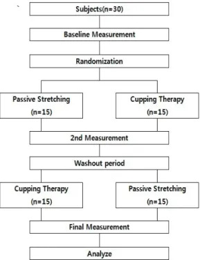
Fig. 1. Study design
each subject and approved by the institutional review board of Sahmyook University in Korea. All subjects provided written informed consent prior to study enrollment.
2. Procedure
After pre-test, subjects were randomly divided into cupping therapy group and passive stretching group by coin-tossing method. Three physical therapists with greater than 3 years' experience conducted stretching and cupping therapy intervention and each of the four testing sessions
under the same environmental conditions. Participants were not blinded to the intervention. Only the testers were blinded.
Cupping therapy was applied to the hamstring muscle for 5 minutes in the cupping therapy group. The passive stretching group was treated with a passive stretching for 10 seconds and repeated 9 times. Then, a post test was conducted after ether cupping therapy or passive stretching, in a manner similar to that of the pretest. Passive range of motion (PROM) (straight leg raising), active range of motion (AROM), and algometer (pain) testing, and MVC assessment using EMG were performed as dependent variables. After 1 week of washout period, following a pretest, subjects were switched to the other group. Then, a posttest was conducted, in a manner similar to that of the pretest (Fig. 1).
3. Interventions
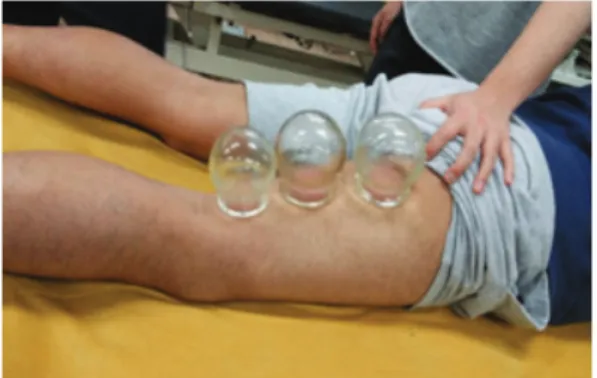
1) Cupping Therapy
Cupping therapy can be applied either with the use of a suction pump or by briefly heating the inside of a glass cup with a flame. Cupping therapy is usually applied with a suction pump but there is a gradual increase in the use of heat for cupping therapy due to outstanding treatment effects and convenience in sterilization. The application time varies based on the reason for treatment and intensity of negative pressure, but the application time is usually 1-10 minutes (Tham et al., 2006). Accordingly, the assessment was conducted in this study after a 5-minute application of cupping therapy with the use of a flame.
In flame-heated cupping therapy, the inside of the glass
cup momentarily develops a vacuum. When the glass cup
is applied to the skin, the negative pressure within the
cup causes myofascial decompression. To apply cupping
therapy to the hamstring muscle in the experiment leg
group, the muscle was divided into 3 areas and 3 cupping
therapy cups were applied to each area (Fig. 2).
Fig. 2. Attachment point of cupping therapy
2) Passive stretching
Subjects lay supine keeping their backs flat throughout the stretch. The leg was passively raised until to the point of “slight discomfort” in the hamstring and held for 10 seconds, followed by a and slow return; this was repeated 9 times (Johnson et al., 2014).
4. Measurement
In order to verify treatment efficacy of cupping therapy and passive stretching, a pre and posttest were conducted.
ROM, algometer, and MVC tests were conducted before and after the intervention.
1) ROM Test
PROM and AROM testing were conducted. PROM was conducted with the subject in supine position on a medical bed. The distance between the greater trochanter and the knee joint was indicated with a straight line for the PROM test, and an electronic goniometer (Dualer IQ, J-tech Medical, USA) was attached to the femoral region on the indicated line. Measurement was conducted after setting the value in the supine position at zero. Examiner performed the passive SLR by keeping the knee in full extension and the ankle in neutral position. Full ankle dorsiflexion was avoided to prevent calf muscle stiffness or pain (gastrocnemius and soleus) from confounding the sensation of hamstring stiffness and pain which would signal the
limit of the SLR test. The examiner would hold the talus and avoid any hip rotation during flexion of the hip as they lifted the subject's lower limb until he or she first complained of stiffness or pain in the region of the thigh, bent his/her knee, or began to swing into a posterior pelvic tilt (noted as movement of the ASIS).
For AROM, the hip joint angle was measured after voluntary straight leg raising. For this measurement, the ankle was held in ankle neutral position and the leg was lifted slowly to avoid generating elasticity from a quick leg lift. This measurement was within a range that would not cause bending of the knee on the measured side or an increase in lumbar lordosis angle.
2) Pain Threshold Test
The pain threshold value based on pressure applied to the hamstring muscle was measured with the use of an electronic algometer (Algometer, J-tech Medical, USA) with the subject in prone position on a medical bed. Three areas 2cm apart from the cupping place on the hamstring muscle were selected as pain points and the average value for the 6 pain points was calculated to obtain the pain index.
3) EMG test
Wireless EMG electrodes were attached to the
semitendinosus (ST) and biceps femoris (BF) with the
subject in prone position on a medical bed. Before
attachment of electrodes, hair at the attachment area was
removed, and the keratin layer was removed to reduce
impedance to the myoelectric signal by rubbing; electrodes
were then attached after cleansing the skin 3-4 times with
a sterilized alcohol swab. EMG electrodes were attached
along the contracting direction of the muscle fibers. The
ST electrode was attached at the midpoint between the
ischial tuberosity (IT) and the halfway point to the medial
condyle in the femoral region. The BF electrode was
attached at the midpoint from the IT to the halfway point
of the lateral condyle in the femoral region (Konrad, 2005; Shenoy et al., 2010). The distance between wireless electrodes was 2-3 cm. Muscle activity during sustained isometric MVC for 5seconds with the knee held in 90°
knee flexion was measured.
5. Data Analysis
Data collected in this study were analyzed with SPSS 18.0 for Windows and a statistically significant level α was set as .05. In order to compare the differences between the 2 groups before experiment for homogeneity ver- ification, an independent t-test was conducted. Normality was verified by conducting a Kolmogorov-Smirnov test for the 2 groups. The differences between the groups were compared with an independent parametric t-test, and a paired t-test was used to compare results for ROM, pain, and EMG before and after intervention in the experiment and control groups.
Ⅲ. Results
There was a statistically significant increase in ROM after intervention in the cupping therapy and passive stretching group for both the PROM and AROM tests.
However, there was no significant difference between the 2 groups for both tests (Table 2, 3).
The pain threshold increased from 56.1 to 63.8 before and after the intervention in the cupping therapy group (p<.05). However, there was no statistically significant difference in the Passive stretching group from 53.3 to 58.2 (p<.05). In a comparison of the 2 interventions, there was no difference between the groups (Table 4).
The muscle activity value of the ST increased from 175.4
㎶ to 214.9㎶ before and after the intervention in the cupping therapy group (p<.05). The muscle activity value of the ST increased from 176.0㎶ to 210.8㎶ before and after the intervention in the passive stretching group
Cupping therapy (n=15)
Passive stretching
(n=15) t p
Before (angle, °) 64.6 ± 9.085 64.8 ± 10.23 -.75 .940
After (angle, °) 76.0 ± 10.65 75.1 ± 11.09 .218 .829
Difference 11.4 ± 8.46 10.2 ± 7.67 .384 .704
t -5.215 -5.180
P .000* .000*
mean ± standard deviation
Table 2. Comparison of Cupping Therapy and Passive stretching on PROM
Cupping therapy (n=15)
Passive stretching
(n=15) t p
Before (angle, °) 55.5 ± 9.26 56.5 ± 9.65 -.280 .728
After (angle, °) 66.8 ± 7.68 66.6 ± 14.34 .35 .972
Difference 12.3 ± 7.04 10.6 ± 12.59 .405 .689
t -5.160 -2.587
P .000* .025*
mean ± standard deviation
Table 3. Comparison of Cupping Therapy and Passive stretching on AROM
Cupping therapy (n=15)
Passive stretching
(n=15) t P
Before Pain
†56.1 ± 11.16 53.3 ± 10.06 .738 .467
After Pain 63.8 ± 12.71 58.2 ± 12.04 1.235 .227
Difference 7.6 ± 12.47 4.8 ± 9.88 .663 .513
t -2.365 -1.918
P .033* .076
†
mean ± standard deviation
Table 4. Comparison of Cupping Therapy and Passive stretching on Pain Threshold
Cupping therapy (n=15)
Passive stretching
(n=15) t p
Before- MVC (㎶)
†175.4 ± 98.94 176.0 ± 58.08 -.202 .984
After- MVC (㎶) 214.9 ± 115.07 210.8 ± 71.75 .118 .072
Difference 39.5 ± 50.50 34.8 ± 39.86 .285 .778
t -3.035 -3.384
P .009* .004*
†
mean ± standard deviation
Table 5. Comparison of Cupping Therapy and Passive stretching on Semitendinosus-EMG
Cupping therapy (n=15)
Passive stretching
(n=15) t P
Before- MVC (㎶)
†115.2 ± 57.40 80.0 ± 44.47 1.877 .907
After- MVC (㎶) 132.5 ± 64.18 128.1 ± 130.6 .116 .908
Difference 17.3 ± 22.87 48.1 ± 126.0 -.932 .359
T -2.938 -1.481
P .011* .161
†