Biomedical Science Letters 2018, 24(4): 305~310 https://doi.org/10.15616/BSL.2018.24.4.305 eISSN : 2288-7415
Crosstalk Signaling between IFN-γ and TGF-β in Microglia Restores the Defective β-amyloid Clearance Pathway in
Aging Mice with Alzheimer's Disease
Go-Eun Choi†,*
Department of Clinical Laboratory Science, College of Health Sciences, Catholic University of Pusan, Busan 46252, Korea
Microglia are emerging as critical regulators of innate immune responses in AD and other neurodegenerative disorders, highlighting the importance of understanding their molecular and cellular mechanisms. We attempted to determine the role of crosstalk signaling between IFN-γ and TGF-β in Aβ clearance by microglia cells. We used in vitro and in vivo mouse models that recapitulated acute and chronic aspects of microglial responses to Aβ peptides. We showed that crosstalk signaling between TGF-β and Smad2 was an important mediator of neuro-inflammation. These findings suggest that microglial TGF-β activity enhances the pathological progression to AD. As TGF-β displays broad regulatory effects on beneficial microglial functions, the activation of inflammatory crosstalk signaling between TGF-β and Smad2 may be a promising strategy to restore microglial functions, halt the progression of Aβ-driven pathology, and prevent AD development.
Key Words: Alzheimer's disease, Microglia, IFN-γ, TGF-β, APPswe transgenic mice
INTRODUCTION
Alzheimer's disease (AD) is a neurodegenerative disorder associated with the accumulation of amyloid β42 (Aβ42).
It triggers inflammation, tau hyper-phosphorylation, and synaptic and neuronal losses in the brain, leading to cogni- tive decline. Microglia are emerging as critical regulators of innate immune responses in AD and other neurodegenera- tive disorders, highlighting the importance of understanding their molecular and cellular mechanisms (Leissring et al., 2003). In fact, accumulating Aβ42 may cause microglia to lose their normal ability to clear toxic proteins and control
inflammation, a detrimental phenotype observed with age- associated Aβ42 accumulation.
Interferon (IFN)-γ is a potent activator of microglia (Ng et al., 1999; Klegeris et al., 2005). IFN-γ level increases in the aged brain, although its endogenous cell source remains unidentified (Lyons et al., 2011). The primary signaling path- ways induced by IFN-γ are signal transduction and activation of transcription-type-1 (STAT1) and Mitogen-activated pro- tein kinase (MAPK)s (Blanchette et al., 2003; Gough et al., 2008).
The Smad pathway is very important for the regulatory and neuroprotective effect of transforming growth factor (TGF)-β (Derynck and Zhang, 2003), as it is involved in
Original Article
Received: October 24, 2018 / Accepted: November 15, 2018
*Professor.
†Corresponding author: Go-Eun Choi. Department of Clinical Laboratory Science, College of Health Sciences, Catholic University of Pusan, Busan 46252, Korea.
Tel: +82-51-510-0563, Fax: +82-51-510-0568, e-mail: [email protected]
○CThe Korean Society for Biomedical Laboratory Sciences. All rights reserved.
○CCThis is an Open Access article distributed under the terms of the Creative Commons Attribution Non-Commercial License (http://creativecommons.org/licenses/by-nc/3.0/) which permits unrestricted non-commercial use, distribution, and reproduction in any medium, provided the original work is properly cited.
the induction of the quiescent phenotype of microglia. Age- related alteration of the TGF-β pathway includes changes in TGF-β release, Smad3 activation, and changes in the microglial response induced by inflammatory stimuli in the hippocampus of aged mice, as well as abolition of TGF-β induced phagocytosis (Tichauer and von Bernhardi, 2012) by aged microglia (Tichauer et al., 2014).
IFN-γ suppresses TGF-β signaling through up-regulation of inhibitory Smad7 (Ulloa et al., 1999), and there is re- ciprocal regulatory interaction between TGF-β- and IFN-γ- activated pathways. Aged animals show increased levels of IFN-γ, directly potentiating inflammatory signaling and further inhibiting the Smad pathway through the induction of Smad7. However, decreased activation of TGF-β and Smad2 due to both age-related changes and increased IFN-γ suppresses the regulatory effect of TGF-β on inflammatory activation. Therefore, several studies have focused on age- related changes in IFN-γ signaling; very little is known about the role of age-related crosstalk signaling between IFN-γ and TGF-β in AD.
In the present study, we attempted to determine the role of crosstalk signaling between IFN-γ and TGF-β in Aβ clearance by microglia cells. We used in vitro and in vivo mouse models that recapitulated acute and chronic aspects of microglial responses to Aβ peptides. Our findings demon- strate that microglial crosstalk signaling between IFN-γ and TGF-β regulates multiple processes of the age-associated responses to Aβ peptides.
MATERIALS AND METHODS Reagents and antibodies
Peptide Aβ was obtained from Bachem (Torrance, CA, USA). Antibodies against Stat1, p-stat1, Smad2, p-smad2, Smad7, α-tubulin, and Aβ were purchased from Santa Cruz Biotechnology (Santa Cruz, CA, USA). Specific small mole- cules such as IFN-γ were purchased from InvivoGen (San Diego, CA, USA).
Animals
APPswe transgenic mice (B6C3-Tg) were purchased from Jackson Laboratories (Bar Harbor, ME, USA). The APPswe
transgene is integrated into a single locus and is indepen- dently under the control of separate mouse prion protein promoter elements, which direct the expression of the trans- gene predominantly to the central nervous system neurons (Borchelt et al., 1997; Jankowsky et al., 2001). Mice were used in pairs of age-matched transgenic or wild-type (WT) littermates and euthanized according to approved institutional procedures. All protocols were approved by the Dong-A University Institutional Animal Care and Use Committee guidelines for the humane care of animals.
Cell isolation, culture, and treatment
Mouse microglial BV-2 cells were cultured in Dulbecco's modified Eagle's medium (DMEM) supplemented with 10%
fetal bovine serum (FBS), penicillin (100 U/mL), and streptomycin (100 g/mL). Cultures were maintained at 37℃
in a humidified incubator with 95% O2 and 5% CO2. Pri- mary microglia were isolated from postnatal C57Bl/6 mouse pups using CD11b magnetic bead separation (OctoMACS, Miltenyi Biotech). The purity of the isolated microglia was assessed by flow cytometric analysis of cells stained for CD11b and F4/80, and was determined to be > 96%. Peptide Aβ, a full-length amyloid beta (Aβ1-42), was dissolved in deionized distilled water at a concentration of 1 mM and incubated at 37℃ for 72 h to induce aggregation (Lou et al., 2011). Confluent microglia BV-2 cells were stimulated with different concentrations of Aβ1-42 (1 μM) in serum-free medium and maintained for 24 h.
ELISA
Supernatants from cultured microglia were used to deter- mine the amount of tumor necrosis factor-alpha (TNF-α), TGF-β and interleukin-1-beta (IL-1β) released. Enzyme- linked immunosorbent assays (ELISA) were performed according to the manufacturer's recommendations (R&D Systems, Minneapolis, MN, USA).
Western blot analysis
Microglia were lysed in a lysis buffer (50 mM Tris-HCl [pH 7.5], 150 mM NaCl, 1% Triton X-100, 5 mM EDTA, 0.5 mM Na3VO4, 50 mM NaF, 1 mM PMSF, and protease inhibitor cocktail [Thermo Fisher Scientific, Waltham, MA,
USA]) for 30 min on ice. Cell debris was removed by cen- trifugation at 20,000 × g for 15 min at 4℃. The protein concentration in the cell lysates was determined using a Bio-Rad protein assay kit. An equal amount of protein from each sample was separated by 8% or 10% sodium dodecyl sulfate polyacrylamide gel electrophoresis (SDS-PAGE) and subsequently transferred onto polyvinylidene difluoride mem- branes (Millipore). After blocking with 5% skim milk for 1 h in TBS-T (0.1% Tween 20 in Tris-buffered saline [TBS]), membranes were incubated with primary antibodies, follo- wed by probing with the respective horseradish peroxidase (HRP)-conjugated secondary antibody (Santa Cruz).
Histological and immunohistochemical analyses
Formalin-fixed (4% paraformaldehyde), paraffin-embedded tissue sections of mouse brain 12 μm thick were deparaf- finized in xylene and rehydrated. Coronal sections were prepared for hematoxylin and eosin (H&E) staining. The sections were treated with a heat-based antigen retrieval method using a citrate solution (pH 6.0, 10 mM). Endogen- ous peroxidase was blocked by incubation in 1.5% hydrogen peroxide in methanol, and nonspecific binding of antibodies was blocked by incubation in 10% donkey serum in 0.02 M PBS and 0.1% Triton X-100 in PBS for 1 h. The slides were then incubated overnight at 4℃ with goat anti-Aβ primary antibody (1:300; Santa Cruz) diluted in the same blocking
solution. After washing, biotinylated rabbit anti-goat IgG antibody (Vectastain Elite ABC kit) was applied for 1 h.
The slides were washed once again, and then avidin-biotin- peroxidase complex (Vectastain Elite ABC kit) was applied for 30 min, followed by peroxidase detection with a mixture of 3,3'-diaminobenzidine (DAB; Vector Laboratories). To determine the specificity of immunostaining, control sections were incubated in solutions in which the primary antibodies were omitted.
Statistical analysis
All data were analyzed using GraphPad Prism v. 4.00 (GraphPad Software Inc., San Diego, CA, USA). Results are presented as means ± standard deviation (SD). Mann- Whitney U test was used for binary and multiple compari- sons, respectively. Significance was defined as P < 0.05.
RESULTS
Increased inflammatory response in microglia from aged mice upon stimulation with Aβ42 and IFN-γ
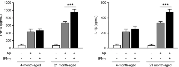
We measured the amounts of TNF-α and IL-1β produced by mouse microglial cells in response to Aβ induction. We found that Aβ (1 μM for 24 h) stimulated both TNF-α and IL-1β production in microglia (Fig. 1). Moreover, we found a significant increase in TNF-α and IL-1β production in the
Fig. 1. Increased inflammatory response in microglia from aged mice upon stimulation with Aβ and IFN-γ. Microglia were incubated with Aβ42 (100 ng/mL) for 24 hours in the absence or presence of IFN-γ (10 mM). The level of TNF-α and IL-1β from the supernatant of culture were determined by ELISA analysis. Data are expressed as means ± SD. ***P < 0.001, Mann-Whitney U test.
microglia of aged mice upon stimulation with Aβ42 and IFN-γ compared to stimulation with or without IFN-γ. We determined the effect of Aβ on IFN-γ signaling in microglia
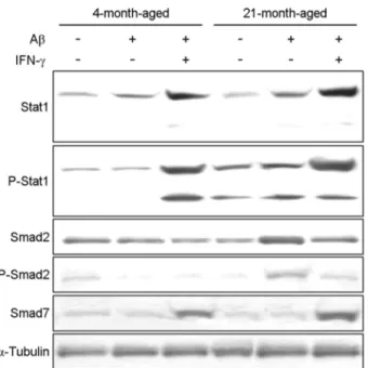
from young (4 months) and aged (21 months) mice by evaluating Stat1, Stat1-phosphorylation, Smad2, Smad2- phosphorylation and Smad7 protein levels using western blot analysis. We determined the level of phosphorylation of Stat1 in young and aged mice in response to Aβ42 stimu- lation with or without IFN-γ, and found a significant increase in the phosphorylation of Stat1 in the microglia of aged mice compared to young mice (Fig. 2). In hence, microglia from aged mice upon stimulation with Aβ42 and IFN-γ showed decreased expression of the phosphorylation of Smad2 com- pared to stimulation without IFN-γ.
Inhibited production of TGF-β expression in aged mice upon stimulation with IFN-γ
We determined the level of TGF-β production in young and aged mice in response to stimulation with or without IFN-γ, and found no significance in TGF-β production in the serum of aged mice upon stimulation with IFN-γ compared to stimulation without IFN-γ (Fig. 3). In contrast, the serum from aged mice showed increased expression of TNF-α and IL-1β compared to young mice. Moreover, we found a sig- nificant increase in TNF-α production in the serum of aged mice upon stimulation with IFN-γ compared to stimulation without IFN-γ. To investigate the effect of TGF-β on the pathogenesis of AD, we determined the level of Aβ accu- Fig. 2. Decreased expression of the phosphorylation of Smad2
in microglia from aged mice upon stimulation with Aβ and IFN-γ. Microglia were incubated with Aβ42 (100 ng/mL) for 1 hours in the absence or presence of IFN-γ (10 mM). Relative protein levels of microglia were determined by western blot and norm- alized to α-tubulin.
Fig. 3. Inhibited production of TGF-β expression in aged mice upon stimulation with IFN-γ. APPswe mice were challenged with IFN-γ (10 mM) to induce acute peritoneal inflammation. The level of TGF-β and TNF-α in serum from mice were determined by ELISA analysis. Data are expressed as means ± SEMs (n = 4 per group). **P < 0.005; Mann-Whitney U test.
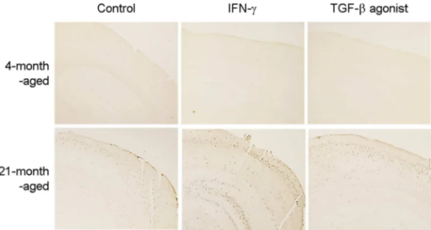
mulation by intraperitoneal administration of the selective agonist for TGF-β. The Aβ accumulation in the brain tissue increased following administration of the agonist (Fig. 4).
DISCUSSION
Microglial activity is essential for defense against the development of neurodegenerative diseases; microglia clear misfolded proteins, elaborate trophic and regenerative factors, and regulate and terminate toxic inflammation. In AD, micro- glia not only lose their capacity to clear Aβ peptides, but also develop a persistent proinflammatory phenotype that fails to resolve, which accelerates neuronal and synaptic injuries.
Inflammatory chemokines such as TNF-α and IL-1β are important for the recruitment of monocytic cells, including macrophages and microglia. The levels of these chemokines were increased by the age of 9 months in APPswe mice, suggesting they have a specific role in the inflammatory response to Aβ peptides (Radde et al., 2006; Darvesh et al., 2012). We demonstrated that APPswe mice showed increa- sed activation of the Stat1 protein in microglia upon stimu- lation with Aβ42 oligomers. This interesting regulation was associated with a robust immune response to Aβ42, which was significantly enhanced upon administration of IFN-γ.
Moreover, the age dependence of Stat1 upregulation and the IFN-γ driven inflammatory gene is highly relevant to the pathogenesis of Aβ.
In this study, we used in vitro and in vivo strategies to
reveal that microglial crosstalk signaling between TGF-β and Smad2 negatively regulates multiple distinct beneficial functions that are critical to suppression of the harmful effects of accumulating Aβ42. Together, our findings in distinct mouse models of Aβ inflammation demonstrated the inhibi- tion of microglial crosstalk signaling between TGF-β and Smad2, regulation of inflammatory responses, and trophic factor generation and signaling.
In conclusion, we showed that crosstalk signaling between TGF-β and Smad2 was an important mediator of neuro- inflammation and that microglial crosstalk signaling between TGF-β and Smad2 induced multiple beneficial functions that are essential to combat the toxic effects of Aβ42 peptides.
These findings suggest that microglial TGF-β activity en- hances the pathological progression to AD. As TGF-β dis plays broad regulatory effects on beneficial microglial func- tions, the activation of inflammatory crosstalk signaling between TGF-β and Smad2 may be a promising strategy to restore microglial functions, halt the progression of Aβ42- driven pathology, and prevent AD development.
ACKNOWLEDGEMENT
This research was supported by a grant from NRF- 2013R1-A1A2061273 and NRF-2016R1D1-A1B03935541.
CONFLICT OF INTEREST
No potential conflict of interest relevant to this article was reported.
Fig. 4. TGF-β agonist increase Aβ accumu- lation in brain tissue of aged mice. APPswe mice were administrated by peritoneal chal- lenge with IFN-γ (10 mM) or TGF-β agonist once every three days for one month. The level of Aβ accumulations in brain from mice were determined by immunohistochemistry. Repre- sentative immunohistochemical staining for Aβ in the brain tissue for each group of mice.
REFERENCES
Blanchette J, Jaramillo M, Olivier M. Signalling events involved in interferon-gamma-inducible macrophage nitric oxide gene- ration. Immunology. 2003. 108: 513-522.
Borchelt DR, Ratovitski T, van Lare J, Lee MK, Gonzales V, Jenkins NA, Copeland NG, Price DL, Sisodia SS. Accelerated amyloid deposition in the brains of transgenic mice coexpres- sing mutant presenilin 1 and amyloid precursor proteins.
Neuron. 1997. 19: 939-945.
Darvesh S, Cash MK, Reid GA, Martin E, Mitnitski A, Geula C.
Butyrylcholinesterase is associated with beta-amyloid pla- ques in the transgenic APPSWE/PSEN1dE9 mouse model of Alzheimer disease. J Neuropathol Exp Neurol. 2012. 71: 2-14.
Derynck R, Zhang YE. Smad-dependent and Smad-independent pathways in TGF-beta family signalling. Nature. 2003. 425:
577-584.
Gough DJ, Levy DE, Johnstone RW, Clarke CJ. IFNgamma signaling-does it mean JAK-STAT? Cytokine Growth Factor Rev. 2008. 19: 383-394.
Jankowsky JL, Slunt HH, Ratovitski T, Jenkins NA, Copeland NG, Borchelt DR. Co-expression of multiple transgenes in mouse CNS: a comparison of strategies. Biomol Eng. 2001. 17: 157 -165.
Klegeris A, Bissonnette CJ, McGeer PL. Modulation of human microglia and THP-1 cell toxicity by cytokines endogenous to the nervous system. Neurobiol Aging. 2005. 26: 673-682.
Leissring MA, Farris W, Chang AY, Walsh DM, Wu X, Sun X, Frosch MP, Selkoe DJ. Enhanced proteolysis of beta-amyloid in APP transgenic mice prevents plaque formation, secondary pathology, and premature death. Neuron. 2003. 40: 1087-1093.
Lyons A, Murphy KJ, Clarke R, Lynch MA. Atorvastatin prevents age-related and amyloid-β-induced microglial activation by blocking interferon-γ release from natural killer cells in the brain. J Neuroinflammation. 2011. 31: 27.
Ng YK, Yong VW, Ling EA. Microglial reaction in some CNS nuclei following nerves transection in BALB/c and interferon- gamma gene knockout mice. Neurosci Lett. 1999. 262: 207 -210.
Radde R, Bolmont T, Kaeser SA, Coomaraswamy J, Lindau D, Stoltze L, Calhoun ME, Jäggi F, Wolburg H, Gengler S, Haass C, Ghetti B, Czech C, Hölscher C, Mathews PM, Jucker M. Abeta42-driven cerebral amyloidosis in transgenic mice reveals early and robust pathology. EMBO Rep. 2006. 7:
940-946.
Tichauer J, Flores B, Soler B, Eugenín-von Bernhardi L, Ramírez G, von Bernhardi R. Age-dependent changes on TGFβ1 Smad3 pathway modify the pattern of microglial cell activation. Brain Behav Immun. 2014. 37: 187-196.
Tichauer J, von Bernhardi R. Transforming growth factor-β stimu- lates β amyloid uptake by microglia through Smad3-dependent mechanisms. J. Neurosci Res. 2012. 90: 1970-1980.
Ulloa L, Doody J, Massague J. Inhibition of transforming growth factor-beta/SMAD signalling by the interferon-gamma/STAT pathway. Nature. 1999. 397: 710-713.
https://doi.org/10.15616/BSL.2018.24.4.305
Cite this article as: Choi GE. Crosstalk Signaling between IFN-γ and TGF-β in Microglia Restores the Defective β-amyloid Clearance Pathway in Aging Mice with Alzheimer's Disease. Biomedical Science Letters.
2018. 24: 305-310.