89
Magnetic Resonance Spectroscopy Findings in Perro de Presa Canario Dogs
with Spongy Degeneration of the Central Nervous System
Sae-Byel Hong*, In Lee**, Yu-Mi Song**, Young-Won Lee* and Ho-Jung Choi*1
*College of Veterinary Medicine, Chungnam National University, Daejeon 34134, Korea **Ian Animal Diagnostic Center, Seoul 06014, Korea
(Received: December 28, 2020 / Revised: January 27, 2021 / Accepted: March 18, 2021)
Abstract : 2-Month-old, three related Perro de Presa Canario dogs were evaluated for similar neurological symptoms like circling, head pressing, depressed mental status, hypermetria, and vocalization. On magnetic resonance imaging (MRI) of the brain, there were large, bilaterally symmetrical lesions with involvement of thalamus, and brainstem that were T2- and FLAIR-hyperintense and T1-iso/hypointense. There was no inclusion of cerebellum. Single-voxel spectroscopy acquisition was located in the thalamus where abnormalities were found in MR images. The results of magnetic resonance spectroscopy (MRS) showed markedly decreased N-acetylaspartic acid value. Euthanasia was performed and lesions consistent with the canine spongy degeneration. Alteration in metabolites in the brain can be determined by MRS, which helps in diagnosing degeneration/leukodystrophy of the central nervous system in dogs. Key words : magenetic resonance spectroscopy, spongy degeneration, leukodystrophy, demyelination, dog.
Introduction
Spongy degeneration of the central nervous system (CNS) results from an astrocytic biochemical abnormality of sev-eral different diseases (1). Histologic findings include separa-tion of the myelin sheath or neuronal cell body, often with accompanied vacuolation of the brain tissue. Spongy degen-eration/leukodystrophy of the brain has been reported as pri-marily affecting the white matter in Labrador retrievers (8), Dalmatians, Silkie terriers, Samoyeds, Shetland sheepdogs, a Scottish terrier, a miniature Poodle (3,4), Domestic short hair kitten, Persian kitten, and Birman kittens (6). Primarily gray matter spongy degeneration has been described in Bull Mas-tiffs (12), Salukis, Malinois/Shepherd mixed-breed dogs, and Cocker spaniels (11).
Spongy white matter degeneration is similar to Canavan’s disease of humans (16). Canavan’s disease in children has a familial incidence and an autosomal recessive mode of inher-itance. Congenital, infantile, and juvenile forms have been recognized (1,8). In the first few months of life, patients develop psychomotor retardation, abnormally decreased mus-cular tone, seizure, megalencephaly, and blindness with optic atrophy, and early death (9). In human medicine, characteris-tic of magnecharacteris-tic resonance imaging (MRI) has been reported as symmetrical, subcortical white matter degenerative lesion primarily within the cerebral hemispheres with lesser involve-ment of the cerebellum and brain stem (8).
Magnetic resonance spectroscopy (MRS) is a useful method for identifying molecular structures that provide metabolic information from brain tissues. The development of improved
techniques for spatial localization and water suppression pro-vides metabolic information obtained from conventional MRI about the tissue under study (3). Brain lesions contain abnormal levels of metabolites, N-acetyl aspartate (NAA), choline (Cho), creatine (Cr), as compared to normal brain tis-sue (2). It has been reported that MRS could detect the met-abolic changes in brain of human patients with Canavan’s disease (7).
In this article, we described the MR imaging features of spongy degeneration of three Perro de Pressa Canario dogs, and MRS results in two of them.
Dog 1
A 2-month-old male Perro de Presa Canario, weighing 3.8-kg was referred with intermittent circling, hiding, lack of sucking, head pressing, depressed mental status, hypermetria and vocalization. Physical and neurologic examination revealed the proprioceptive deficit of all limbs and absent menace response. The results of complete blood count and serum bio-chemistry were within normal range. There were no signifi-cant findings in abdominal and thoracic radiography.
Brain conventional MRI (1.5 Tesla unit, Magnetom Essenza, Siemens, Germany) was performed at another animal medi-cal imaging center. Scanning parameters were slice thickness 3.0 mm, and field of view 84 × 112 mm in transverse plane, 105 × 140 mm in sagittal plane, 104 × 130 mm in dorsal plane. The dorsal plane slices were obtained parallel to the hard pal-ate (14). The transverse slices were obtained at right angles to the dorsal plane. Conventional MRI including T2-weighted (TR/TE 5550/71 ms on transverse, 3020/81 ms on sagittal, 4070/77 ms on dorsal plane), T1-weighted (TR/TE 510/13 ms on transverse, 430/14 ms on sagittal plane), and FLAIR (TR/TE/TI 8000/77/2400 ms on transverse) were obtained.
1Corresponding author.
MR images of the brain showed large, bilaterally symmet-rical lesions with T2 and FLAIR hyperintensity and T1 iso/ hypointensity at the level of the thalamus, midbrain, and pons. There were paired areas of focal increased signal inten-sity in several nucleus areas, especially, caudate nucleus, hypothalamus, rostral colliculus, oculomotor nucleus, pon-tine reticular nucleus, caudal cerebellar peduncle and vestib-ular nucleus (Fig 1). There was no contrast enhancement on post-contrast T1-weighted images (Fig 2). The distinction between the gray and white matter was not clear in the pro-ton density-weighted images and subcortical-periventricular white matter leukodystrophy was also observed.
A cisternal cerebrospinal fluid collection was performed under aseptic conditions after the MRI examination. Results of cytologic and protein analyses of cerebrospinal fluid (CSF) were normal, and antibody titer to canine distemper virus, West Nile virus, bartonella spp., blastomyces dermatitids, coc-cidiodes spp., cryptococcus spp., histoplasma capsulatum, bor-relia burgdorferi, neospora spp., and toxoplasma gondii was
negative.
The euthanasia was requested, and a necropsy was per-formed. The result of histopathologic examination was spongy degeneration (Fig 4).
Dog 2 and 3
A year later, the same parent Perro de Presa Canario of Dog 1 gave birth to 12 puppies. At the age of 2 month-old, two puppies were relatively small compared to the other lit-ter mates. One of them was male weighing 3.8-kg, (Dog 2), and the other was female weighing 3.8-kg (Dog 3). They were referred with head pressing, and vocalization three days before the visit. They had been lethargic and anorexic since they were born.
Physical examination confirmed hypermetria, cerebellar ataxia, circling, head pressing, vocalization, and tremor. Neu-rologic examination identified that the mental was depressed to the stuporous, left side of the oculovestibular test was Fig 1. (A-F) T2-weighted transverse images at different levels of the brain of Dog 1. Multiple, bilateral, symmetrical T2 hyperintense lesions were found at the level of thalamus, hypothalamus, and brainstem (white arrows). Lesions include several nucleus areas, like (A) caudate nucleus, (B) thalamic and hypothalamic region (thalamic & hypothalamic nuclei), (C) midbrain (oculomotor nucleus, ros-tral colliculus), (D, E) pons (pontine, pontine reticular nucleus), and (F) medulla oblongata (vestibular, facial motor, pyramid).
Fig 2. (A-B) T1-weighted pre and post-contrast transverse images of Dog 1. There was no contrast enhancement. (C) In T2-weighted mid-sagittal image, note the multifocal T2 hyperintense lesion in thalamus and brain stem (white arrow).
delayed, lack of menace responses and vision bilaterally, with normal pupillary light reflex, and also moderate postural test reaction deficits characterized by slower than normal initia-tion of hopping with thoracic and pelvic limbs. Blood exam-ination of two dogs showed increased creatine kinase of 1,015 U/L, 662 U/L (reference range 99-436 U/L), phospho-rus of 7.6 mg/dL, 7.1 mg/dL (reference range 2.6-6.2 mg/ dL), potassium of 5.51 mmol/L, 5.28 mmol/L (reference range 2.7-5 mmol/dL), respectively and decreased total pro-tein level of 5.1 g/dL in both dogs (reference range 5.4-7.4 d/ dL). There was no significant finding in thoracic radiogra-phy. Considering they were puppies with the same parents as dog 1 diagnosed with spongy degeneration a year ago and had similar clinical symptoms, they suspected of suffering from the same disease.
Combined brain MRI (1.5 Tesla unit, Vantage ElanTM,
Canon Medical Systems, Japan) and localized proton MRS using a 16 channel, the small size coil (1.5T Receive-only 16
channel Flex SPEEDER small, Canon Medical Systems, Japan) was performed in Chungnam National University Animal Hospital. The dogs were positioned in sternal recum-bency. Conventional MR sequences including T2-weighted (TR/TE 3300/84 ms on transverse (Dog 2), TR/TE 5300/104 ms on transverse (Dog 3), 3900/108 ms on sagittal, 4350/108 ms on dorsal plane), T1-weighted (TR/TE 725/15 on trans-verse, 500/15 on sagittal, 560/15 on dorsal plane), and FLAIR (TR/TE/TI 9000/120/2700 on transverse plane) images were obtained from the brain of dog 2 and 3. MRI findings were similar to the results of dog 1 previously described.
A two-dimensional MRS sequence using point-resolved spectroscopy (TR/TE 3900/108 ms; thickness 3.4 mm; a field of view 92 × 90 mm (Dog 2), 110 × 100 mm (Dog 3)) was used for the square volume of interest (VOI) selection. Sin-gle-voxel spectroscopy acquisition with 10 mm3 volume was
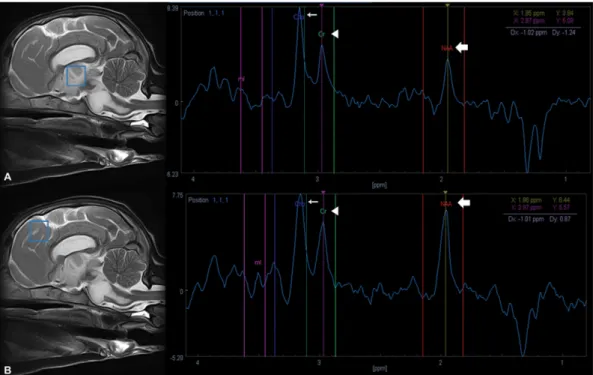
located in thalamus where abnormalities were found in the previous MRI while omitting bone structures and ventricles. Fig 3. Detection of markedly decreased N-acetylasparate (NAA) peak (thick arrow) on MR spectroscopic images in brain lesion at thalamus in Dog 2 (A) compared with brain parenchyma at frontoparietal area which appeared to be unaffected (B). Cr = creatine (arrowhead), Cho = Choline (thin arrow).
The metabolite concentrations of voxels corresponding to the specific regions at the sagittal plane were measured in each peak area of the spectra. Next, metabolite ratios (NAA/Cr, Cho/Cr, and NAA/Cho) were calculated for the major metab-olites in each brain region. On MRS of the brain, our patients showed markedly decreased NAA in the thalamic lesion (Fig 3) and NAA/Cho was also lowest value. The quantitative analyses and the normal range of metabolite concentrations (2) summarized in Table 1. Both dogs were euthanized after all examinations.
Discussion
MRS is an imaging technique that provides specific bio-chemical data on diverse metabolites in a noninvasive way. MRS technique is now routinely used in humans to catch pathological changes associated with the brain diseases. MRS data are usually presented as line spectra, the area under each peak representing the relative concentration of nuclei detected for a given metabolite. The major detectable metabolites analyzed on MRS are NAA which is produced in mitochondria of the neurons and transported into the neuro-nal cystoplasm, Cr as ubiquitous compounds linked to energy metabolism, and Cho which is a metabolic marker of mem-brane density and integrity. In the clinical study, creatine value is supposed to be constant and is used for calculating metabolite ratios (NAA/Cr and Cho/Cr) (15). The normal range of metabolites levels varies slightly depending on the anatomic part of the brain and age. A large amount of NAA exists in normal brain parenchyma and appears to be the highest peak in the normal spectrum. As the brain matures, the concentration of NAA increases and the concentration of Cho decreases. In the elderly, there is a normal decline in the level of NAA (13,15).
The usefulness of NAA as an axonal marker is supported by the loss of NAA in many white matter diseases, including leukodystrophy. The only disease with high levels of NAA is Canavan’s disease in humans (15). It is known that the dis-ease is caused by a mutation in the gene that encodes the aspartoacylase enzyme (5). This enzyme’s main role is con-verting N-acetylaspartic acid to aspartate and acetate in the brain. When converting the N-acetylaspartic acid is blocked because of aspartoacylase enzyme deficiency, then N-acetyl-aspartic acid accumulates in the white matter and
compli-brain lesion. To our knowledge, papers of MRS study in dogs with spongy/leukodystrophy degeneration have not been reported in veterinary literature. The generalized reduction of NAA in the areas with the abnormal signal on T2-weighted images was observed in present patients, unlike what was reported in human patients with Canavan’s disease. We do not have a definitive reason to explain this discrepancy. A potential explanation for this is that the disease in canine can result in a decrease in NAA value because it causes a neuro-nal loss or dysfunction. There is another possibility that the etiology of the disease in dogs might be different from that of humans.
In human and animal brain tissue, N-acetyl aspartate exists only in neurons, making it helpful in the evaluation of neu-ron density. The MRS peak of NAA is normally higher than spectroscopic peaks corresponding to creatine and choline (2). Although the normal range of MRS in each part of the brain, frontoparietal, parietal, temporal, occipital thalamus, and the cerebellum has been studied in dogs, no intensive research has been conducted on the brainstem. In human medicine, it is known that the NAA value is the highest in brainstem, just like any other brain part (10). Further research on brainstem MRS figures is needed in dogs.
There are some reports of leukodystrophy/spongy degener-ation of CNS in dogs diagnosed by histopathology and MRI. The disease typically begins within the first 6 months of life and progressively worsens. Previous reports assumed the dis-ease to be hereditary, a degenerative condition similar to the Canavan’s disease in children, a rare leukodystrophy (8). Clin-ical signs are variable, including seizures, depressed mental status, tremors, ataxia, dysphagia, paraparesis, and tetraparesis (4). In our cases the three related Perro de Presa Canario dogs showed head pressing, hypermetria and also depressed. These were similar symptoms to those of previous reports (8).
In canine spongy degeneration of the CNS, MRI disclosed symmetrical, hyperintense lesions on T2-weighted images, which correlated with degenerative white matter lesions detected during gross and histologic examination (4). Lesions are low signal on T1-weighted images and high signal on T2-weighted images, implying increased water substance and decreased myelin in the white matter in these sites (8). In our cases, dogs also showed bilaterally symmetric, hyperintensi-ties at thalamus, midbrain, and pons on T2-weighted images were identified and no involvement of the cerebellum. Histo-logic lesions include loss of myelin in areas of spongy degen-eration; preservation of axons, nerve cells, and oligodendroglia; the increase of astroglia in both white and gray matter, scar-city of fibrillary gliosis; and lack of vascular reaction and inflammation.
NAA/Cr 0.75 00.83
NAA/Cho 0.45 00.60
Conclusion
The present study describes MR imaging features and MRS result of spongy degeneration in three puppies from the same parents. MR spectroscopy, noninvasive diagnostic examina-tion, can give important physiological and metabolic informa-tion, complementing morphological findings from conventional MRI in the clinical setting.
Acknowledgment
This work was supported by research fund of Chungnam National University.
References
1. Adachi M, Schneck L, Cara J, Volkb W. Spongy degeneration of the central nervous system (Van Bogaert and Bertrand type; Canavan’s disease). Hum Pathol 1973; 4: 331-347. 2. Choi S, Song Y, Lee K, Lee Y, Choi H. Multi-voxel magnetic
resonance spectroscopy of cerebral metabolites in healthy dogs at 1.5 Tesla. J Vet Sci 2016; 17(2): 217-224.
3. Coates JR, Kline KL. Congenital and inherited neurologic disorders in dogs and cats. In: JD Bonagura (ed.), Kirt’s Current Veterinary Therapy XII. 12th ed. Philadelphia: Saunders WB 1995: 1111-1120.
4. Dewey CW. Encephalopathies: disorders of the brain. Dewey CW(ed): A practical Guide to Canine and Feline Neurology, Ames, IA, Iowa State Press 2003; 141-236.
5. Hagenfeldt L, Bollgren I, Venizelos N. N-Acetylasparticaciduria due to aspartoacylase deficiency-a new aetiology of childhood leukodystrophy. J Inherited Metab Dis 1987; 10: 135-141. 6. Jones BR, Alley MR, Shimada A, Lyon M. An
encephalo-myelopathy in related Birman kittens. N Z Vet J 1992; 40: 160-163.
7. Karimzadeh P, Jafari N, Nejad Biglari H, Ahmadabadi F,
Nemati H, Nasehi MM, Ghofrani M, Mollamohammadi M. The clinical features and Diagnosis of Canavan’s disease: A case series of Iranian Patients. Iran J Child Neurol 2014; 8(3): 66-71.
8. Mariani CL, Clemmons RM, Graham JP, Phillips LA, Chrisman CL. Magnetic resonance imaging of spongy degeneration of the central nervous system in a Labrador Retriever. Vet Radiol Ultrasound 2001; 42(4): 285-290.
9. Marks HG, Caro PA, Wang ZY, Detre JA, Bogdan AR, Gusnard DA, Zimmerman RA. Use of computed tomography, magnetic resonance imaging, and localized 1H magnetic resonance spectroscopy in Canavan’s disease: A case report. Ann Neurol 1991; 30: 106-110.
10. Mascalchi M, Brugnoli R, Guerrini L, Belli G, Nistri M, Politi LS, Gavazzi C, Lolli F, Argenti G, Villari N. Single-voxel long TE 1H-MR spectroscopy of the Normal brainstem and cerebellum. J Magn Reson Imaging 2002; 16: 532-537. 11. Morales C, Bernardini M, Pumarola M, Siso S. Familial
spongy degeneration in Cocker Spaniel dogs. J Vet Intern Med 2001; 15: 72.
12. Morrison JP, Schatzberg SJ, De Lahunta A, Ross JT, Bookbinder P, Summers BA. Oligodendroglial dysplasia in two Bullmastiff dogs. Vet Pathol 2006; 43: 29-35.
13. One K, Kitagawa M, Ito D, Tanaka N, Watari T. Regional variations and age-related changes detected with magnetic resonance spectroscopy in the brain of healthy dogs. Am J Vet Res 2014; 75(2): 179-186.
14. Ratai EM, Gilberto GR. Handbook of Clinical Neurology, Vol. 135 (3rd series): Clinical magnetic resonance spectroscopy of the central nervous system 2016: 93-116.
15. Soares DP, Law M. Magnetic resonance spectroscopy of the brain: review of metabolites and clinical applications. Clin Radiol 2009; 64: 12-21.
16. Summers BA, Cummings JF, de Lahunta A. Degenerative disease of the central nervous system. Veterinary neuropathology. Mosby: St Louis. 1995: 208-350.