Redox Regulation of Lipopolysaccharide-mediated Sulfiredoxin
Induction, Which Depends on Both AP-1 and Nrf2
*
Received for publication, March 25, 2010, and in revised form, August 31, 2010Published, JBC Papers in Press, September 7, 2010, DOI 10.1074/jbc.M110.126839 Hojin Kim, Yuyeon Jung, Bong Soo Shin, Hyeryeon Kim, Hyunsook Song, Soo Han Bae, Sue Goo Rhee,
and Woojin Jeong1
From the Department of Life Science, Division of Life and Pharmaceutical Sciences, and the Center for Cell Signaling and Drug Discovery Research, Ewha Womans University, Seoul 120-750, Korea
Sulfiredoxin (Srx) is an enzyme that catalyzes the reduction of cysteine sulfinic acid of hyperoxidized peroxiredoxins and exerts a protective antioxidant role. Here we investigated the regulatory mechanism of Srx induction by lipopolysaccharide (LPS) in mouse macrophages. LPS up-regulated Srx expression on the transcriptional level. The promoter region of the Srx gene contained putative NF-B and AP-1 (activator protein-1) sites, and the proximal site of three AP-1 sites was embedded within the antioxidant response element (ARE), a cis-acting element for Nrf2 (nuclear factor erythroid 2-related factor). Mutational analysis of the Srx promoter revealed that Srx induction is dependent on AP-1 sites and ARE but not on NF-B sites. Con-sistently, both transcription factors, AP-1 and Nrf2, were required for LPS-mediated Srx induction, as revealed by chro-matin immunoprecipitation using antibodies specific for c-Jun and c-Fos and little Srx induction in Nrf2-null bone marrow-derived macrophages. Among mitogen-activated protein kinases that mediate the signal transduction by LPS, JNK played a major role in Srx induction. Moreover, chemical antioxidants, such as N-acetylcysteine and butylated hydroxyanisole, and the NADPH oxidase inhibitor diphenyleneiodonium inhibited Srx induction as well as generation of reactive oxygen species, both of which were also suppressed in Nox2 (NADPH oxidase 2)-de-ficient bone marrow-derived macrophages. These results sug-gest that LPS-mediated Srx induction is dependent on both AP-1 and Nrf2, which is regulated by Nox2-derived reactive oxy-gen species.
Peroxiredoxins (Prxs)2are a family of thiol-dependent per-oxidases that reduce H2O2and alkyl hydroperoxides and are involved in many cellular functions including proliferation, cell cycle, apoptosis, and differentiation as well as cellular
protec-tion against oxidative stress (1–3). There are six mammalian Prx isoforms, which are distributed in most cellular compart-ments, including cytosol, mitochondria, nucleus, endoplasmic reticulum, and peroxisomes. They have been divided into three subgroups, designated 2-Cys Prxs (Prx I–IV), atypical 2-Cys Prx (Prx V), and 1-Cys Prx (Prx VI). Under highly oxidizing condi-tions, their peroxidatic active site cysteine residue undergoes oxidation to sulfinic acid (Cys-SO2H), resulting in loss of per-oxidase activity (4, 5). However, the hyperoxidation of Prxs was revealed to be reversible in cells (6, 7), and Srx was subsequently identified as an enzyme responsible for the reversal of hyper-oxidized Prx in mammals as well as yeast (8 –11). Srx catalyzes transfer of␥-phosphate of ATP to sulfinic cysteine of Prx, and the resulting sulfinic phosphoryl ester is reduced by thiol equiv-alents, such as thioredoxin and glutathione (11–14). Srx can reduce the sulfinic forms of 2-Cys Prxs among six mammalian isoforms (10) and also has deglutathionylation activity (15, 16). Macrophages play essential roles in inflammation and host defense against bacterial infection. LPS, an integral component of the outer membrane of Gram-negative bacteria, is one of the potent activators of macrophages and a key mediator of endotoxic shock (17, 18). Stimulation of macrophages with LPS leads to acti-vation of intracellular signaling pathways that culminate in the induction of inflammatory molecules as well as production of ROS that are utilized for the killing of engulfed pathogens (19 –21). LPS induces expression of proinflammatory molecules and antioxidant enzymes by binding to Toll-like receptor 4, which triggers phos-phorylation of several kinases, including IB kinase (IKK), phos-phoinositide 3-kinase, and MAPKs, leading to activation of tran-scription factors, including NF-B and AP-1 (activator protein-1) (22–24). In stimulated macrophages, ROS are mainly generated by the Nox2 (NADPH oxidase 2) enzyme complex, which consists of two transmembrane flavocytochrome b components (gp91phox and p22phox) and four cytosolic components (p47phox, p67phox, p40phox, and Rac proteins) (25). Given that ROS can cause damage to various cellular components, including nucleic acids, mem-brane lipids, and proteins, cells are equipped with antioxidant enzymes, which are often induced in response to oxidative stress. For instance, Prx enzymes were up-regulated and exerted a pro-tective antioxidant role in macrophages exposed to LPS (26 –30). Recent studies demonstrated that Srx is induced by several stimuli, including metabolic activation, synaptic activity, and hyperoxia, and contributes to the protective response against oxidative insults (31).
Here we show that exposure of LPS to mouse BMM and RAW264.7 cells led to activation of Nrf2 (nuclear factor eryth-*This work was supported by Bio R&D Program Grant
M10642040002-07N4204-00210, Grant R01-2008-000-11321-0 and National Core Research Center Program Grant R15-2006-020 through the Korea Science and Engi-neering Foundation funded by the Ministry of Education, Science, and Technology, and by funds from the Brain Korea 21 Scholars Program (to Y. J. and H. K.).
1To whom correspondence should be addressed: Science Bldg. C, Rm. 207, 11-1 Daehyun-dong, Seodaemun-gu, Seoul 120-750, Korea. Tel.: 82-2-3277-4495; Fax: 82-2-3277-3760; E-mail: [email protected].
2The abbreviations used are: Prx, peroxiredoxins; Srx, sulfiredoxin; SrxP, Srx promoter; NF-B, nuclear factor-B; ARE, antioxidant response element; BMM, bone marrow-derived macrophage; Nox, NADPH oxidase; ROS, reac-tive oxygen species; NAC, N-acetylcysteine; DPI, diphenyleneiodonium; BHA, butylated hydroxyanisole; BSO,L-buthionine-sulfoximine; GCLC, glu-tamate-cysteine ligase catalytic subunit; TBH, tert-butylhydroquinone.
at Ewha Medical Library on October 24, 2016
http://www.jbc.org/
roid 2-related factor) in addition to NF-B and AP-1, of which both AP-1 and Nrf2 were required for up-regulation of Srx expression by LPS. Among the MAPKs activated by LPS, JNK was the major MAPK mediating Srx induction. In addition, chemical antioxidants, Nox inhibitor, and Nox2 deficiency decreased LPS-mediated Srx induction as well as ROS produc-tion, suggesting that Nox2-derived ROS may contribute to reg-ulation of Srx induction in macrophages exposed to LPS. EXPERIMENTAL PROCEDURES
Reagents and Antibodies—LPS (from Escherichia coli 0127: B8), cycloheximide, actinomycin D, SP600125, SB202190, U0126, N-acetylcysteine (NAC), diphenyleneiodonium (DPI), butylated hydroxyanisole (BHA), and L
-buthionine-sulfoxi-mine (BSO) were obtained from Sigma; 5-(and-6-)chloro-methyl-2⬘,7⬘-dichlorodihydrofluorescein diacetate was from Molecular Probes. Normal rabbit IgG was from Invitrogen; rab-bit polyclonal antibodies to Srx were prepared as described pre-viously (9); mouse monoclonal antibody (mAb) to-actin was from Abcam; mAbs to tubulin, p65, and ERK2 and rabbit poly-clonal antibodies to p65, c-Jun, c-Fos, JNK1, and p38 were from Santa Cruz Biotechnology, Inc. (Santa Cruz, CA); a mAb to the FLAG epitope was from Sigma; and a rat antibody to the hemagglutinin epitope (HA) was from Roche Applied Science; rabbit polyclonal antibodies to phospho-c-Jun, phospho-JNK, phospho-p38, and phospho-ERK were from Cell Signaling Technology; horseradish peroxidase-conjugated goat antibod-ies to rabbit or mouse IgG were from Amersham Biosciences.
Cell Isolation and Culture—Breeding pairs of Nrf2 knock-out (Nrf2⫺/⫺) and Nox2 knock-out (Nox2⫺/⫺) mice were obtained from RIKEN BioResource Center (Tsukuba, Japan) and Jackson Laboratory (Bar Harbor, ME), respectively. All ani-mal experiments were approved by the Aniani-mal Care and Use Committee of Ewha Womans University. Bone marrow cells were obtained by flushing femurs of male C57BL/6 mice (8 –10 weeks of age) with␣-minimal essential medium (Invitrogen) using a sterile 21-gauge syringe and cultured in ␣-minimal essential medium supplemented with 10% fetal bovine serum (FBS), 100 units/ml penicillin, and 100g/ml streptomycin for 1 day. BMM cells were differentiated from nonadherent bone marrow cells by culture in␣-minimal essential medium containing 10% FBS and 30 ng/ml recombinant macrophage colony-stimulating factor (R&D Systems). After 2 days, non-adherent cells, including lymphocytes, were washed out, and adherent cells were used as BMM cells. RAW264.7 cells were cultured in Dulbecco’s modified Eagle’s medium supple-mented with 10% FBS, 100 units/ml penicillin, and 100 g/ml streptomycin.
RNA Isolation, Reverse Transcription, and Real-time PCR Analysis—Cells that had been stimulated with LPS (100 ng/ml) for 16 h were harvested, and total RNA was isolated with the use of the TRIzol reagent (Invitrogen) and quantified by measure-ment of absorbance at 260 nm. Reverse transcription was per-formed with 2 g of total RNA and M-MLV reverse tran-scriptase (Promega) for 1 h at 42 °C followed by 10 min at 70 °C, and the resulting cDNAs were subjected to real-time PCR anal-ysis with primers (sense and antisense, respectively) for Srx (5 ⬘-AGC CTG GTG GAC ACG ATC-3⬘ and 5⬘-AGG AAT AGT
AGT AGT CGC CA-3⬘), -actin (5⬘-ACC CTA AGG CCA ACC GTG-3⬘ and 5⬘-GCC TGG ATG GCT ACG TAC-3⬘), Nrf2 (5⬘-TCT CCT CGC TGG AAA AAG AA-3⬘ and 5⬘-AAT GTG CTG GCT GTG CTT TA-3⬘), and NQO1 (NAD(P)H: quinone oxidoreductase 1) (5⬘-TTC TCT GGC CGA TTC AGA G-3⬘ and 5⬘-GGC TGC TTG GAG CAA AAT AG-3⬘). PCRs were performed with an ABI Prism 7300 sequence detec-tion system and SYBR Green PCR Master Mix (Applied Biosystems).
Cloning and Mutagenesis of Promoter Region of Murine Srx Gene—The 5⬘-flanking region of the Srx gene was obtained from mouse genomic DNA by PCR with a forward primer (5 ⬘-GCC TCG AGT CGA ATG GAA TAT TAC AGA GAC G-3⬘) containing the XhoI site (underlined) and a reverse primer (5⬘-CCA AGC TTA CCT CTT CCT TGG TGG CCA G-3⬘) containing the HindIII site (underlined). The amplified product was purified and digested with XhoI and HindIII. The digested fragment was cloned into promoterless pGL3-basic plasmid to generate SrxP-795. A series of 5⬘-end deletion mutants, SrxP-485, SrxP-344, SrxP-148, SrxP-94, SrxP-68, and SrxP-29, were generated using the individual forward primers, which are annealed to sequences that are 485, 344, 148, 94, 68, and 29 base pairs upstream of the transcription start site, respectively, and the common reverse primer. Individual mutants of three AP-1 sites were generated using a QuikChange XL site-directed mutagenesis kit (Stratagene) with complementary primers con-taining a 3-base pair mismatch that changes TGA and TGC sequences (indicated in italic type in Fig. 2) to CAG and CAT, respectively. The resulting single AP-1 site mutants were used as templates to generate double or triple mutants in which two or three AP-1 sites were mutated, respectively.
Transfection and Luciferase Reporter Assay—RAW264.7
cells were transfected for 24 h using Lipofectamine 2000 rea-gent (Invitrogen) according to the manufacturer’s instructions with 1.2g of luciferase reporter plasmid and 0.4 g of pRL-SV40 (internal control) unless otherwise stated. In the ectopic expression experiments, HA-c-Jun, p65, IKK, FLAG-JNK1, and FLAG-p38 were cotransfected at various concentrations. Equal amounts of plasmid DNA were adjusted with the respec-tive empty vectors. A dual luciferase assay was subsequently performed with a kit (Promega). The activity of firefly luciferase was normalized by that of the Renilla enzyme and was then expressed as -fold increase relative to the normalized value for control cells.
Chromatin Immunoprecipitation—RAW264.7 cells grown in
15-cm dishes were stimulated by LPS (100 ng/ml) for 1 h, washed with 1⫻ phosphate-buffered saline (PBS), and fixed by adding 27 ml of 1⫻ PBS containing 1% formaldehyde. The dishes were rocked for 10 min at room temperature, and the cross-linking reaction was stopped by adding 3 ml of 1.25M
glycine (final 0.125M) and rocking for 5 min. The cells were
washed twice with ice-cold 1⫻ PBS, scraped in 1⫻ PBS con-taining protease inhibitors, and harvested. The cells were resus-pended in 3 ml of lysis buffer (1⫻ PBS, 1% Nonidet P-40, 0.5% sodium deoxycholate, 0.1% SDS, 1 mMEDTA, and protease
inhibitors) and then sonicated using a Branson digital sonifier on power setting 25% for 40 rounds of 1 s; all samples were kept on ice at all times. Following sonication, a portion of the
at Ewha Medical Library on October 24, 2016
http://www.jbc.org/
cated solution was uncross-linked for analysis of proper shear-ing of genomic DNA. The extracts were clarified by centrifuge at 10,000 rpm for 15 min at 4 °C and were aliquoted for the immunoprecipitation. One aliquot was set aside to serve as an input control. Other aliquots were incubated with antibodies specific for c-Jun and c-Fos or normal rabbit IgG overnight with rotation. Immune complexes were precipitated by incubating with the salmon sperm DNA/protein A-agarose for 1 h at 4 °C with rotation. The resins were washed with lysis buffer three times and resuspended in elution buffer (1% SDS and 0.1M
NaHCO3). The immunoprecipitated samples were eluted from the resins by shaking for 15 min and were incubated at 65 °C for 4 h to reverse the formaldehyde cross-links. The resulting DNA sample was incubated with proteinase K (0.1 mg/ml) in the buffer containing 40 mMTris-HCl (pH 8.0) and 10 mMEDTA at 50 °C for 90 min and was subsequently purified with QIAEX II resin (Qiagen). The immunoprecipitated DNA was quantified by performing PCR with primers (5⬘-GAG GGC CTG AGT CAC CAC-3⬘ and 5⬘-CTG ACC TAG CTG CCC ACT G-3⬘). RESULTS
Transcriptional Induction of Srx Gene by LPS in Mouse BMM
and RAW264.7 Cells—Expression of the Srx gene by LPS was
investigated in RAW264.7 macrophage cells. Srx protein was considerably induced by LPS (Fig. 1A). A time course experi-ment showed that up-regulation of Srx protein by LPS reached the maximum level after 8 h and was sustained until 48 h (Fig. 1B). In addition, a dose-dependent increase of Srx protein expression was observed with a maximum at concentration more than 100 ng/ml LPS (Fig. 1C). To elucidate which level of Srx expression was induced by LPS, we treated RAW264.7 cells with either cycloheximide, which inhibits protein de novo syn-thesis, or actinomycin D, which inhibits cellular transcription. LPS-mediated Srx induction was blocked by pretreatment with
both cycloheximide and actinomycin D, suggesting that LPS-mediated Srx induction was regulated on the transcriptional level (Fig. 1D). Indeed, Srx mRNA was up-regulated by LPS, as revealed in real-time PCR (Fig. 1E). During incubation with cycloheximide, the protein level of Srx slightly decreased, sug-gesting that Srx protein is unstable. We estimated the half-life of Srx protein by measuring the remaining amount of Srx pro-tein in a time course in the presence of cycloheximide. In this manner, the half-life of Srx protein was determined to be⬃5.3 h (Fig. 1F). In primary mouse BMM cells, Srx expression was also increased by LPS treatment at the protein and the mRNA levels (Fig. 1, G and H). These results suggest that the Srx gene is induced by LPS on the transcriptional level in mouse BMM and RAW264.7 cells.
Identification of Potential cis-Acting Elements in Srx
Promoter—A BLAST search of mouse genome sequences at the
University of California Santa Cruz Genome Bioinformatics Web site revealed that the gene for Srx is located at chromo-some 2qG3 and comprises two exons. The nucleotide sequence of the 5⬘-flanking region of the Srx gene is shown in Fig. 2. The transcription start site was assumed to be a 5⬘-end of the Srx mRNA sequence with the longest 5⬘-untranslated region (accession number BC049957) and was localized 217 base pairs upstream of the translational start site. Putative transcription factor binding sites were predicted by TFSEARCH (available on the World Wide Web). A number of cis-acting sequences were identified, including, but not limited to, NF-B, AP-1, and ARE sites (Fig. 2).
LPS-mediated Induction of Srx Promoter Activity Depends on AP-1 Sites and ARE but Not NF-B Sites—In order to
charac-terize the 5⬘-flanking region of the mouse Srx gene as its pro-moter, the 1015-base pair DNA fragment containing the upstream sequence of the translation initiation codon was FIGURE 1. Induction of the Srx gene by LPS in RAW264.7 and BMM cells. A, RAW264.7 cells were treated with 100 ng/ml LPS for 16 h, and then total cell lysates were subjected to Western blot analysis with antibodies against Srx and-actin. Band intensities for Srx were quantified by TINA software and normalized by those for-actin. A representative from three independent experiments is shown. B–D, RAW264.7 cells were treated with 100 ng/ml LPS for the indicated times (B), with the indicated concentrations of LPS for 16 h (C) or with 0.5g/ml cycloheximide (CHX) or 1 g/ml actinomycin D (ActD) for 1 h before 100 ng/ml LPS was added for another 8 h (D). The -fold induction of Srx was calculated as described in A. E, total RNA was extracted from the cells treated with 100 ng/ml LPS for 12 h and used in a real-time PCR to quantify the level of Srx mRNA. The relative levels of Srx mRNA were normalized to those of-actin mRNA. Data are means⫾ S.D. (error bars) of values from three independent experiments. F, RAW264.7 cells were cultured in the presence of 0.5g/ml cycloheximide for the indicated times. The amount of remaining Srx protein was quantified as described in A and fitted in a nonlinear regression (right). G and H, murine BMM cells obtained as described under “Experimental Procedures” were exposed to 100 ng/ml LPS for 16 h (G) or for 12 h (H). The -fold induction of protein and mRNA of Srx was calculated as in A and E, respectively.
at Ewha Medical Library on October 24, 2016
http://www.jbc.org/
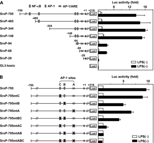
cloned into pGL3-basic, resulting in the SrxP-795 construct (numbering refers to the transcriptional start site). To identify the minimal promoter region that is necessary for Srx induction by LPS, we first carried out a functional deletion analysis (Fig. 3A). RAW264.7 cells were transfected with serially 5⬘-deleted Srx promoter-luciferase reporters and then were exposed to LPS. Induction of luciferase activity of SrxP-795 by LPS was about 14-fold, and the induction fold was preserved until the 5⬘-flanking sequence was deleted to 148 base pairs upstream of the transcriptional start site. However, the deletion of addi-tional nucleotides markedly decreased induction of promoter activity, as shown in 94 and 68. Moreover, in SrxP-29, the promoter activity almost disappeared as in pGL3-basic. These results indicate that the sequence 148 base pairs upstream of the transcriptional start site that contains AP-1 sites and the ARE is the minimal region required for Srx gene transcription in both basal and LPS-stimulated conditions.
Three AP-1 sites were located 45, 87, and 102 base pairs upstream of the transcriptional start site. The proximal, central, and distal AP-1 sites were designated AP-1(A), AP-1(B), and AP-1(C), respectively (Fig. 2). The proximal AP-1 site is over-lapped with two AREs that are positioned in the opposite direc-tion. To confirm the functional role of AP-1 sites and AREs in LPS-induced promoter activity, mutational analysis of the Srx promoter was performed (Fig. 3B). As shown in Fig. 2, the first three nucleotides of the proximal AP-1 site are common and conserved in two oppositely positioned AREs. Because it is expected that the mutation of these nucleotides leads to in-activation of AREs as well as the proximal AP-1 site, three
AP-1 sites were changed by site-di-rected mutagenesis as follows: A (TGAGTCA) to mtA (CAGGTCA), B (TGCGTCA) to mtB (CAT-GTCA), and C (TGAGTCA) to mtC (CAGGTCA), respectively (the nucleotides to be mutated are in italic type). Individual mutation of the proximal and central AP-1 sites significantly decreased induction of promoter activity by LPS, whereas the mutation of the distal one slightly decreased it. Double muta-tion of the proximal and central AP-1 sites resulted in no response to LPS exposure like triple mutation of three AP-1 sites. When the proxi-mal AP-1 site was mutated, how-ever, additional mutation of the dis-tal one led to a significant further decrease in LPS-induced promoter activity. These results suggest that AREs as well as the proximal and central AP-1 sites are primarily required for Srx promoter activity induced by LPS, and the distal one is required for full promoter activity.
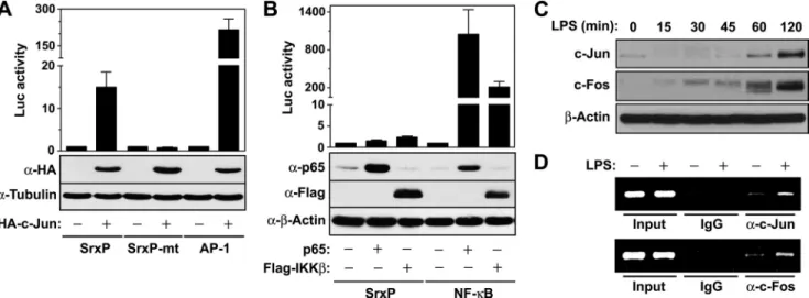
LPS-mediated Srx Induction Is Mediated by Nrf2 as Well as AP-1— Transcription factor AP-1 is a collective term referring to dimeric transcription factors composed of Jun, Fos, or acti-vating transcription factor and primarily acts as a het-erodimer of Jun and Fos (32). To examine the regulatory role of the AP-1 transcription factor on Srx promoter activity, we transfected cells with expression vector for c-Jun along with various luciferase reporters. Expression of c-Jun enhanced the luciferase activity under the control of the wild-type Srx promoter (SrxP) but not under the control of the mutant Srx promoter (SrxP-mt), where all three AP-1 sites were mutated (Fig. 4A). As a positive control, c-Jun expression promoted AP-1-dependent luciferase activity. To investigate the possibility that NF-B is implicated in LPS-mediated Srx induction, we tested the effects of expression of two NF-B pathway intermediates, IKK and p65, on Srx promoter activity. Expression of neither IKK nor p65 enhanced Srx promoter activity, whereas they strongly promoted the NF-B-dependent activity (Fig. 4B). Exposure of LPS to macrophages also promoted the expression of major compo-nents of transcription factor AP-1, c-Jun, and c-Fos (Fig. 4C), which were recruited to AP-1 sites of the Srx promoter, as revealed by the chromatin immunoprecipitation assay with antibodies specific for c-Jun or c-Fos (Fig. 4D), suggesting that AP-1 is involved in LPS-mediated Srx induction.
In a mutational study of Srx promoter, it was suggested that LPS-mediated Srx induction is dependent on ARE as well as the AP-1 response element, based on the assumption that the mutation of the proximal AP-1 site leads to inactivation of not only the AP-1 site but also ARE because it was embedded within FIGURE 2. Nucleotide sequence of the mouse Srx promoter containing potential AP-1 and NF-B sites as
well as ARE. Nucleotides are numbered relative to the transcription start site (⫹1), shown in boldface type. The
translation initiation codon (ATG) is in boldface type. The DNA sequences homologous to NF-B consensus motifs are underlined. The boxed region and arrow indicate AP-1 response element and ARE, respectively. The nucleotides to be mutated in this study are in italic type. The arrowhead indicates the 5⬘-end of a series of
deletion mutants. at Ewha Medical Library on October 24, 2016
http://www.jbc.org/
the ARE. To confirm the role of the Nrf2-ARE system in LPS-mediated Srx induction, it was examined whether Nrf2 regu-lates Srx induction by LPS. It has been demonstrated that LPS stimulation of human monocytes induces the expression of NQO1 and heme oxygenase-1, which are regulated by Nrf2 (33, 34), indicating that LPS also activates Nrf2 in macrophages. Indeed, mRNA expression of Nrf2 and NQO1 was quite induced by LPS stimulation in wild-type BMMs, whereas there was no response to LPS in Nrf2-deficient BMMs (Fig. 5, A and
B). In addition, the LPS-induced expression of Srx mRNA was almost abolished in Nrf2-null BMMs (Fig. 5C). These results suggest that LPS stimulation of macrophages induces and acti-vates Nrf2, which mediates Srx expression.
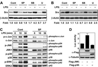
JNK Is a Major MAPK Contributing to LPS-mediated Srx Induction—LPS stimulation of Toll-like receptor 4 activates MAPKs that mediate the activation of transcription factors AP-1 and Nrf2. To investigate which MAPK is a key player in transcriptional induction of Srx by LPS, we examined the effects of MAPK inhibitors on LPS-mediated Srx induction. In RAW264.7 and BMM cells exposed to LPS, the level of Srx protein was significantly decreased by the JNK inhibitor
SP600125, whereas it was slightly changed by the p38 inhibitor SB202190 or the ERK kinase inhibi-tor U0126 (Fig. 6, A and B). The JNK inhibitor SP600125 strongly sup-pressed the phosphorylation of JNK as well as its downstream target pro-tein c-Jun but slightly affected the activities of p38 and ERK, suggest-ing that SP600125 specifically inhibits JNK activity (Fig. 6C). In addition, the ectopic expression of JNK protein increased Srx promoter activity, but p38 expression slightly decreased it (Fig. 6D). These results suggest that JNK is a major MAPK contributing to LPS-mediated Srx induction in macrophages.
Redox Regulation of
LPS-medi-ated Srx Induction—In response
to LPS stimulation, phagocytes, in-cluding macrophages, generate ROS that mediate the production of immunoregulatory and proinflam-matory cytokines, such as tumor necrosis factor␣ and interleukin-1 (19 –21, 35), and one of the impor-tant sources of ROS is Nox enzyme (36, 37). The intracellular ROS level was increased by about 10-fold at 30 min following LPS stimulation of RAW264.7 cells, but such ROS production almost disappeared in the cells that had been pretreated with antioxidant BHA or Nox inhibitor DPI (Fig. 7A). To explore whether ROS are involved in LPS-mediated Srx induction, we examined the effects of antioxi-dants NAC and BHA and Nox inhibitor DPI on LPS-medi-ated Srx induction. LPS-induced Srx protein expression was significantly decreased by NAC, BHA, and DPI (Fig. 7B). In addition, NAC, BHA, and DPI decreased the Srx promoter activity induced by LPS, whereas the glutathione depletion agent BSO significantly increased it, as revealed by the lucif-erase reporter assay (Fig. 7C).
In phagocytic cells, Nox2 is expressed at a high level. To test the role of Nox2 in production of ROS that are involved in LPS-medi-ated Srx induction, we examined Nox2 dependence on ROS pro-duction and Srx inpro-duction by LPS using Nox2-null mice. When wild-type BMM cells were stimulated with LPS, their ROS level was increased by about 8-fold at 30 min following LPS treatment, but such ROS generation was strongly suppressed in Nox2-defi-cient cells (Fig. 7D). In addition, LPS-induced expressions of pro-tein and mRNA of Srx in Nox2-deficient BMM cells were signifi-cantly reduced in comparison with those observed in wild type cells (Fig. 7, E and F). These results suggest that Srx induction is in part regulated by ROS produced through Nox2 in the macro-phages stimulated with LPS.
FIGURE 3. Identification of the cis-acting elements that are responsible for LPS-mediated Srx induction. A, the indicated mouse Srx promoter fragments were cloned into pGL3-basic plasmid. The locations of putative NF-B, AP-1, and ARE sites are indicated. RAW264.7 cells were transfected with a series of luciferase reporter constructs and pRL-SV40 (internal control) for 24 h and then exposed to 100 ng/ml LPS for 24 h. The luciferase activities were measured with a dual luciferase assay system as described under “Experimental Procedures.” The -fold induction relative to the control was determined. Data are means⫾ S.D. (error bars) of values from three independent experiments. B, three AP-1 sites (A, B, and C) were disrupted by site-directed mutagenesis, as described under “Experimental Procedures.” The luciferase activities of mutant Srx promoters were deter-mined as in A.
at Ewha Medical Library on October 24, 2016
http://www.jbc.org/
DISCUSSION
Here we demonstrated that stimulation of macrophages with LPS induces the up-regulation of Srx in mRNA and protein levels, which depends on both AP-1 and Nrf2. Recent studies showed that Srx is induced by several stimuli through either AP-1 or Nrf2. Srx induction by metabolic stimulation (glucose/ cAMP) in pancreatic beta cells, by synaptic activity in rat neu-rons, and by tumor promoter 12-o-tetradecanoylphorbol-13-acetate in mouse epidermal cells was mediated by AP-1 (38 – 40). In cortical neurons, Srx was up-regulated by the treatment with Nrf2 activators, such as 3H-1,2-dithiole-3-thione (41). Nrf2-mediated induction of Srx by cigarette smoke or hyper-oxia was also shown in the lung (42, 43).
Analysis of the mouse Srx promoter sequence revealed two sequences (GGGGAGGCCC and GGGGAGTTCC) resem-bling the NF-B site with the consensus sequence GGGRN-NYYCC (where N represents any base, R is purine, and Y is pyrimidine) (44), three putative AP-1 sites of the sequence TGAGTCA or TGCGTCA, and two sequences (TCACCCT-GAGTCAGCG and TCAGGGTGAATTTGCA) positioning in
opposite directions that resemble an ARE consensus sequence TMA-nnRTGAYnnnGCRwwww (essen-tial nucleotides are shown in capital letters, and the core sequence is in boldface type) (45, 46). A serial dele-tion study of the Srx promoter showed that putative NF-B sites are not required for LPS-mediated Srx induction and identified the activity-inducible region between ⫺148 and ⫹218 relative to potential transcription start site. No necessity of NF-B for Srx induction was fur-ther confirmed by little induction of Srx promoter activity by expression of two NF-B pathway intermediates, IKK and p65. Mutational study of three AP-1 sites revealed that the cen-tral site as well as the proximal site is essential, and the distal one is required for full promoter activity. Although Wei et al. (40) showed that both proximal and distal AP-1 sites are impor-tant for tumor promoter-induced Srx promoter activity, they did not pay attention to the central AP-1 site. Also, two major components of AP-1, c-Jun and c-Fos, were induced and recruited to the AP-1 site of the Srx promoter in response to LPS. These results suggest that LPS-mediated Srx induction requires AP-1.
The consensus sequence TGAGTCA recognized by AP-1 is often embedded within AREs (47). It was demonstrated that LPS stimulation of human monocytes induces the expression of NQO1 and HO-1, which are regulated by Nrf2 (33, 34). In this study, Nrf2 was induced and activated in mouse macrophages stimulated with LPS. Given that the proximal AP-1 site of the Srx promoter is also embedded within AREs and that its first three nucleotides, TGA, correspond to the core and essential FIGURE 4. AP-1 is responsible for LPS-mediated Srx induction. A, HEK293 cells were transfected with c-Jun expression construct along with SrxP-795 (SrxP), SrxP-795mtABC (SrxP-mt), or AP-1-dependent luciferase reporter for 48 h. The luciferase activities were measured as in Fig. 3A, and the expression of HA-tagged c-Jun was evaluated by immunoblot analysis with anti-HA antibody. The -fold induction relative to the control was determined. Data are means⫾ S.D. (error bars) of values from three independent experiments. B, HEK293 cells were transfected with expression vectors for p65 or IKK along with SrxP-795- or NF-B-dependent luciferase reporter for 48 h. The luciferase activities were measured as in A, and the expression of p65 and IKK was evaluated by immunoblot with anti-p65 and anti-FLAG antibodies. C, RAW264.7 cells were treated with LPS (100 ng/ml) for the indicated times, and the expression of c-Jun and c-Fos were analyzed by immunoblot with antibodies against c-Jun and c-Fos, respectively. D, RAW264.7 cells were treated or not with LPS (100 ng/ml) for 1 h and were cross-linked with formaldehyde. Soluble chromatin was subjected to immunoprecipitation with antibodies against c-Jun (top), c-Fos (bottom), or normal IgG. PCR analysis of the positive control (input) indicates that the soluble chromatin samples have equal amounts of chromatin fragments containing the Srx promoter. A representative of two experiments was shown.
FIGURE 5. Induction of Nrf2 and its target genes in BMM cells exposed to LPS. BMM cells prepared from wild-type (Nrf2⫹/⫹) or Nrf2 knock-out mice (Nrf2⫺/⫺) were treated with LPS (100 ng/ml) for the indicated times. Total RNA was extracted from the cells and used in real-time PCR to quantify the mRNA levels of Nrf2 (A), NQO1 (B), and Srx (C). Relative levels of individual mRNA were normalized to those of-actin mRNA. Data are means⫾ S.D. (error bars) of values from three independent experiments.
at Ewha Medical Library on October 24, 2016
http://www.jbc.org/
nucleotides of AREs positioning in forward and reverse direc-tions, respectively (see Fig. 2), mutation of these nucleotides within the proximal AP-1 site (TGAGTCA3 CAGGTCA; the changed nucleotides are shown in boldface type) might lead to inactivation of AREs as well as the AP-1 site. Mutation of the proximal AP-1 site resulted in a partial decrease of the LPS-induced promoter activity, suggesting that ARE is in part involved in LPS-mediated Srx induction. In Nrf2-deficient macrophages, however, the mRNA level of Srx was never induced by LPS treatment like NQO1, a target of Nrf2. This discrepancy is probably caused by two possibilities. One is incomplete inactivation of AREs by mutation of the proximal AP-1 site. The other is the Nrf2-dependent AP-1 activity. Glu-tamate-cysteine ligase catalytic subunit (GCLC) catalyzes the formation of␥-glutamylcysteine from glutamate and cysteine, the first step of glutathione biosynthesis (48). The human GCLC promoter contains the ARE and the AP-1 site (49, 50), and tert-butylhydroquinone (TBH) leads to induction of human GCLC, which is mediated by Nrf1 and Nrf2 (51–53). TBH also induces the expression of rat GCLC, although its pro-moter lacks ARE, suggesting that AP-1 appears to be essential for TBH-mediated induction of rat GCLC (54, 55). However, it was demonstrated that Nrf1 and Nrf2 regulate rat GCLC pro-moter activity despite the absence of ARE by modulating the expression of key AP-1 components (56). The basal protein and mRNA levels and nuclear binding activities of c-Jun and c-Fos were lower in Nrf1- or Nrf2-deficient cells, which exhibited a blunted response to TBH (56). Therefore, Nrf2 may regulate FIGURE 6. Roles of MAPKs in LPS-mediated Srx induction. A and
B, RAW264.7 (A) or wild-type BMM (B) cells were pretreated with medium (Cont), 10MSP600125 (SP), 10MSB202190 (SB), or 10MU0126 (U) for 30 min following stimulation with 100 ng/ml LPS for 16 h, and then total cell lysates were subjected to Western blot analysis with antibodies against Srx and-actin. The -fold induction of Srx was analyzed as in Fig. 1A. A represent-ative from two independent experiments was shown. C, RAW264.7 cells were pretreated with medium or 10MSP600125 for 30 min following stimulation with 100 ng/ml LPS for the indicated times, and then total cell lysates were subjected to Western blot analysis with antibodies to phospho-c-Jun and c-Jun, to JNK and JNK1, to p38 and p38, or to phospho-ERK and phospho-ERK2 (top and bottom of each pair of images, respectively). D, HEK293 cells were transfected with the construct for expression of JNK or FLAG-p38 along with pSrxP-795 for 48 h. The luciferase activities were measured as in Fig. 3A, and the expression of FLAG-tagged JNK and p38 was evaluated by immunoblot analysis with anti-FLAG antibody. The -fold induction relative to the control was determined. Data are means⫾ S.D. (error bars) of values from three independent experiments.
FIGURE 7. Involvement of Nox2-derived ROS in LPS-mediated Srx induction. A, RAW264.7 cells were preincubated in medium without phenol red in the absence or presence of 5MDPI or 100MBHA for 30 min and then were treated with LPS (1g/ml) for the indicated times. The cells were immediately incubated with 2.5M5-(and-6-)chloromethyl-2⬘,7⬘-dichlorodihydrofluorescein diacetate for 5 min, and DCF fluorescence was then visualized with a confocal laser-scanning microscope (left). Relative fluorescence intensity per cell was measured by averaging the values for five groups of cells in each image and is presented as means⫾ S.D. (error bars) (right). B, RAW264.7 cells were exposed to LPS (100 ng/ml) for 16 h after pretreatment with 2 mMNAC, 5MDPI, or 100 MBHA for 30 min, and then the induction level of Srx protein was analyzed as in Fig. 1A. The result is representative of two independent experiments. C, RAW264.7 cells were transfected with pSrxP-795 and pRL-SV40 (internal control) for 24 h. The cells were pretreated with 100MBSO, 2 mMNAC, 5MDPI or 100MBHA following exposure to LPS (100 ng/ml) for 24 h. The luciferase activities were measured as in Fig. 3A. Data are means⫾ S.D. of values from three independent experiments. D–F, BMM cells were prepared from wild-type (Nox2⫹/⫹) or Nox2 knock-out mice (Nox2⫺/⫺). The cells were incubated in medium without phenol red in the presence of LPS (1g/ml) for the indicated times, and intracellular ROS levels were measured as in A (D). The cells were exposed to LPS (100 ng/ml) for 16 h (E) or for 12 h (F). Induction levels of protein (E) and mRNA (F) of Srx were calculated as in Fig. 1, A and E, respectively.
at Ewha Medical Library on October 24, 2016
http://www.jbc.org/
LPS-mediated Srx induction by modulating the AP-1 activity as well as through its cis-acting element ARE.
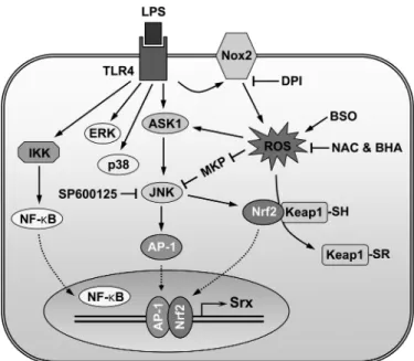
Binding of a wide range of ligands to their receptors leads to production of ROS, which regulate their signal transduction pathways (2, 57). Srx induction was regulated by LPS-induced ROS, as revealed by its inhibition by antioxidants, Nox inhibi-tor, and Nox2 deficiency. AP-1 activity can be regulated by the reversible S-glutathiolation of a conserved cysteine residue (58), and the reversible redox regulation by thioredoxin and redox factor 1 (59). It can also be regulated through the JNK pathway. JNK is a member of the MAPK superfamily of serine/ threonine kinases. All MAPKs are activated through tyrosine and threonine phosphorylation at their activation loops by MAPK kinases and are inactivated by dephosphorylation of the same sites by MAPK phosphatases (60). One of the upstream kinases of JNK is the ASK-1 (apoptosis-signal regulating kinase-1), which is maintained in an inactive state by bound reduced thioredoxin. Oxidation of thioredoxin by ROS releases ASK-1, permitting its activation (61– 63). Several protein-tyrosine phosphatases are transiently inactivated in cells exposed to growth factors through oxidation of their catalytic cysteine with low pKa(64, 65). Members of the MAPK phosphatase subgroup of protein-tyrosine phosphatases, JNK phosphatases are also inhibited by oxidation of their catalytic cysteine by tumor necrosis factor␣-induced ROS (66). Nrf2 is a transcrip-tion factor that mediates expression of phase II detoxifying or antioxidant enzymes (67, 68). The cysteine-rich Keap1 (Kelch-like ECH-associated protein 1) is a cytoplasmic repressor of Nrf2 that binds to Nrf2, retains it in the cytoplasm, and pro-motes its proteasomal degradation (69). The phosphorylation of Nrf2 by several kinases, including MAPKs, phosphoinositide 3-kinase, and protein kinase C, leads to its release from Keap1-mediated repression (70, 71). In addition, phase II enzyme inducers and prooxidants can cause oxidation or covalent mod-ification of several critical cysteine residues of Keap1, resulting in the activation of Nrf2 (72, 73). In our study, JNK was a major MAPK mediating Srx induction by LPS. Thus, LPS-mediated Srx induction is probably modulated by redox-dependent reg-ulation of AP-1 and Nrf2, which is mediated through the JNK pathway regulated via ASK-1 activation and/or JNK phospha-tase inactivation by ROS as well as oxidative modification of Keap1 as described in the legend to Fig. 8.
Activated macrophages produce ROS through the Nox enzyme complex, which is composed of membrane-bound fla-vocytochrome b558, consisting of gp91
phox
and p22phox, and cytosolic regulatory subunits p67phox, p47phox, p40phox, and the small GTPase Rac (25). Several gp91phox (known as Nox2) homologues, termed Nox1, Nox3, Nox4, and Nox5, have been identified in nonphagocytic cells (74, 75). Lee et al. (76) reported that Nox2 is a major isozyme expressed in BMM cells, whereas Nox1 is expressed at a low level, and the expression of Nox3 and Nox4 is undetectable. In the present study, ROS pro-duction as well as Srx inpro-duction was strongly suppressed in Nox2-deficient BMM cells stimulated with LPS, suggesting that Nox2-derived ROS might contribute to the regulation of LPS-mediated Srx induction, although we cannot rigorously rule out the possibility of the involvement of other Nox isozymes. Meanwhile, LPS-mediated ROS generation in macrophages is
dependent on Rac1 (19). Also, several reports showed that the activation of JNK and p38 was mediated through the PI3K/ Rac1/p21-activated kinase signaling pathway (77–79). Thus, Rac1 may be involved in LPS-mediated Srx induction through its roles in the PI3K/Rac1/p21-activated kinase signaling path-way as well as in Nox activation.
We here described a redox-dependent regulation mecha-nism of LPS-induced Srx expression that is mediated by both AP-1 and Nrf2. Srx is an enzyme responsible for the recovery of catalytically inactive hyperoxidized Prxs, which are accumu-lated during the catalysis removing ROS under oxidative stress (11). Thus, it seems to play a protective antioxidant role against oxidative stress. Indeed, Nrf2 activator-mediated Srx induction protects cortical neurons against oxidative stress (41). Srx translocates from the cytoplasm to mitochondria in response to oxidative insult, and its mitochondria-targeted expression sup-presses apoptosis by protecting mitochondria from oxidative damage (80). The physiological role of the LPS-dependent Srx induction is probably for self-defense. Treatment of macro-phages with LPS results in the extracellular production of large amounts of H2O2, which can diffuse freely across biological membranes and impose oxidative stress on the cells. Prxs are responsible for the elimination of H2O2. During their peroxi-dase function, Prxs are expected to undergo hyperoxidation. Longer exposure of RAW264.7 cells to LPS results in a little hyperoxidation of Prxs (data not shown), although Srx is induced. This suggests that the elevated levels of Srx are insuf-ficient to counteract the hyperoxidation. Therefore, it is likely that the defect of Srx expression leads to oxidative damage on phagocytic cells under inflammatory conditions.
REFERENCES
1. Rhee, S. G., Kang, S. W., Chang, T. S., Jeong, W., and Kim, K. (2001) IUBMB Life 52,35– 41
2. Rhee, S. G., Kang, S. W., Jeong, W., Chang, T. S., Yang, K. S., and Woo, H. A. (2005) Curr. Opin. Cell Biol. 17, 183–189
3. Immenschuh, S., and Baumgart-Vogt, E. (2005) Antioxid. Redox Signal. 7, 768 –777
FIGURE 8. The proposed model for redox regulation of LPS-induced Srx
expression, which is mediated by both AP-1 and Nrf2.
at Ewha Medical Library on October 24, 2016
http://www.jbc.org/
4. Yang, K. S., Kang, S. W., Woo, H. A., Hwang, S. C., Chae, H. Z., Kim, K., and Rhee, S. G. (2002) J. Biol. Chem. 277, 38029 –38036
5. Rabilloud, T., Heller, M., Gasnier, F., Luche, S., Rey, C., Aebersold, R., Benahmed, M., Louisot, P., and Lunardi, J. (2002) J. Biol. Chem. 277, 19396 –19401
6. Woo, H. A., Chae, H. Z., Hwang, S. C., Yang, K. S., Kang, S. W., Kim, K., and Rhee, S. G. (2003) Science 300, 653– 656
7. Woo, H. A., Kang, S. W., Kim, H. K., Yang, K. S., Chae, H. Z., and Rhee, S. G. (2003) J. Biol. Chem. 278, 47361– 47364
8. Biteau, B., Labarre, J., and Toledano, M. B. (2003) Nature 425, 980 –984 9. Chang, T. S., Jeong, W., Woo, H. A., Lee, S. M., Park, S., and Rhee, S. G.
(2004) J. Biol. Chem. 279, 50994 –51001
10. Woo, H. A., Jeong, W., Chang, T. S., Park, K. J., Park, S. J., Yang, J. S., and Rhee, S. G. (2005) J. Biol. Chem. 280, 3125–3128
11. Rhee, S. G., Jeong, W., Chang, T. S., and Woo, H. A. (2007) Kidney Int. Suppl., S3–S8
12. Jeong, W., Park, S. J., Chang, T. S., Lee, D. Y., and Rhee, S. G. (2006) J. Biol. Chem. 281,14400 –14407
13. Jo¨nsson, T. J., Murray, M. S., Johnson, L. C., and Lowther, W. T. (2008) J. Biol. Chem. 283,23846 –23851
14. Roussel, X., Be´chade, G., Kriznik, A., Van Dorsselaer, A., Sanglier-Cianfe-rani, S., Branlant, G., and Rahuel-Clermont, S. (2008) J. Biol. Chem. 283, 22371–22382
15. Findlay, V. J., Townsend, D. M., Morris, T. E., Fraser, J. P., He, L., and Tew, K. D. (2006) Cancer Res. 66, 6800 – 6806
16. Park, J. W., Mieyal, J. J., Rhee, S. G., and Chock, P. B. (2009) J. Biol. Chem.
284,23364 –23374
17. Ulevitch, R. J., and Tobias, P. S. (1995) Annu. Rev. Immunol. 13, 437– 457 18. Miyake, K. (2004) Trends Microbiol. 12, 186 –192
19. Sanlioglu, S., Williams, C. M., Samavati, L., Butler, N. S., Wang, G., Mc-Cray, P. B., Jr., Ritchie, T. C., Hunninghake, G. W., Zandi, E., and En-gelhardt, J. F. (2001) J. Biol. Chem. 276, 30188 –30198
20. Hsu, H. Y., and Wen, M. H. (2002) J. Biol. Chem. 277, 22131–22139 21. Matsuzawa, A., Saegusa, K., Noguchi, T., Sadamitsu, C., Nishitoh, H.,
Na-gai, S., Koyasu, S., Matsumoto, K., Takeda, K., and Ichijo, H. (2005) Nat. Immunol. 6,587–592
22. Takeda, K., and Akira, S. (2004) Semin. Immunol. 16, 3–9 23. Akira, S., Uematsu, S., and Takeuchi, O. (2006) Cell 124, 783– 801 24. Kawai, T., and Akira, S. (2007) Semin. Immunol. 19, 24 –32
25. Minakami, R., and Sumimotoa, H. (2006) Int. J. Hematol. 84, 193–198 26. Bast, A., Fischer, K., Erttmann, S. F., and Walther, R. (2010) Biochim.
Biophys. Acta. 1799,402– 410
27. Abbas, K., Breton, J., Picot, C. R., Quesniaux, V., Bouton, C., and Drapier, J. C. (2009) Free Radic. Biol. Med. 47, 794 – 802
28. Li, L., Kaifu, T., Obinata, M., and Takai, T. (2009) J. Biochem. 145, 425– 427
29. Yang, C. S., Lee, D. S., Song, C. H., An, S. J., Li, S., Kim, J. M., Kim, C. S., Yoo, D. G., Jeon, B. H., Yang, H. Y., Lee, T. H., Lee, Z. W., El-Benna, J., Yu, D. Y., and Jo, E. K. (2007) J. Exp. Med. 204, 583–594
30. Li, L., Shoji, W., Takano, H., Nishimura, N., Aoki, Y., Takahashi, R., Goto, S., Kaifu, T., Takai, T., and Obinata, M. (2007) Biochem. Biophys. Res. Commun. 355,715–721
31. Soriano, F. X., Baxter, P., Murray, L. M., Sporn, M. B., Gillingwater, T. H., and Hardingham, G. E. (2009) Mol. Cells 27, 279 –282
32. Barnes, P. J., and Karin, M. (1997) N. Engl. J. Med. 336, 1066 –1071 33. Rushworth, S. A., Chen, X. L., Mackman, N., Ogborne, R. M., and
O’Connell, M. A. (2005) J. Immunol. 175, 4408 – 4415
34. Rushworth, S. A., MacEwan, D. J., and O’Connell, M. A. (2008) J. Immunol.
181,6730 – 6737
35. Asehnoune, K., Strassheim, D., Mitra, S., Kim, J. Y., and Abraham, E. (2004) J. Immunol. 172, 2522–2529
36. Forman, H. J., and Torres, M. (2002) Am. J. Respir. Crit. Care Med. 166, S4 –S8
37. Cross, A. R., and Segal, A. W. (2004) Biochim. Biophys. Acta 1657, 1–22 38. Glauser, D. A., Brun, T., Gauthier, B. R., and Schlegel, W. (2007) BMC Mol.
Biol. 8,54
39. Papadia, S., Soriano, F. X., Le´veille´, F., Martel, M. A., Dakin, K. A., Hansen, H. H., Kaindl, A., Sifringer, M., Fowler, J., Stefovska, V., McKenzie, G.,
Craigon, M., Corriveau, R., Ghazal, P., Horsburgh, K., Yankner, B. A., Wyllie, D. J., Ikonomidou, C., and Hardingham, G. E. (2008) Nat. Neurosci.
11,476 – 487
40. Wei, Q., Jiang, H., Matthews, C. P., and Colburn, N. H. (2008) Proc. Natl. Acad. Sci. U.S.A. 105,19738 –19743
41. Soriano, F. X., Le´veille´, F., Papadia, S., Higgins, L. G., Varley, J., Baxter, P., Hayes, J. D., and Hardingham, G. E. (2008) J. Neurochem. 107, 533–543 42. Singh, A., Ling, G., Suhasini, A. N., Zhang, P., Yamamoto, M.,
Navas-Acien, A., Cosgrove, G., Tuder, R. M., Kensler, T. W., Watson, W. H., and Biswal, S. (2009) Free Radic. Biol. Med. 46, 376 –386
43. Bae, S. H., Woo, H. A., Sung, S. H., Lee, H. E., Lee, S. K., Kil, I. S., and Rhee, S. G. (2009) Antioxid. Redox Signal. 11, 937–948
44. Miyamoto, S., and Verma, I. M. (1995) Adv. Cancer Res. 66, 255–292 45. Wasserman, W. W., and Fahl, W. E. (1997) Proc. Natl. Acad. Sci. U.S.A. 94,
5361–5366
46. Nioi, P., McMahon, M., Itoh, K., Yamamoto, M., and Hayes, J. D. (2003) Biochem. J. 374,337–348
47. Li, Y., and Jaiswal, A. K. (1992) J. Biol. Chem. 267, 15097–15104 48. Lu, S. C. (1999) FASEB J. 13, 1169 –1183
49. Moinova, H. R., and Mulcahy, R. T. (1998) J. Biol. Chem. 273, 14683–14689
50. Mulcahy, R. T., Wartman, M. A., Bailey, H. H., and Gipp, J. J. (1997) J. Biol. Chem. 272,7445–7454
51. Wild, A. C., Moinova, H. R., and Mulcahy, R. T. (1999) J. Biol. Chem. 274, 33627–33636
52. Chan, K., Han, X. D., and Kan, Y. W. (2001) Proc. Natl. Acad. Sci. U.S.A. 98, 4611– 4616
53. Chen, L., Kwong, M., Lu, R., Ginzinger, D., Lee, C., Leung, L., and Chan, J. Y. (2003) Mol. Cell. Biol. 23, 4673– 4686
54. Yang, H., Wang, J., Huang, Z. Z., Ou, X., and Lu, S. C. (2001) Biochem. J.
357,447– 455
55. Yang, H., Zeng, Y., Lee, T. D., Yang, Y., Ou, X., Chen, L., Haque, M., Rippe, R., and Lu, S. C. (2002) J. Biol. Chem. 277, 35232–35239
56. Yang, H., Magilnick, N., Lee, C., Kalmaz, D., Ou, X., Chan, J. Y., and Lu, S. C. (2005) Mol. Cell. Biol. 25, 5933–5946
57. Thannickal, V. J., and Fanburg, B. L. (2000) Am. J. Physiol. Lung Cell Mol. Physiol. 279,L1005–L1028
58. Abate, C., Patel, L., Rauscher, F. J., 3rd, and Curran, T. (1990) Science 249, 1157–1161
59. Xanthoudakis, S., and Curran, T. (1992) EMBO J. 11, 653– 665 60. Davis, R. J. (2000) Cell 103, 239 –252
61. Saitoh, M., Nishitoh, H., Fujii, M., Takeda, K., Tobiume, K., Sawada, Y., Kawabata, M., Miyazono, K., and Ichijo, H. (1998) EMBO J. 17, 2596 –2606
62. Gotoh, Y., and Cooper, J. A. (1998) J. Biol. Chem. 273, 17477–17482 63. Tobiume, K., Saitoh, M., and Ichijo, H. (2002) J. Cell. Physiol. 191,
95–104
64. Lee, S. R., Kwon, K. S., Kim, S. R., and Rhee, S. G. (1998) J. Biol. Chem. 273, 15366 –15372
65. Meng, T. C., Fukada, T., and Tonks, N. K. (2002) Mol. Cell 9, 387–399 66. Kamata, H., Honda, S., Maeda, S., Chang, L., Hirata, H., and Karin, M.
(2005) Cell 120, 649 – 661
67. Itoh, K., Chiba, T., Takahashi, S., Ishii, T., Igarashi, K., Katoh, Y., Oyake, T., Hayashi, N., Satoh, K., Hatayama, I., Yamamoto, M., and Nabeshima, Y. (1997) Biochem. Biophys. Res. Commun. 236, 313–322
68. Motohashi, H., O’Connor, T., Katsuoka, F., Engel, J. D., and Yamamoto, M. (2002) Gene 294, 1–12
69. Itoh, K., Wakabayashi, N., Katoh, Y., Ishii, T., Igarashi, K., Engel, J. D., and Yamamoto, M. (1999) Genes Dev. 13, 76 – 86
70. Zipper, L. M., and Mulcahy, R. T. (2000) Biochem. Biophys. Res. Commun.
278,484 – 492
71. Huang, H. C., Nguyen, T., and Pickett, C. B. (2002) J. Biol. Chem. 277, 42769 – 42774
72. Dinkova-Kostova, A. T., Holtzclaw, W. D., Cole, R. N., Itoh, K., Wakaba-yashi, N., Katoh, Y., Yamamoto, M., and Talalay, P. (2002) Proc. Natl. Acad. Sci. U.S.A. 99,11908 –11913
73. Wakabayashi, N., Dinkova-Kostova, A. T., Holtzclaw, W. D., Kang, M. I., Kobayashi, A., Yamamoto, M., Kensler, T. W., and Talalay, P. (2004) Proc.
at Ewha Medical Library on October 24, 2016
http://www.jbc.org/
Natl. Acad. Sci. U.S.A. 101,2040 –2045
74. Geiszt, M., and Leto, T. L. (2004) J. Biol. Chem. 279, 51715–51718 75. Lambeth, J. D. (2004) Nat. Rev. Immunol. 4, 181–189
76. Lee, N. K., Choi, Y. G., Baik, J. Y., Han, S. Y., Jeong, D. W., Bae, Y. S., Kim, N., and Lee, S. Y. (2005) Blood 106, 852– 859
77. Coso, O. A., Chiariello, M., Yu, J. C., Teramoto, H., Crespo, P., Xu, N.,
Miki, T., and Gutkind, J. S. (1995) Cell 81, 1137–1146
78. Bagrodia, S., De´rijard, B., Davis, R. J., and Cerione, R. A. (1995) J. Biol. Chem. 270,27995–27998
79. Hsu, H. Y., and Twu, Y. C. (2000) J. Biol. Chem. 275, 41035– 41048 80. Noh, Y. H., Baek, J. Y., Jeong, W., Rhee, S. G., and Chang, T. S. (2009) J. Biol.
Chem. 284,8470 – 8477
at Ewha Medical Library on October 24, 2016
http://www.jbc.org/
Bae, Sue Goo Rhee and Woojin Jeong
Hojin Kim, Yuyeon Jung, Bong Soo Shin, Hyeryeon Kim, Hyunsook Song, Soo Han
Depends on Both AP-1 and Nrf2
doi: 10.1074/jbc.M110.126839 originally published online September 7, 2010 2010, 285:34419-34428.
J. Biol. Chem.
10.1074/jbc.M110.126839 Access the most updated version of this article at doi:
Alerts:
When a correction for this article is posted •
When this article is cited •
to choose from all of JBC's e-mail alerts Click here
http://www.jbc.org/content/285/45/34419.full.html#ref-list-1
This article cites 79 references, 43 of which can be accessed free at
at Ewha Medical Library on October 24, 2016
http://www.jbc.org/