저작자표시-비영리-변경금지 2.0 대한민국 이용자는 아래의 조건을 따르는 경우에 한하여 자유롭게 l 이 저작물을 복제, 배포, 전송, 전시, 공연 및 방송할 수 있습니다. 다음과 같은 조건을 따라야 합니다: l 귀하는, 이 저작물의 재이용이나 배포의 경우, 이 저작물에 적용된 이용허락조건 을 명확하게 나타내어야 합니다. l 저작권자로부터 별도의 허가를 받으면 이러한 조건들은 적용되지 않습니다. 저작권법에 따른 이용자의 권리는 위의 내용에 의하여 영향을 받지 않습니다. 이것은 이용허락규약(Legal Code)을 이해하기 쉽게 요약한 것입니다. Disclaimer 저작자표시. 귀하는 원저작자를 표시하여야 합니다. 비영리. 귀하는 이 저작물을 영리 목적으로 이용할 수 없습니다. 변경금지. 귀하는 이 저작물을 개작, 변형 또는 가공할 수 없습니다.
Master's Thesis
Eriodictyol induced anticancer and
apoptotic effects in human pancreatic
cancer cells.
Ui Hyeon Oh
Department of Biotechnology
GRADUATE SCHOOL
JEJU NATIONAL UNIVERSITY
011 i.I 9 = E| = a 9J 3LL fl| =L %L All I Oil ^|
5Le±EEL9L ^ii=^L= EEr= EiE±L[r.
jz|EEf ZJ 7fl ±
Ei] E EE
o| =i±Tg O|i+ ^i^T+I-fi =iiJ2_± 7||=6T]T
2020 L± 1 JE
S29|E±e] o|++ ^i^Tit-fi =¥Tg O[±6T]+
xplFt» i+E ffl++JJ
Eriodictyol induced anticancer and
apoptotic effects in human pancreatic
cancer cells.
Ui Hyeon Oh
(Supervised by Professor Jae Hoon Kim)
A thesis submitted in partial fulflllment of the requirement For the degree of Master of Science
January, 2020
This thesis has been examined and approved.
74c7-Chairperson of the supervising committee
Professor Tatsya Unno, Ph.D., College of Applied Life Sciences, Jeju National University
gon`. \{rw` Oho
Professor So Mi Kin, Ph.D., College of Applied Life Sciences, Jeju National University
__i;`e noon,, kTm
Professor Jae Hoon Kim, Ph.D., College of Applied Life Sciences, Jeju National University
Department of Biotechnology
GRADUATE SCHOOL JEJU NATIONAL UNIVERSIT
i
CONTENTS
CONTENTS... i
LIST OF FIGURES...ii
ABSTRACT...1
1. INTRODUCTION...2
2. MATERIALS AND METHODS...5
2.1. Cell culture and reagents…………..……….………...……….5
2.2. Cell viability assay………....….……….5
2.3. Cell cycle assay………...…...6
2.4. Apoptosis detection………...…….……...6
2.5. Western blot assay ……….……….….……...…...…...7
2.6. Statistical analysis……….………....…………..…….7
3. RESULTS ……….………...…..8
3.1. Eriodictyol reduced the cell viability of pancreatic cancer cells. 3.2. Eriodictyol induced cell cycle S phase arrest 3.3. Eriodictyol induced apoptosis in pancreatic cancer cells 3.4. Eriodictyol inhibit the proliferation of pancreatic cancer cells via the MAPK and FAK/AKT signaling pathway
4. DISCUSSION...16
5. REFERENCES...18
ii
LIST OF FIGURES
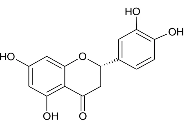
Figure 1. Structure of eriodictyol, 5, 7, 3', 4-Tetrahydroxyflavanone
Figure 2. The effects of eriodictyol on the viability in human pancreatic cancer cells.
Figure 3. The eriodictyol suppressed colony formation in Panc-1 cells.
Figure 4. The eriodictyol suppressed colony formation in SNU-213 cells.
Figure 5. Effects of eriodictyol on the cell cycle of human pancreatic cancer cells.
Figure 6. Effects of eriodictyol on the cell cycle-related proteins in human pancreatic cancer
cells
Figure 7. Effects of eriodictyol on the apoptosis of human pancreatic cancer cells.
Figure 8. Effects of eriodictyol on the apoptosis-related proteins in human pancreatic cancer
cells.
Figure 9. Effects of eriodictyol on phosphorylation level of MAPK in pancreatic cancer
cells.
Figure 10. Effects of eriodictyol on phosphorylation level of FAK and AKT in pancreatic
1
ABSTRACT
Despite many studies over the past several decades, pancreatic cancer has known as one of the most vulnerable cancers. Apoptosis is considered one of the well-known treatment options for various cancer treatment research methods. Eriodictyol, a plant-derived flavonoid found in citrus fruits has been known to have diverse biological activities such as anti-oxidant, anti-cancer, and anti-inflammatory properties. In this study, we have investigated the anticancer property of eriodictyol and its mechanisms of action in pancreatic cancer cells. Eriodictyol induced apoptosis in both SNU213 and Panc-1 cells. Also, we found that eriodictyol has reduced the level of phosphorylation in AKT, and ERK. Furthermore, eriodictyol has increased the phosphorylation level of JNK. These results can help to discover the molecular mechanisms of eriodictyol-induced cell death in pancreatic cancer cell lines and potentially contribute to the development of candidate reagents for pancreatic cancer.
2
1. INTRODUCTION
Cancer is the second leading cause of death in the United States, and pancreatic cancer is a deadly disease with a five-year survival rate of less than eight percent [1]. It is expected to be the second major cause of cancer deaths by 2030 [2].Despite efforts improve the treatment of this disease, surgery followed adjuvant chemotherapy is still an effective treatment for Pancreatic cancer. However, only 20 percent of pancreatic cancer patients are eligible for surgery/chemotherapy [3]. Gemcitabine and Fluorouracil (5FU) are primary chemotherapy drugs for pancreatic cancer patients. But the pancreatic cancer prognosis is still not good because of drug resistance [4,5]. Therefore, it is urgently needed to find new target that are more sensitive than current chemotherapy drugs.
Flavonoid is a major class of plant secondary metabolism that are found in vegetable foods and beverages such as fruits, vegetables, tea, and coffee [6,7]. Flavonoids, such as kaempferol, quercetin, naringenin, hesperetin, catechin, curcumin, and eriodictyol, etc. have physiological functions in various cancers as well as antioxidants [8], anti-inflammation [9], antidiabetic [10], and antibacterial agents [11]. Flavonoids have been reported to enhance the drug bioavailability and reduce chemo-resistance of cancer and interfere with the cell cycle, induce apoptosis, inhibit angiogenesis [12–15].
Eriodictyol, 5, 7, 3', 4-Tetrahydroxyflavanone, is a flavanone substance commonly found in fruits such as Lemon (Citrus limon BURM. f.) [16], Sutil Lemon (Citrus aurantiifolia) [17], and Bergamot orange (Citrus bergamia Risso) [18]. Recent studies showed that eriodictyol possesses
3
anti-melanogenesis in murine melanoma cells [19]. It is reported that eriodictyol has a neuroprotective effect in lipopolysaccharide (LPS)-induced neuroinflammation diseases [20]. Eriodictiol also showed anticancer effects in cancer cells. A previous study has shown that eriodictyol reduced cell proliferation in dimethyl hydrazine-induced colorectal cancer cells [21]. Besides, it is reported mitochondrial apoptosis and G2/M cell cycle arrest induction in liver and lung cancer cells [22,23].
But the mechanism of eriodictyol in pancreatic cancer cells has not been reported so far. Therefore, we identified the anti-cancer effects of eriodictyol and mechanism for human pancreatic cancer cell states. We first evaluated the effects of eriodictyol on the viability, colony formation, cell cycle, and cell apoptosis in pancreatic cancer cells. Next, the cell cycle and apoptosis-related proteins activation were examined by western blot.
4
Figure 1. Structure of eriodictyol, 5, 7, 3', 4-Tetrahydroxyflavanone.
O
O
H
OH
O
OH
O
H
5
2. MATERIALS AND METHODS
2.1 Cell culture and reagents
We obtained human embryonic kidney cells 293T, human pancreatic cancer cells Panc-1, and SNU-213 from Korean Cell Line Bank (KCLB, Seoul, Korea). The 293T and Panc-1 cells were cultured in DMEM supplemented with 10% fetal fetal bovine serum (Gibco-BRL, Gaithersburg, MD, USA), 100 U/mL penicillin, 100 µg/ml streptomycin (Invitrogen, Carlsbad, CA, USA), and 5% CO2 at 37℃. SNU-213 cells were cultured in RPMI-1640 supplemented with 10% fetal bovine serum (Gibco-BRL, Gaithersburg, MD, USA), 100U/mL penicillin, 100 µg/ml streptomycin (Invitrogen, Carlsbad, CA, USA), and 5% CO2 at 37℃. Eriodictyol (Cat. No. 020056) were purchased Indofine Chemical Company
(Hillsborough, NJ, USA).
2.2 Cell viability assay
We assessed cell viability using the WST-1 (2- (4-iodophenyl) -3- (4-nitrophenyl) -5- (2,4-disulfophenyl) 2H-tetrazolium) solution (Boechringer Mannheim, Mannheim ,Germany). Cells were seeded 2.5× cells/well in 24 well plates. After cells were incubated for 24 h, they were treated Eriodictyol in DMEM with concentrations of 5, 10, 25, 50, and 100 µM for 72 h at 37℃. Each well was added to a final concentration of 10% WST-1 solution after 15 min incubation at room temperature. The absorbance was measured at 450 nm using Multiskan Spectrum microplate reader (Thermo Scientific, Finland).
6
2.3 Colony formation assay
The Panc-1 cells and SNU-213 cells were seeded in 60mm dishes in each 200 cells/well. After 24 h, different concentrations of eriodictyol (0, 25, 50 μM) were added and the cells continued to be maintained at 37°C with a humidified atmosphere of 5% CO2 for 10–14 days. The colonies fixed with 4% paraformaldehyde and then stained with 0.1% crystal violet solution for 15 min and washed with sterile deionized water. Visible colonies were then stained manually counted.
2.4 Cell cycle
Propidium iodide (PI) staining assay was used to analyze the cell cycle distribution. Panc-1 and SNU-213 cells were seeded at a density of 1.2×10 in 60mm dish. After cells incubated overnight for attachment, Cells were treated with 0.5% DMSO or 25, 50 μM of eriodictyol for 48 h in media containing 5% FBS. At the end of incubation, cells were collected by trypsin-EDTA and centrifugation, washed twice with cold PBS and then fixed in cold 70% ethanol at -20℃ overnight. The fixed cells were centrifuged and stained for 15 min with propidium iodide (PI)/RNase Staining Buffer (BD Pharmingen, USA) and subjected to analysis on a flow cytometer (LSRFortessa, BD).
2.5 Detection of apoptosis
For apoptosis analysis, 293T, Panc-1, and SNU-213 were seeded in 6-well plates. After 24 h, they were treated eriodictyol for 72 h. After collecting the cells, we incubated the cells with annexin V-FITC and propidium (PI) (FITC
7
Annexin V apoptosis detection kit, BD pharmingen, USA). The apoptotic cells were detected by flow cytometry (LSRFortessa, BD).
2.6. Western blot assay
Cells were lysed in M-PER lysis buffer (Thermo science, Bonn, Germany) containing protease inhibitor cocktail (Roche), 2 mM sodium vanadate, 30 mM sodium pyrophosphate, and 100 mM sodium fluoride. After total protein quantification, proteins were separated by 10% SDS-PAGE (sodium dodecyl sulfate-polyacrylamide gel electrophoresis) and transferred to nitrocellulose membranes (Amersham Bioscience, Little Chalfont, Buckinghamshire, UK). Membranes were blocked with 5% skim milk in TBST. Primary antibodies, such as cleaved PARP, phosphorylation FAK (Tyr397), phosphorylation AKT (Ser473), phosphorylation JNK (Thr183/Tyr185), phosphorylation p38
(Thr180/Tyr182), phosphorylationERK1/2 (Thr202/Try204), p21Waf1/Cip1, and GAPDH (Cell signaling technology, USA), were diluted 1:1000 in TBST and incubated overnight at 4°C. Secondary antibodies (Merck Millipore, Germany) were diluted in TBST 1:4000 and incubated for 1h. We detected the protein bands using the ECL kit (Biosesang).
2.7. Statistical analysis
Error bars represent ±SEM. We did statistical analysis by one-way ANOVA, and student’s t-test. P< 0.05 was considered to indicate significant differences.
8
3. RESULTS
3.1 Eriodictyol reduce the cell viability of pancreatic cancer cells
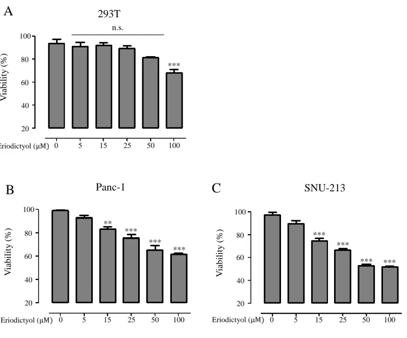
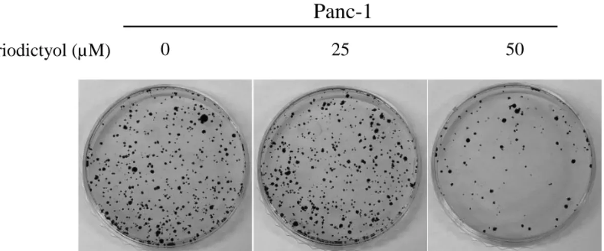
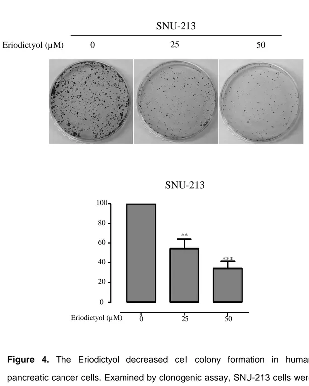
We examined cell proliferation of pancreatic cancer cells using the eriodictyol. Human embryonic kidney cells 293T, human pancreatic cancer cells Panc-1, and SNU-213 were treated with eriodictyol at different concentrations for 72 h. When we measured Cell viability using WST-1, we identify that the viability rate of pancreatic cancer cells were decreasing by the dose-dependent manner. Eriodictyol decreases the viability rate of about 33% from 50 μM compared to control in Panc-1 cells. SNU-213 cells were declined viability rate of about 44% from 50 μM compared to control. However, no significant effect was given to 293T cells (Figure 2A, 2B, and 2C). Additionally, to check the long-term viability of pancreatic cancer cells, measurements were made using the colony formulation assay. Eriodictyol has significantly decreased colony formation in pancreatic cancer cells. Eriodictyol decreases colony formation rate by 73% from 50 μM compared to control in Panc-1 cells (Figure 3). SNU-213 cells were declined colony formation rate by 72% from 50 μM compared to control (Figure 4).
9
Figure 2. The effects of eriodictyol on the viability in human pancreatic cancer
cells. (A-C) human embryonic kidney cells 293T, human pancreatic cancer cells Panc-1, and SNU-213 were treated with various concentrations (0, 5, 15, 25, 50, and 100 μM) of eriodictyol for 72 h. Cell viability was determined by WST-1 assay. Graphs showed mean ±SEM, *P< 0.05; **P< 0.01; ***P<0.001 to control group. 100 80 60 40 20 0 5 15 25 50 100 Eriodictyol (μM)
SNU-213
V iabili ty ( % ) *** *** *** *** 100 80 60 40 20 n.s.293T
0 5 15 25 50 100 Eriodictyol (μM) V ia bili ty ( % ) *** 100 80 60 40 20 0 5 15 25 50 100 Eriodictyol (μM)Panc-1
V iabili ty ( % ) ** *** *** ***A
B
C
10
Figure 3. The Eriodictyol decreased cell colony formation in human
pancreatic cancer cells. Examined by clonogenic assay, Panc-1 cells were treated with 0, 25, and 50 μM of Eriodictyol for 24 h. Eriodictyol significantly attenuated clonogenic assay in Panc-1. Graphs showed mean ±SEM, *P< 0.05; **P< 0.01; ***P<0.001 to control group.
Panc-1
0 25 50 Eriodictyol (µM)Panc-1
100 80 60 40 20 0 0 25 50 Eriodictyol (µM) **11
Figure 4. The Eriodictyol decreased cell colony formation in human
pancreatic cancer cells. Examined by clonogenic assay, SNU-213 cells were treated with 0, 25, and 50 μM of eriodictyol for 24 h. Eriodictyol significantly attenuated clonogenic assay in dose-dependent manner in SNU-213. Graphs showed mean ±SEM, *P< 0.05; **P< 0.01; ***P<0.001 to control group.
SNU-213
0 25 50 Eriodictyol (µM)SNU-213
0 25 50 Eriodictyol (µM) 100 80 60 40 20 0 ** ***12
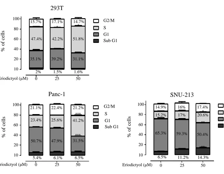
3.2 Eriodictyol induce cell cycle arrest
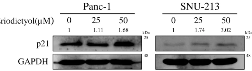
To examined cell cycle of pancreatic cancer cells using the eriodictyol. The cell cycle was detected by flow cytometry following eriodictyol treatment for 48 h. Eriodictyol increases the S phase about 17% from 50 μM compared to control in Panc-1 cells. SNU-213 cells were increased the sub-G1 about 8% from 50 μM compared to control. Whereas, eriodictyol is showed a slight difference in 293T cells (Figure 5). Furthermore, we detected the expression level of cyclin-dependent kinase inhibitor proteins p21 (Waf1/cip1) to explore the mechanism accounting for cell cycle arrest. Eriodictyol treatment has increased the expression of p21 in pancreatic cancer cells (Figure 6).
13
Figure 5. Effects of eriodictyol on the cell cycle of human pancreatic cancer
cells. Cell cycle analysis of eriodictyol-treated 293T, Panc-1, SNU-213 cells. Cells were incubated with 0, 25, and 50 μM of eriodictyol for 48 h. Following cells were fixed, stained with PI. Stained cells were detected by flow cytometry. The percentage indicated cells in sub G1, G0/G1, S, and G2/M phage of the cell cycle. Graphs showed mean ±SEM, *P< 0.05; **P< 0.01; ***P<0.001 to control group. 100 80 60 40 20 10 0 25 50
293T
% of c ell s Eriodictyol (μM) G2/M S G1 Sub G1 * * 15.7% 17.1% 14.7% 47.4% 42.2% 51.8% 35.1% 39.2% 31.1% 2% 1.5% 1.6% 0 25 50 100 80 60 40 20 10 % of c ell sPanc-1
*** *** Eriodictyol (μM) G2/M S G1 Sub G1 21.1% 22.4% 21.2% 23.4% 25.6% 41.2% 50.7% 47.9% 31.5% 5.4% 6.1% 6.5% 100 80 60 40 20 10 % of c ell s 0 25 50 ** **SNU-213
Eriodictyol (μM) * G2/M S G1 Sub G1 14.9% 16% 17.4% 15.2% 17% 20.6% 65.3% 59.3% 50.4% 6.5% 11.2% 14.3%14
Figure 6. Effects of eriodictyol on the cell cycle related proteins in human
pancreatic cancer cells. Western blot analysis of cell cycle-related proteins after Panc-1 and SNU-213 cells treated with 0, 25, and 50 μM of eriodictyol for 48 h. The cell lysates were analyzed by SDS-PAGE, following by western blot analysis using antibodies of p21 and GAPDH.
p21
Panc-1
SNU-213
Eriodictyol(µM)
0 25 50 0 25 50 GAPDH kDa kDa 25 25 48 48 1 1.74 3.02 1 1.11 1.6815
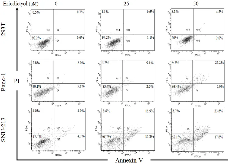
3.3 Eriodictyol induced apoptosis in pancreatic cancer cells
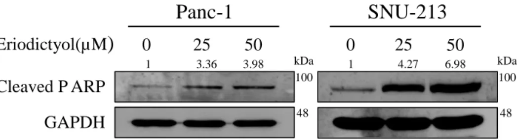
In order to examine the apoptotic effects of eriodictyol in pancreatic cancer cells, we performed flow cytometry after treatment of eriodictyol for 72 h. Panc-1 and SNU-213 cells were treated with 25 μM and 50 μM of eriodictyol. Panc-1 cells have increased apoptosis about 20% from 50 μM compared to control. SNU-213 increased apoptosis about 33% from 50uM compared to control. However, eriodictyol is showed a slight difference in 293T cells (Figure 7). Additionally, we have determined that eriodictyol affects apoptosis pathways in pancreatic cancer cells. Eriodictyol increased cleavage PARP in pancreatic cancer cells (Figure 8).
16
Figure 7.Effects of Eriodictyol on the apoptosis of human pancreatic cancer cells. Representative flow cytometry dot plots with Annexin V and PI staining. Apoptosis analysis of eriodictyol treated 293T, Panc-1, and SNU-213 cells. Cells were incubated with 0, 25, and 50 μM of eriodictyol for 72 h. Following the cells were collected, stained with Annexin V-FITC and PI. Stained cells were detected by flow cytometry. Graphs showed mean ±SEM, *P< 0.05; **P< 0.01; ***P<0.001 to control group.
17
Figure 8.Effects of eriodictyol on the apoptosis-related proteins in human pancreatic cancer cells.Western blot analysis of apoptosis-related proteins after Panc-1 and SNU-213 treated with 0, 25, and 50 μM of eriodictyol for 48 h. The cell lysates were analyzed by SDS-PAGE, following by western blot analysis using antibodies of cleaved PARP and GAPDH. Graphs showed mean ±SEM, *P< 0.05; **P< 0.01; ***P<0.001 to control group.
Eriodictyol(µM
)
0 25 50 0 25 50 Cleaved P ARP GAPDHPanc-1
SNU-213
kDa kDa 100 100 48 48 1 3.36 3.98 1 4.27 6.9818
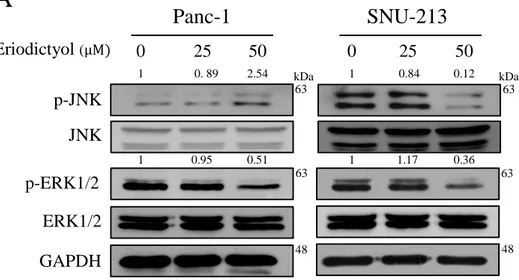
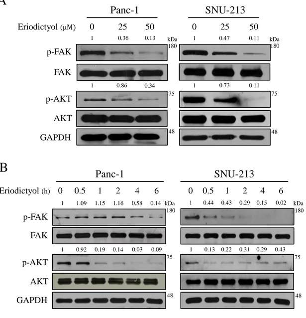
3.4 Eriodictyol inhibit the proliferation of pancreatic cancer cells via the MAPK and FAK/AKT signaling pathway
To elucidate the mechanisms for pancreatic cancer cells of eriodictyol, we were performed western blot analysis after treated with eriodictyol for 48 h. We examined the phosphorylation level of FAK/AKT and MAPK in Pancreatic cancer cells. The results confirmed that eriodictyol inhibited the phosphorylation level of ERK1/2 in Panc-1 and SNU-213 cells. Eriodictyol has increased the phosphorylation level of JNK in Pacn-1whereas SNU-213 cells have decreased the phosphorylation level of JNK (Figure 9). Also, our results showed that eriodictyol inhibits the phosphorylation level of FAK and AKT in Panc-1 and SNU-213 cells. In addition, the phosphorylation level of FAK and AKT were decreased in a time-dependent manner in Panc-1 and SNU-213 cells after 50μM eriodictyol treatment (Figure 10).
19
Figure 9. Effects of eriodictyol on phosphorylation level of MAPK in
pancreatic cancer cells. (A) Panc-1 and SNU-213 cells were treated with 0, 25, and 50 μM of eriodictyol for 48 h. (B) Panc-1 and SNU-213 cells were treated with 50 μM of eriodictyol for 0, 24, 30, 36, 42, 48 h. The cell lysates were analyzed by SDS-PAGE, following by western blot analysis using antibodies of p-JNK, JNK, p-ERK1/2, ERK1/2 and GAPDH. The relative band intensities were measured using Image J software.
63 63 63 63
Panc-1
50 25 0 1 0. 89 2.54 1 0.84 0.12 kDa 48 GAPDH JNK p-JNK Eriodictyol (μM) p-ERK1/2 ERK1/2 1 0.95 0.51 1 1.17 0.36SNU-213
50 25 0 kDa 48A
20
Figure 10. Effects of eriodictyol on phosphorylation level of FAK and AKT in
pancreatic cancer cells. (A) Panc-1 and SNU-213 cells were treated with 0, 25, and 50 μM of eriodictyol for 48 h. (B) Panc-1 and SNU-213 cells were treated with 50 μM of eriodictyol for 0, 0.5, 1, 2, 4, 6h. The cell lysates were analyzed by SDS-PAGE, following by western blot analysis using antibodies of p-FAK, FAK, p-AKT, AKT and GAPDH. The relative band intensities were measured using Image J software.
75 180 180 75
Panc-1
50 25 0 1 0.36 0.13 1 0.86 0.34 1 0.47 0.11 1 0.73 0.11 kDa 48 GAPDH FAK AKT p-AKT p-FAK Eriodictyol (μM)SNU-213
50 25 0 kDa 48A
75 180 180 75Panc-1
SNU-213
1 0.5 0 2 4 6 0 0.5 1 2 4 6 kDa 1 0.92 0.19 0.14 0.03 0.09 1 1.09 1.15 1.16 0.58 0.14 1 0.44 0.43 0.29 0.15 0.02 1 0.13 0.22 0.31 0.29 0.43 Eriodictyol (h) 48 kDa 48 GAPDH FAK AKT p-AKT p-FAKB
21
4. DISCUSSION
In this study, we showed that eriodictyol can selectively affect pancreatic cancer cells without harming the normal cells. Dual-specificity protein phosphatases (DUSPs) constitute a family of protein phosphatase characterized by the ability to dephosphorylate a tyrosine residue and serine/threonine residues [24]. The expression of DUSP28 was reported to induce chemo-resistance in pancreatic cancer cells. Also, DUSP28 was highly expressed in Panc-1 and SNU-213 was weakly expressed under the same conditions [25]. Similarly, we found that SNU-213 cells were more sensitive effects than Panc-1 cells. Eriodictyol triggered cell cycle arrest and induced cell apoptosis through FAK/AKT/MAPK signaling pathways.
Apoptosis is considered as one of the imperative processes which allow the body to eliminate harmful cells from the body [26]. A previous study reported that eriodictyol induced apoptosis through bax and cleavage of PARP in Hep-G2 cells [22]. In our study, we found that eriodictyol induced apoptosis through cleavage of PARP in pancreatic cancer cells.
FAK and PI3K/AKT signaling pathway is a cancer survival pathway that plays a central role in diverse cellular functions, including proliferation, survival, growth, and metastasis [27,28]. PI3K stimulated by FAK can stimulate AKT, which inhibits apoptosis by regulating cell death proteins. Indeed, FAK activation of PI3K/AKT signaling pathway was reported to be associated with resistance to apoptosis induced by TRAIL and UV irradiation [29,30]. Similarly, we found that eriodictyol has reduced the level of phosphorylation in FAK and AKT.
22
In addition to apoptosis, cell cycle arrest is another mechanism by which anticancer agents induce anticancer effects. If the cancer cells are arrested in any phase of the cell cycle, they are unable to complete cell division and tumor growth is halted [31]. The results of the present study revealed that eriodictyol caused S phase cell cycle arrest in Panc-1 cells. However, the previously reported study has demonstrated that A549 and Hep-G2 cells treated with eriodictyol showed G2/M phase cell cycle arrest [22,23]. We hypothesize that the results of eriodictyol in tumor cell cycle arrest may be due to the different cell lines and mechanisms.
MAPK signaling including JNK, ERK, and p38 are mediators of cellular responses to extracellular signals [32,33]. A previous study found that cell cycle progression of the MDA-MB-231 and Panc-1 cells were arrested through JNK after treatment with natural compounds [34]. In our study found that treatment with eriodictyol increased the phosphorylation level of JNK in Panc-1 cells. Therefore, we expect that the upregulation of p21 by JNK can result in cell cycle arrest at S phase in Panc-1 cells. However, eriodictyol reduced the level of JNK phosphorylation in SNU-213 cells and no effect on the cell cycle progression.
Therefore, further study is required to reveal the mechanism of eriodictyol for SNU-213 cells. In conclusion, this study found that eriodictyol inhibited proliferation and induces apoptosis in pancreatic cancer cells. Also, this result demonstrated the molecular mechanisms of eriodictyol-induced apoptosis in pancreatic cancer cells and potentially will contribute to the development of candidate reagents for pancreatic cancer.
23
6. REFERENCE
1. Siegel RL, Miller KD, Jemal A. Cancer Statistics , 2018. CA-A CANCER J Clin. 2018;68: 7–30. doi:10.3322/caac.21442
2. Rahib L, Smith BD, Aizenberg R, Rosenzweig AB, Fleshman JM, Matrisian LM. Projecting Cancer Incidence and Deaths to 2030 : The Unexpected Burden of
Thyroid , Liver , and Pancreas Cancers in the United States. Cancer Res. 2014;14: 0155. doi:10.1158/0008-5472.CAN-14-0155
3. Chand S, Hayer KO, Blanco FF, Winter JM, Brody JR. The Landscape of Pancreatic Cancer Therapeutic Resistance Mechanisms. Int J Biol Sci. 2016;12: 273–282. doi:10.7150/ijbs.14951
4. Bullock A, Stuart K, Jacobus S, Abrams T, Wadlow R, Miksad R. Capecitabine and oxaliplatin as first and second line treatment for locally advanced and metastatic pancreatic ductal adenocarcinoma. J Gastrointest Oncol. 2017;8: 945–952. doi:10.21037/jgo.2017.06.06
5. Ono H, Basson MD, Ito H. PTK6 promotes cancer migration and invasion in pancreatic cancer cells dependent on ERK signaling. PLoS One. 2014;9: 1–7.
doi:10.1371/journal.pone.0096060
6. Zamora-ros R, Touillaud M, Rothwell JA, Romieu I, Scalbert A. Measuring exposure to the polyphenol metabolome in observational epidemiologic studies: current tools and applications and their limits1–3. Am J Clin Nutr. 2014;100: 11–26. doi:10.3945/ajcn.113.077743.1
7. Tripoli E, Guardia M La, Giammanco S, Majo D Di, Giammanco M. Citrus flavonoids: Molecular structure, biological activity and nutritional properties: A review. Food Chem. 2007;104: 466–479. doi:10.1016/j.foodchem.2006.11.054
24
8. Xu D, Hu MJ, Wang YQ, Cui YL. Antioxidant activities of quercetin and its complexes for medicinal application. Molecules. 2019;24.
doi:10.3390/molecules24061123
9. He Y, Yue Y, Zheng X, Zhang K, Chen S, Du Z. Curcumin, inflammation, and chronic diseases: How are they linked? Molecules. 2015;20: 9183–9213.
doi:10.3390/molecules20059183
10. Alkhalidy H, Moore W, Wang Y, Luo J, McMillan RP, Zhen W, et al. The flavonoid kaempferol ameliorates streptozotocin-induced diabetes by suppressing hepatic glucose production. Molecules. 2018;23.
doi:10.3390/molecules23092338
11. Gomes FMS, da Cunha Xavier J, dos Santos JFS, de Matos YMLS, Tintino SR, de Freitas TS, et al. Evaluation of antibacterial and modifying action of catechin antibiotics in resistant strains. Microb Pathog. 2018;115: 175–178.
doi:10.1016/j.micpath.2017.12.058
12. Amawi H, Jr CRA, Tiwari AK. Cancer chemoprevention through dietary flavonoids : what ’ s limiting ? Chin J Cancer. 2017; 1–13.
doi:10.1186/s40880-017-0217-4
13. Sak K. Chemotherapy and Dietary Phytochemical Agents. Chemother Res Pract. 2012;2012: 1–11. doi:10.1155/2012/282570
14. Maru GB. Understanding the molecular mechanisms of cancer prevention by dietary phytochemicals: From experimental models to clinical trials. World J Biol Chem. 2016;7: 88. doi:10.4331/wjbc.v7.i1.88
15. Biology C, Ravishankar D, Rajora AK, Greco F, Osborn HMI. Flavonoids as prospective compounds for anti-cancer therapy Divyashree. Int J Biochem Cell Biol. 2013;45: 2821–2831. doi:10.1016/j.biocel.2013.10.004
16. MIYAKE Y, YAMAMOTO K, OSAWA T. Isolation of Eriocitrin(Eriodictyol 7-rutinoside) from Lemon Fruit(Citrus limon BURM. f.) and Its Antioxidative
25
Activity. Food Sci Technol Int Tokyo. 1997;3: 84–89.
doi:10.3136/fsti9596t9798.3.84
17. Brito A, Ramirez JE, Areche C, Sepúlveda B, Simirgiotis MJ. HPLC-UV-MS profiles of phenolic compounds and antioxidant activity of fruits from three citrus species consumed in Northern Chile. Molecules. 2014;19: 17400–17421.
doi:10.3390/molecules191117400
18. Mandalari G, Bennett RN, Bisignano G, Saija A, Dugo G, Lo Curto RB, et al. Characterization of flavonoids and pectins from bergamot (Citrus bergamia Risso) peel, a major byproduct of essential oil extraction. J Agric Food Chem. 2006;54: 197–203. doi:10.1021/jf051847n
19. Imen MB, Chaabane F, Nadia M, Soumaya KJ, Kamel G, Leila CG. Anti-melanogenesis and antigenotoxic activities of eriodictyol in murine
melanoma (B16-F10) and primary human keratinocyte cells. Life Sci. 2015;135: 173–178. doi:10.1016/j.lfs.2015.06.022
20. He P, Yan S, Zheng J, Gao Y, Zhang S, Liu Z, et al. Eriodictyol Attenuates LPS-Induced Neuroinflammation, Amyloidogenesis, and Cognitive
Impairments via the Inhibition of NF-κB in Male C57BL/6J Mice and BV2 Microglial Cells. J Agric Food Chem. 2018;66: 10205–10214.
doi:10.1021/acs.jafc.8b03731
21. Kalaiyarasu T, Manju V. Effect of eriodictyol on preneoplastic lesions, oxidative stress and bacterial enzymes in 1,2-dimethyl hydrazine-induced colon
carcinogenesis. Toxicol Res (Camb). 2017;6: 678–692.
doi:10.1039/c7tx00074j
22. Fang Wang, Yu-Hang Wang, Jing-Jing Wang H-LX and C-MW.
Eriodictyol-induced anti-cancer and apoptotic effects in human hepatocellular carcinoma cells are associated with cell cycle arrest and modulation of
26
apoptosis-related proteins. Bangladesh J Pharmacol. 2016; 285–291.
doi:https://doi.org/10.3329/bjp.v11i2.25549
23. Zhang Y, Zhang R, Ni H. Eriodictyol exerts potent anticancer activity against A549 human lung cancer cell line by inducing mitochondrial-mediated apoptosis , G2 / M cell cycle arrest and inhibition of m-TOR / PI3K / Akt signalling pathway. Basic Res Journals. 2019.
doi:https://doi.org/10.5114/aoms.2019.85152
24. Motiwala T, Jacob ST. Role of Protein Tyrosine Phosphatases in Cancer. Prog Nucleic Acid Res Mol Biol. 2006;81: 297–329.
doi:10.1016/S0079-6603(06)81008-1
25. Lee J, Hun Yun J, Lee J, Choi C, Hoon Kim J. Blockade of dual-specificity phosphatase 28 decreases chemo-resistance and migration in human pancreatic cancer cells. Sci Rep. 2015;5: 1–12. doi:10.1038/srep12296 26. Koff JL, Ramachandiran S, Bernal-mizrachi L. A Time to Kill : Targeting
Apoptosis in Cancer. Int J Mol Sci. 2015;16: 2942–2955.
doi:10.3390/ijms16022942
27. Hay N. The Akt-mTOR tango and its relevance to cancer. Cancer Cell. 2005;8: 179–183. doi:10.1016/j.ccr.2005.08.008
28. Kanteti R, Mirzapoiazova T, Riehm JJ, Dhanasingh I, Mambetsariev B, Wang J, et al. Focal adhesion kinase a potential therapeutic target for pancreatic cancer and malignant pleural mesothelioma. Cancer Biol Ther. 2018;19: 316–327.
doi:10.1080/15384047.2017.1416937
29. Lane D, Matte I, Laplante C, Garde-granger P, Rancourt C, Piché A.
Osteoprotegerin ( OPG ) activates integrin , focal adhesion kinase ( FAK ), and Akt signaling in ovarian cancer cells to attenuate TRAIL-induced apoptosis. J Ovarian Res. 2013;6: 82. doi:10.1186/1757-2215-6-82
27
30. Chan P, Lai J, Cheng C, Tang M, Chiu C, Chen H. Suppression of Ultraviolet Irradiation-induced Apoptosis by Overexpression of Focal Adhesion Kinase in Madin-Darby Canine Kidney Cells *. J Biol Chem. 1999;274: 26901–26906.
doi:10.1074/jbc.274.38.26901
31. Apoptosis SBI, Cells GC. Schisandrin B Induces Apoptosis and Cell Cycle Arrest of Gallbladder Cancer Cells. Molecules. 2014;19: 13235–13250.
doi:10.3390/molecules190913235
32. Wu J, Wang J, Su Q, Ding W, Li T, Yu J, et al. Traditional Chinese medicine Astragalus polysaccharide enhanced antitumor effects of the angiogenesis inhibitor apatinib in pancreatic cancer cells on proliferation, invasiveness, and apoptosis. Onco Targets Ther. 2018;11: 2685–2698.
doi:10.2147/OTT.S157129
33. Kim A, Im M, Yim NH, Kim T, Ma JY. A novel herbal medicine, KIOM-C, induces autophagic and apoptotic cell death mediated by activation of JNK and reactive oxygen species in HT1080 human fibrosarcoma cells. PLoS One. 2014;9. doi:10.1371/journal.pone.0098703
34. Xie J, Yu H, Song S, Fang C, Wang X, Bai Z, et al. Pu-erh tea water extract mediates cell cycle arrest and apoptosis in MDA-MB-231 human breast cancer cells. Front Pharmacol. 2017;8: 1–9. doi:10.3389/fphar.2017.00190
28