http://dx.doi.org/10.3350/cmh.2016.22.1.18 Clinical and Molecular Hepatology 2016;22:18-75
Review
Corresponding author : KASL (Committee chair: Kwan Sik Lee) Room A1210 MapoTrapalace, 53 Mapo-daero, Mapo-gu, Seoul 04158, Korea Tel: +82-2-703-0051, Fax: +82-2-703-0071
E-mail: [email protected]
* Clinical Practice Guidelines Committee of KASL for the Management of Chronic Hepatitis B
Kwan Sik Lee (Committee Chair, Yonsei University College of Medicine), Si Hyun Bae (Catholic University of Korea), Won Hyeok Choe (Konkuk University College of Medicine), Moon Seok Choi (Sungkyunkwan University School of Medicine), Woo Jin Chung (Keimyung University School of Medicine), Chang Wook Kim (Catholic University of Korea), Hyung Joon Kim (Chung-Ang University College of Medicine), Ja Kyung Kim (Yonsei University College of Medicine), Ji Hoon Kim (Korea University College of Medicine), Suk Bae Kim (Dankook University Medical College), Yoon Jun Kim (Seoul National University College of Medicine), Jae Sung Ko (Seoul National University College of Medicine), Byung Seok Lee (Chungnam National University College of Medicine), Jung Il Lee (Yonsei University College of Medicine), Young-Suk Lim (University of Ulsan College of Medicine), Won Young Tak (Kyungpook National University School of Medicine), Jong Eun Yeon (Korea University College of Medicine), Ki Tae Yoon (Pusan National University School of Medicine).
Received : Feb. 18, 2016 / Accepted : Feb. 22, 2016
PREAMBLE
Aims
The clinical practice guidelines for the management of chronic hepatitis B (CHB) were first presented in 2004 by the Korean As-sociation for the Study of the Liver (KASL), and were revised in 2007 and 2011. The American Association for the Study of Liver Diseases (AASLD) published their guidelines in 2015, the Europe-an Association for the Study of the Liver (EASL) in 2012, the AsiEurope-an Pacific Association for the Study of the Liver (APASL) in 2015 and the World Health Organization (WHO) in 2015. These guidelines
carry some variations due to ethnic differences and different med-ical environments. Therefore, there is a demand for Korean prac-tice guidelines which reflect medical pracprac-tice in Korea. Problems with emergence of drug resistant mutation are eminent in Korea and the KASL updated their guidelines regarding the manage-ment of antiviral resistant mutation in 2014.
In 2015, the objective of this manuscript was to update the rec-ommendations for management of CHB, including epidemiology, prevention, natural history, diagnosis, treatment, monitoring, drug resistance mutations and treatment of special populations dis-cussed herein based on current evidences or if, evidences lack, on expert opinions after deliberation.
KASL clinical practice guidelines: management of
chronic hepatitis B
The Korean Association for the Study of the Liver (KASL)*
Keywords: Chronic hepatitis B; Mangement; KASL guidelines
Abbreviations:
AAR, AST/ALT ratio; AASLD, American Association for the Study of Liver Diseases; ALP, alkaline phosphatase; ALT, alanine aminotransferase; anti-HAV, hepatitis A virus antibody; HBc, hepatitis B core antibody; HBe, hepatitis B e antibody; anti-HBs, hepatitis B surface antibody; APASL, Asian Pacific Association for the Study of the Liver; APR, Antiretroviral Pregnancy Registry; APRI, aspartate aminotransferase-platelet ratio index; ASPRI, age-spleen-aminotransferase-platelet ratio index; AST, aspartate aminotransferase; BCP, basal core promoter; CBC, complete blood count; CHB, chronic hepatitis B; CK, creatine kinase; CPGRC, Clinical Practice Guideline Revision Committee; cpm, copies/mL; CTP, Child-Turcotte-Pugh; DAA, direct-acting agent; DMARD, disease-modifying antirheumatic drug; EASL, European Association for the Study of the Liver; GGT, gamma-glutamyl transpeptidase; GRADE, Grading of Recommendations, Assessment, Development and Evaluation; HAART, anti-retroviral therapy; HBcAg, hepatitis B core antigen; HBeAg, hepatitis B e antigen; HBIG, hepatitis B immunoglobulin; HBsAg, hepatitis B surface antigen; HBV, Hepatitis B virus; HCC, hepatocellular carcinoma; HCV, hepatitis C virus; HDV, hepatitis D virus; HIV, human immunodeficiency virus; IU, international unit; peginterferon, pegylated interferon; KASL, Korean Association for the Study of the Liver; NA, nucleos(t)ide analog; PC, precore; PT, prothrombin time; qHBsAg, quantity of HBsAg; R-CHOP, rituximab plus cyclophosphamide, doxorubicin, vincristine, and prednisone; SR, success rate; IQR, interquartile range; TACE, transcatheter arterial chemoembolization
Target population
The main targets of this guideline are patients both newly diag-nosed with CHB and those being followed up or treated for CHB. This guideline is also intended to facilitate management of pa-tients under the following special circumstances: malignancy, transplantation, kidney dysfunctions, co-infection with other vi-ruses, pregnancy, and children.
Intended users
This revised CHB guideline is designed as a resource for all Ko-rean clinicians caring for patients with CHB. It also provides physi-cians undertaking training courses with practical information on the management of CHB.
Developer and funding
The CHB Clinical Practice Guideline Revision Committee (CP-GRC) comprising 17 hepatologists and 1 pediatrician was formed with support from the KASL. All of the required funding was pro-vided by the KASL. Each member of the CHB-CPGRC collected and evaluated evidence, and contributed to writing the manu-script.
Conflicts of interest of the CHB-CPGRC members are summa-rized in Conflicts of interest.
Evidence collection
Relevant evidences obtained from a comprehensive literature search using MEDLINE (up to 2015) were systematically reviewed and selected. The languages were limited to English and Korean.
In addition to published articles, abstracts of important meetings published before 2015 were also evaluated. The following search terms were used: “hepatitis B”, “hepatitis B virus”, “HBV”, “chronic hepatitis”, and other key words related to clinical ques-tions (see below). These clinical quesques-tions covered a variety of pertinent topics ranging from epidemiology, natural course, pre-vention, diagnosis, treatment, antiviral resistance, and special sit-uations.
Levels of evidence and grades of recommendation
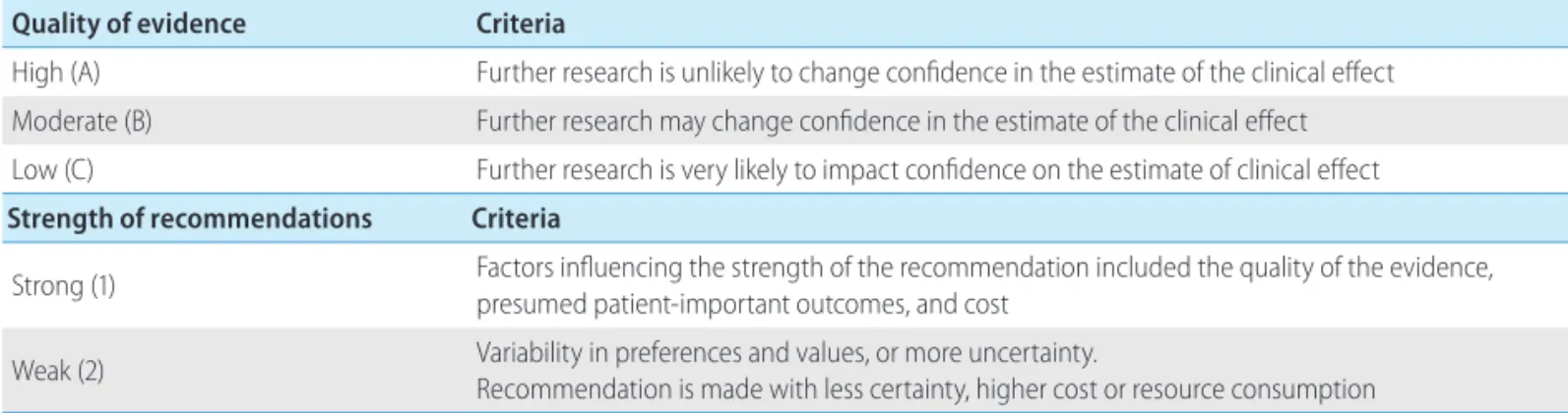
The evidence and recommendations were graded according to Grading of Recommendations, Assessment, Development and Evaluation (GRADE) system with minor modifications (Table 1). The levels of evidence were determined as the possibility of change in the estimate of clinical effect by further research, and were described as high (A), moderate (B) or low (C). The grades of recommendation were either strong (1) or weak (2), as determined by the quality of evidence as well as patient-important outcomes and socioeconomic aspects.
List of the clinical questions
The committee considered the following questions as key com-ponents to be covered in this guideline.
1. How does this guideline differ from previous guidelines? 2. What is the updated knowledge on the epidemiology? 3. What is the updated knowledge on the natural course of
CHB?
4. How should the infection be prevented? 5. How are patients evaluated prior to treatment?
Table 1. Grading of Recommendations, Assessment, Development and Evaluation (GRADE)
Quality of evidence Criteria
High (A) Further research is unlikely to change confidence in the estimate of the clinical effect
Moderate (B) Further research may change confidence in the estimate of the clinical effect
Low (C) Further research is very likely to impact confidence on the estimate of clinical effect
Strength of recommendations Criteria
Strong (1) Factors influencing the strength of the recommendation included the quality of the evidence, presumed patient-important outcomes, and cost
Weak (2) Variability in preferences and values, or more uncertainty.
Recommendation is made with less certainty, higher cost or resource consumption
NOTE. Of the quality levels of evidence, we excluded “very low quality (D)” from the guidelines for convenience. This was originally included in the GRADE system and indicates that the estimate of effect is highly uncertain.
6. When should treatment be considered? 7. What are the goals and endpoints of treatment?
8. What are the optimal first-line treatments for different disease status?
9. How should the treatment be monitored? 10. When can we consider stopping treatment? 11. What are the predictors of a treatment response? 12. What are the definitions of treatment failure? 13. How should we manage drug-resistant CHB patients? 14. What are the definitions of recurrence after treatment
comple-tion and how should these be managed?
15. How should we manage the following special groups: - acute hepatitis B
- liver transplantation
- chemotherapy/immunosuppression - chronic kidney disease
- coinfection [with hepatitis C virus (HCV), hepatitis D virus (HDV), and/or human immunodeficiency virus (HIV)]
16. How can we reduce vertical transmission in pregnant CHB pa-tients?
17. What is the optimal management of CHB in children?
Review of the manuscript
Drafts of the revised guideline were thoroughly reviewed at separate meetings of the committee. A revised manuscript was reviewed at a meeting of an external review board, and at a sym-posium open to all KASL members, and was modified further prior to publication. The external review board comprised of 18 special-ists in CHB who are members of the KASL. The final manuscript was endorsed by the board of executives of the KASL.
Release of the guidelines
The revised CHB guidelines of KASL were released on Novem-ber 26, 2015 (http://www.kasl.org).
Plan for updates
Updates or full revision will be planned when major new evi-dence regarding the diagnosis and/or treatment of CHB becomes available. Detailed plans for updates will be posted on the KASL website at a later date.
EPIDEMIOLOGY
Hepatitis B virus (HBV) infection, as a causative factor of liver disease of 240 million patients globally and death of 600 thou-sand patients annually,1 is a major cause of acute and chronic hepatitis, liver cirrhosis, and hepatocellular carcinoma. It has been recognized as an important public health problem in Korea since the 1970s2 and was designated a third-class communicable dis-ease by law in 1982 and is now the target of a national vaccina-tion program as a second-class communicable disease.3
The prevalence of HBV infection in the Korean population as es-timated by positivity rates for hepatitis B surface antigen (HBsAg) was 8–9% for males and 5–6% for females before commercial-ization of an HBV vaccine in the early 1980s;4 thereafter, the prev-alence of HBV infection tended to decline gradually due to the ini-tiation of a vaccination program for newborn infants in 1991 and a national vaccination program in 1995. For example, in 2006 the prevalence of HBV among children aged 4 to 6 years had de-creased to 0.2%.5 Nevertheless, according to the 2005 National Health and Nutrition Examination Survey, the HBsAg positivity rate was 4.0% at 2009.6 The Ministry of Health and Welfare re-ported that the HBsAg positivity rate was 3.4% for males and 2.6% for females, with 3.0% of the total population being infect-ed with HBV in 2012.7 Positivity rates for HBsAg among pregnant females, which represents a major infection route for hepatitis B, declined steadily after 2004, as did the positivity rates among fe-males in the childbearing period.7 Given that HBsAg is detected in approximately 70% of patients with chronic hepatitis or cirrhosis,8 and in 65–75% of HCC patients,9,10 it can be concluded that CHB infection is a matter of importance for public health in Korea. Most Korean CHB patients are infected with HBV genotype C2,11 and tend to have lower hepatitis B e antigen (HBeAg) seroconver-sion rates, more rapid progresseroconver-sion to cirrhosis and HCC, reduced efficacy of interferon treatment, and are subject to higher rates of relapse after antiviral treatment, compared to those infected with other HBV genotypes.12,13
Natural history
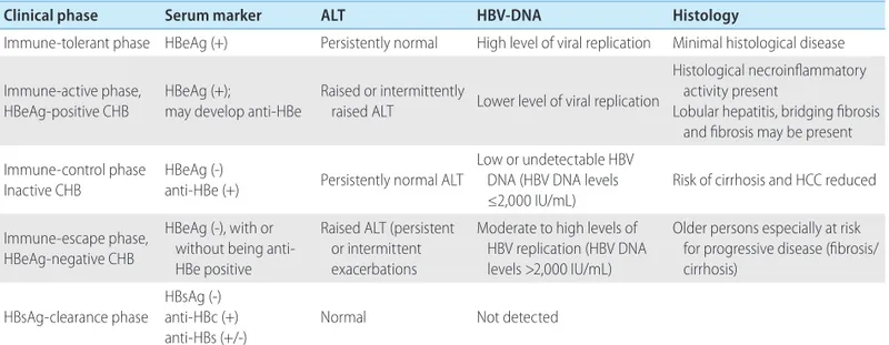
The progression of CHB may be divided into the following five clinical phases: the immune-tolerant phase, immune-active phase, immune-control phase, immune-escape phase, and HBsAg-clear-ance phase. Individual patients do not necessarily experience these clinical phases in a continuous manner, and clinical phases are not always correlated with criteria or indications of antiviral
therapy.14,15 HBV DNA positivity indicates an acute or chronic HBV infection, and negativity indicates resolution of infection. For this reason, the WHO decided to delete the term ‘hepatitis B carrier.’ The natural history of CHB is outlined below (Table 2).
1. Immune-tolerant phase
In cases of perinatal infection, the immune-tolerant phase is characterized by HBeAg positivity, high levels of serum HBV DNA (generally ≥107 IU/mL), normal levels of aspartate aminotrans-ferase/alanine aminotransferase (AST/ALT), and mild or no liver necroinflammation.16-19 Elevation of ALT level was detected in 16% of patients in the immune-tolerant phase during 5 years of follow up.19
This phase may continue for more than three decades in pa-tients infected with HBV genotype C, which is common among Korean patients, and the rate of spontaneous HBeAg loss is very low.20 Therefore, many females infected with this genotype are in the HBeAg-positive immune-tolerant phase when they are of childbearing age. The absence of, or only mild histologic liver damage, despite high levels of HBV DNA, is attributed to immune tolerance to HBV.21
2. Immune-active HBeAg-positive CHB
Most patients in the immune-tolerant phase will experience im-mune responses to HBV as they grow older, and finally reach the immune-active phase, which is characterized by HBeAg positivity, lower serum HBV DNA levels, and increased or fluctuating ALT levels.22,23 Histologic findings in this phase include moderate-to-severe liver inflammation and, in some patients, rapid progression
of fibrosis.24 Such changes are due to enhancement of hepatitis B core antigen (HBcAg) or HBeAg-specific cytotoxic T-lymphocyte activity and the resulting destruction of infected hepatocytes.25 Sustained HBV DNA suppression occasionally accompanies HBeAg seroconversion.
Once HBeAg seroconversion occurs, the natural course of the disease may have one of three clinical features: (1) repeated HBeAg reversion and seroconversion, (2) inactive state, or (3) HBeAg-negative CHB.26,27 Typically, 10–40% of patients who ex-perience seroconversion revert to HBeAg positivity and then expe-rience recurrence of seroconversion at least once with progression of hepatitis activity.24,28,29 In particular, reversion frequently occurs in patients with HBV genotype C, and the rate decreases with age.20 Hepatic decompensation, which occurs in 5% of patients with acute exacerbation, may be fatal.30
3. Immune-control inactive CHB
Most patients who seroconvert during the immune-active phase progress to the immune-control phase, which is characterized by HBeAg negativity, persistent normal ALT levels, and HBV DNA levels of <2,000 IU/mL.31-33 Typical histologic findings in this phase are mild liver inflammation and fibrosis;31 however, patients who have suffered from previous severe inflammation and fibrosis may continue to experience moderate-to-severe inflammation and fibrosis. This may result in even biochemical and histologic tests not being useful for differentiating these patients from those with cirrhosis who require antiviral treatment.32
This phase persists for a long time in most patients, but with a relatively good prognosis; however, an estimated 20% of them
Table 2. Natural course of chronic hepatitis B (CHB)
Clinical phase Serum marker ALT HBV-DNA Histology
Immune-tolerant phase HBeAg (+) Persistently normal High level of viral replication Minimal histological disease Immune-active phase,
HBeAg-positive CHB HBeAg (+);may develop anti-HBe Raised or intermittently raised ALT Lower level of viral replication
Histological necroinflammatory activity present
Lobular hepatitis, bridging fibrosis and fibrosis may be present Immune-control phase
Inactive CHB HBeAg (-)anti-HBe (+) Persistently normal ALT
Low or undetectable HBV DNA (HBV DNA levels ≤2,000 IU/mL)
Risk of cirrhosis and HCC reduced Immune-escape phase,
HBeAg-negative CHB
HBeAg (-), with or without being anti-HBe positive
Raised ALT (persistent or intermittent exacerbations
Moderate to high levels of HBV replication (HBV DNA levels >2,000 IU/mL)
Older persons especially at risk for progressive disease (fibrosis/ cirrhosis)
HBsAg-clearance phase HBsAg (-)anti-HBc (+) anti-HBs (+/-)
Normal Not detected
will reactivate to the HBeAg-negative or HBeAg-positive immune-active phase, and might experience recurring periods of reactiva-tion and inactivareactiva-tion throughout their lives, which can lead to cir-rhosis or HCC.34,35 This is why the ALT levels of patients in the immune-control phase must be measured at least every 6 months for life because currently there are no predictors of which patients will remain in the inactive phase and which will revert to HBeAg-negative active hepatitis.15
4. Immune-escape HBeAg-negative CHB
Approximately 20% of patients who experience HBeAg sero-conversion during their immune-active phase maintain HBeAg negativity and hepatitis B e antibody (anti-HBe) positivity but progress to the immune-escape phase, with findings of HBV DNA levels ≥2,000 IU/mL, increased ALT levels, and active liver necro-inflammation.26 These patients show HBeAg negativity since they harbor HBV variants in the precore (PC) or basal core promoter (BCP) regions of HBV DNA, resulting in failure to produce HBeAg.36-38 HBeAg-negative CHB is associated with low rates of prolonged spontaneous disease remission, and most patients in this phase will experience persistent hepatocellular inflammation and progress to hepatic fibrosis and cirrhosis.38-40 Severe fluctua-tions of HBV DNA and ALT levels can make it difficult to differen-tiate these patients from those in the immune-control phase.41 Accordingly, for the first year after a patient is diagnosed as being in the immune-control phase, HBV DNA and ALT levels should be
measured every 3 months to identify HBeAg-negative CHB pa-tients who require antiviral treatment.15,42
5. HBsAg-clearance phase
Patients in the immune-control phase subsequently progress to the HBsAg clearance phase at a rate of 1–2% annually.41,43,44 Ac-cording to Liaw’s data, HBsAg loss occurs in 1.9% of CHB pa-tients, and 0.8% of those with chronic HBV infection regardless of gender and virus genotype, with age being the only known in-fluencing factor.45,46 It has been reported that Korean patients ex-perience a relatively low rate of HBsAg loss (0.4% annually).47 HBV DNA is not detectable in the serum during this phase, while hepatitis B core antibody (anti-HBc) with or without hepatitis B surface antibody (anti-HBs) is detectable. HBsAg loss is associated with a reduced risk of cirrhosis but a sustained, significant risk of HCC development.34,43,48-53
Risk factors that influence the natural history of CHB
The accumulated incidence of cirrhosis developing from CHB is generally reported to be 8–20%.54,55 In Korea, the reported annual and 5-year accumulated incidences of cirrhosis are 5.1% and 23%, respectively, while those for HCC are 0.8% and 3%.54 The risk fac-tors for hepatitis B progressing to cirrhosis or HCC can be divided into demographic, environmental, social, and viral factors (Table 3).56
Regarding demographic factors, the risk of developing HCC is
Table 3. Risk factors associated with the development of hepatocellular carcinoma (HCC) and/or cirrhosis in persons with chronic hepatitis B Increased risk of HCC Increased risk of cirrhosis
Demographic
Male sex 3+ +
Increasing age >40 years 3+ 3+
Family history of HCC 3+ +
Social and environmental
Alcohol + +
Aflatoxin 3+ Unknown
Smoking + +
Coffee Decreased risk of HCC Slower progression of liver fibrosis
Viral factor
Genotype C 3+ 2+
HBV DNA >2,000 IU/mL 3+ 3+
BCP mutation 3+ +
BCP, basal core promoter; HBV, hepatitis B virus. Modified from McMahon.56
three- to fourfold higher for males than for females, and the risk of HCC and cirrhosis is low among those younger than 40 years, then increases exponentially with increasing age after the fourth decade of life.33,57-59 Those with a family history of HCC also have a higher risk of contracting HCC.60,61 Environmental and social risk factors for progression to cirrhosis or HCC are alcohol consump-tion, exposure to aflatoxin,62 and smoking.63 It is suggested that obesity, metabolic syndrome, and fatty changes in histologic tests increase the risk of CHB patients progressing to hepatic fibrosis or HCC.64-67 Many epidemiological research studies have found that coffee exerts protective effects against the development of hepat-ic fibrosis and HCC.68-72
Viral factors that may influence the progression of CHB patients to cirrhosis or HCC include high levels of serum HBV DNA (≥20,000 IU/mL), genotype C, BCP variants, and coinfection with other viruses.57,59,73-75 According to the Taiwanese Risk Evaluation of Viral Load Elevation and Association Liver Disease/Cancer-Hep-atitis B Virus (REVEAL-HBV) study, the risk of developing HCC during the study period among subjects aged at least 40 years was significantly higher in those with an HBV DNA level of ≥104 copies/mL (cpm) at the start of observation and 105 cpm 11 years later than among those with an entry HBV DNA level of <104 cpm. Likewise, the incidence of cirrhosis was significantly associated with HBV DNA levels higher than 104 cpm at study entry.58,59 If the HBV DNA level decreased during the follow-up period, the risk of developing HCC or cirrhosis decreased. Subse-quent research highlighted the clinical importance of careful eval-uation of patients with an HBV DNA level >2,000 IU/mL who are older than 40 years (especially those HBeAg positive) for the de-velopment of fibrosis 57 and HCC.74,75 Therefore, intervention with antiviral therapy should be performed when appropriate, as rec-ommended by established practice guidelines.56
Unlike HCV infection, the HBV genotype exerts a profound ef-fect on the clinical outcome but—with the exception of interfer-on—little effect on the treatment outcome.76 Eight HBV geno-types have been identified, and that with the worst prognosis is genotype C, which is the most common in Korean CHB patients.77 Genotype C is associated with delayed natural seroconversion and rapid progression to liver cirrhosis and HCC. Therefore, it is an in-dependent risk factor for HCC development. According to a cohort study in Alaska, patients infected with A-, B-, and D-genotype hepatitis B typically experience seroconversion from HBeAg to an-ti-HBe before the age of 20 years, whereas in those infected with the C genotype this occurs at a mean age of 47 years.20 This im-plies that those infected with genotype C would on average
expe-rience a much longer period of infection with high HBV viral loads, and may in part explain why the risks of HCC and cirrhosis are so high in patients infected with genotype C.
Two important genetic mutations of HBV that affect the natural history of CHB infection are the BCP and PC mutations.42,45,75,77-79 BCP mutations are A1762T and G1764A mutations in the HBV BCP regions, and multiple cross-sectional and prospective studies have indicated that they increase the risks of cirrhosis and HCC.42,45,77,78 According to the results of the REVEAL-HBV study, 359 and 1,149 individuals without and with BCP mutations, re-spectively, developed HCC among a population of 100,000.80 PC mutation typically appears near the time of HBeAg seroconver-sion. The mutation results in an amino-acid change that creates a stop codon at site 1896 on the HBV genome, which results in the virus being able to transcribe hepatitis B core protein but not HBeAg.45 Patients infected with PC mutants are characterized by HBeAg negativity and HBeAg positivity, but high levels of HBV DNA.81,82 However, the observed effects of PC mutants on the natural history of CHB have been inconsistent; a recent analysis of the role of PC in the prospective population-based REVEAL-HBV study revealed the opposite to the findings of cross-sectional clin-ic-based studies—that the presence and absence of the PC muta-tion decreased and increased, respectively, the subsequent annual incidence of HCC (269 and 996 per 100,000, respectively).80
PREVENTION
Because HBV infection is endemic in Korea, any person at high risk of liver disease or has suspected liver disease is recom-mended to have their HBsAg and anti-HBs statuses checked.14 CHB patients can transmit virus to others, and hence they should be counseled regarding how to modify their lifestyle so as to pre-vent HBV transmission. Epidemiologic studies found that the daily consumption of 40–80 g of alcohol is associated with liver dam-age and the progression of liver disease,83-88 and a long-term prospective cohort study of patients with chronic HBV infection showed that alcohol consumption increases the risks of liver cir-rhosis and HCC development.57,59 No data are available on the threshold level of alcohol consumption required to significantly increase the risks of liver cirrhosis and HCC in patients with chronic HBV infection. In the general population, a daily alcohol intake of 24 g in males and 12 g in females significantly increas-es the risk of liver cirrhosis.89 So, abstinence or a very limited consumption of alcohol is recommended in patients with chronic
HBV infection.89 According to a long-term prospective study of patients with chronic HBV infection, smoking also increases the risks of liver cirrhosis and HCC, and so non-smoking is recom-mended in patients with chronic HBV infection.57,59,90
Vertical infection is the most important route of HBV trans-mission. Following initiation of the HCV vaccination program, the HBsAg positivity rate among pregnant females was 3.32% (308/9281) and the vertical transmission rate was 1.59% (4/252) in 2014. Therefore, the vaccination program is effective for control of vertical transmission.91 HBV immunoglobulin and vaccination after delivery can prevent 90-95% of vertical transmission to newborns from HBsAg-positive mothers.92-94 Therefore, such in-fants should receive 0.5 mL HBIG and scheduled HBV vaccina-tion within 12 hours of birth and after. Adding immunoglobulin is more effective than vaccination only. The introduction of HBV vaccination did not result in the rate of HBV infection among newborns differing between breast- and formula-feeding HB-sAg-positive mothers (0% vs. 3%, respectively).95
In patients negative for HBsAg and anti-HBs, vaccination is rec-ommended. Isolated anti-HBc positive patients negative for HB-sAg and anti-HBs should consider vaccination, especially if liver function results are abnormal. As HBV is endemic in Korea, pa-tients negative for HBsAg and anti-HBs should be vaccinated,92,93 particularly the household members and sexual partners of pa-tients with chronic HBV infection, as such persons are at increased risk of HBV infection.96,97 Patients with chronic HBV infection are not candidates for vaccination because of its lack of effectiveness. Sexual partners who have not been tested for HBV serologic markers, have not completed the full immunization series, or who are negative for anti-HBs should use barrier protection methods, such as condoms. The three doses constituting the hepatitis B vaccine series administered intramuscularly at 0, 1, and 6 months induce a protective antibody response (anti-HBs >10 mIU/mL) in >90% of recipients. Most non-responders (44–100%) subse-quently respond to a further three-dose revaccination.92,93
Although serologic testing for anti-HBs is not necessary after routine vaccination in immunocompetent adults, post-vaccination testing of anti-HBs status is recommended in some subjects, such as newborns of HBV-infected mothers or 9-18 months old young infants whose family members has CHB. Healthcare workers, dial-ysis patients, workers in dialdial-ysis units and operation rooms, im-munocompromised subjects (e.g., HIV infection, hematopoietic stem cell transplants, patients with chemotherapy), and sexual partners of patients with chronic HBV infection should be tested 1-2 months after their completion of the HBV immunization
se-ries.92,93 While anti-HBs levels can decline or disappear over several decades, vaccinated subjects remain protected against HBV infec-tion and there is no need for booster vaccinainfec-tion in immunocompe-tent individuals. However, an anti-HBs level of <10 mIU/mL in di-alysis patients indicates an increased risk of HBV infection, and so a booster vaccination is needed if annual testing reveals an anti-HBs level of <10 mIU/mL.92 This also applies to immunocompro-mised patients.92,93 A person without protective anti-HBs exposed to HBV-contaminated blood or body fluids should receive hepatitis B immunoglobulin (HBIG, 0.06 mL/kg) and hepatitis B vaccine as soon as possible; preferably within 24 h, otherwise postexposure prophylaxis should be initiated within 7 days for percutaneous ex-posure or within 14 days for sexual exex-posure.98
Coinfection with hepatitis A in HBV carriers increases the risk of mortality by 5.6- to 29-fold.99 Therefore, hepatitis A vaccination is recommended for persons negative for the protective hepatitis A virus antibody (anti-HAV).100
[Recommendations]
1. HBV vaccination is recommended for persons negative for HBsAg and anti-HBs. (A1)
2. Abstinence from alcohol and smoking is recommended for patients with chronic HBV infection. (A1)
3. Newborns of HBV-infected mothers should receive HBIG and hepatitis B vaccine at delivery and complete the recom-mended vaccination series. (A1)
4. Hepatitis A vaccine should be given to patients with chronic HBV infection negative for anti-HAV. (A1)
DIAGNOSIS AND INITIAL EVALUATION
CHB is defined as the presence of HBsAg for longer than 6 months. The initial evaluation of CHB patients should include a thorough history-taking and physical examination, with empha-sis on risk factors such as alcohol consumption or drug use, HAV, HCV, HDV coinfection, and family history of HBV infection and HCC. The causal relationship between HBV infection and liver disease has yet to be established. Appropriate longitudinal long-term follow-up is crucial for patients with CHB. Serologic tests, virologic tests, biochemical tests and/or liver biopsy are used to assess HBV replication and the degree of liver injury in patients with CHB.
Antigen/antibody test
HBsAg immunoassay is a necessary and accurate test for di-agnosis of CHB. By definition, patients who remain positive for HBsAg for longer than 6 months have progressed to chronic infec-tion. Quantitative measurement of HBsAg is now possible and the combination of HBsAg quantification and HBV DNA level is an in-tegral component of monitoring the response to antiviral therapy. Serologic tests, including anti-HBs and anti-HBc, can assist in screening of populations for HBV infection and differentiating among acute, chronic, and past infections. In acute HBV infec-tion, HBsAg appears 1-10 weeks after exposure to HBsAg and disappears 4-6 months after recovering from HBV infection.101 Acute HBV infection is diagnosed by being HBsAg positive and anti-HBc IgM positive. Anti-HBc IgM is the only marker present during the window period, the interval between disappearance of HBsAg and appearance of anti-HBs.
Anti-HBc typically persists for life, but IgM anti-HBc is detect-able for 6 months, and anti-HBc is detectdetect-able thereafter in pa-tients with resolved acute HBV infection. IgM anti-HBc can be de-tected at low levels during chronic HBV infection.93 Persistently positive anti-HBc is shown when anti-HBs titer from the past HBV infection becomes undetectable over time or in cases with occult hepatitis B infection.102-105 Measurement of the serum HBV DNA level might be helpful in these settings. Patients with these sero-logic patterns should be followed with repeated testing of HBsAg, anti-HBs, and anti-HBc for 3–6 months. Patients who recover from HBV infection will test negative for HBsAg and positive for anti-HBs and anti-HBc. Patients who respond adequately to hepa-titis B vaccines will test negative for HBc and positive for anti-HBs, since anti-HBc emerges only after HBV infection and persists for life.
Laboratory tests for patients with CHB should include HBeAg and anti-HBe. HBeAg positivity generally indicates a high level of viral replication, and anti-HBe positivity a low level. Serum HBV DNA and AST/ALT levels are important parameters in HBeAg-negative patients. HBeAg-negative, anti-HBe-positive patients with a normal ALT level and an HBV DNA level of <2,000 IU/mL (<10,000 cpm) may be in the inactive phase. These patients usually have mild or no liver necroinflammation and no or slow progression of fibrosis, but some patients with severe liver damage during the immune-active phase may pres-ent with a cirrhotic liver. HBeAg-negative CHB patipres-ents have an elevated ALT and an HBV DNA level of >2,000 IU/mL. HBe-neg-ative CHB is associated with viral mutants in the PC and/or BCP
regions that are unable to produce or produce only low levels of HBeAg.40 They have severe liver necroinflammation with a low rate of prolonged spontaneous disease remission and a high risk of subsequent complications, such as decompensated cirrhosis and HCC.106
Acute hepatitis A co-infection in chronic hepatitis B patients can result in increased icteric manifestation, longer recovery time, and increased risk of fulminant hepatic failure. Underlying chronic liver disease is an important risk factor for fulminant he-patic failure and death in patients with acute HAV infec-tion.106-108 Therefore, CHB patients younger than 50 years should undergo testing for IgG anti-HAV, and all patients with a nega-tive immune status for hepatitis A should receive HAV vaccine. Laboratory tests should include tests for coinfection with HCV and/or HIV in those at risk.
Serum HBV DNA test
Serum HBV DNA testing provides a direct measure of the level of viral replication. This quantification is essential for characteriz-ing the status of infection, diagnoscharacteriz-ing the disease, makcharacteriz-ing the decision to treat, and subsequent monitoring of patients. It is also important for predicting the risks of cirrhosis and HCC. Therefore, it should be applied to all patients diagnosed with CHB. The intro-duction of the international unit (IU) (1 IU is equivalent to 5.6 HBV DNA copies) as a recommended reporting unit for HBV DNA has facilitated standardized reporting and comparison of serum HBV DNA levels.109 The methods used to quantify HBV DNA levels have evolved rapidly. Real-time PCR-based assays have been intro-duced and demonstrate both high sensitivity and a broad linear range (10–108 IU/mL) of quantification.110 The same test should be specified each time when monitoring HBV DNA levels for a given patient in clinical practice to ensure consistency.
HBV genotypes
HBV genotypes appear to influence the progression of dis-ease, risk of HCC, and response to therapy (including interferon therapy).75,111,112 Some studies in Asia have suggested that geno-type C is associated more frequently with HBV reactivation, se-vere liver disease, and HCC than is genotype B.111,113-115 The spe-cific genotype has also been shown to affect the response to interferon therapy, with the rate of an antiviral response to pe-gylated interferon (peginterferon) therapy being higher for gen-otypes A and B than for gengen-otypes C and D.116 In CHB,
examina-tion of genotyping is recommended selectively to help identify patients who might be at greater risk of disease progression, and routinely to determine the most appropriate candidates for peginterferon therapy.117 However, genotyping is recommended as being unnecessary in Korea because Korean patients are al-most exclusively infected with genotype C.
Biochemical test
Assessments of the severity of liver disease should include biochemical markers such as AST, ALT, gamma-glutamyl trans-peptidase (GGT), alkaline phosphatase (ALP), prothrombin time (PT), and serum albumin. A progressive decline in the serum al-bumin level and prolongation of the PT, often accompanied by a decrease in the platelet count, are characteristically observed after cirrhosis develops. The serum ALT level has been common-ly used in assessments of liver disease and as an important cri-terion for defining which patients are candidates for therapy.118 The ALT level is usually higher than that of AST, but the ratio may be reversed when the disease progresses to cirrhosis. HBV-infected patients with normal or mildly elevated ALT levels have been thought to have mild-to-no or significant necroinflamma-tion on liver biopsy, respectively. However, there is no correla-tion between the degrees of liver cell necrosis and ALT level.119 ALT activity might also be affected by other factors such as body mass index, gender, abnormal lipid and carbohydrate me-tabolism, and uremia.119,120 Therefore, relying solely on the find-ing of elevated ALT as a prerequisite for treatment candidacy has limitations. Data from clinical studies have shown that the true normal level of ALT is significantly lower than the previously established limits: 40 IU/mL for males and 30 IU/mL for females. Moreover, data from cohort studies indicate that the upper limit of normal (ULN) ALT and AST levels should be decreased to 30 IU/mL for males and 19 IU/mL for females.119,120 Clinical stud-ies have shown that patients with ALT levels of 40–45 IU/mL have a high risk of significant liver disease and mortality from complications.121 According to the treatment algorithm for CHB suggested by Keefee et al., serum ALT levels of 30 and 19 IU/mL for males and females, respectively, should be used as the ULN levels when deciding to commence treatment.117 Further pro-spective studies are needed to clarify this issue.
A recent prospective study in Korea involving 2,000 liver donors suggested that healthy serum ALT values should be 33 IU/L for males and 25 IU/L for females.122 Ninety thousand males and 40,000 females aged 35 to 59 years in the prospective NHS
co-hort exhibited upper limits of AST and ALT values for prediction of liver diseases of 31 IU/L and 30 IU/L, respectively.121
Liver biopsy
A liver biopsy is recommended for determining the degree of necroinflammation and fibrosis in patients with elevated ALT, an HBV DNA positive or both, because liver histology is useful when deciding whether or not to commence treatment. A liver biopsy is invasive but the rate of serious complications is very low (1/4,000-10,000).123 Several recent clinical studies found that 12–43% of patients with persistent normal ALT levels had histologic evidence of significant fibrosis or inflammation in a biopsy, particularly those older than 35-40 years.116-121,124 A ret-rospective study of the relationship between ALT level and fi-brosis in CHB patients reported similar results: of the 59 pa-tients with persistent normal ALT levels, 18% had stage 2 fibrosis and 34% had grade 2 or 3 inflammation, with 37% of all patients with persistent normal ALT levels having significant fibrosis and inflammation.125 Subgroup analysis also demon-strated that most of the patients with fibrosis had high normal ALT levels. These results indicate that the ALT level in CHB pa-tients with high normal ALT levels should be interpreted in con-junction with the serum HBV DNA level, age, and liver histology results when deciding to commence treatment. Therefore, in HBsAg-positive patients with HBV DNA levels of ≥20,000 IU/mL and normal ALT levels, a liver biopsy should be considered in those older than 35 years since they are less likely to be in the immune-tolerance phase of infection. Treatment should be con-sidered if a liver biopsy reveals fibrosis at stage 2 or greater and/or necroinflammation. When deciding whether to com-mence treatment in this patient population, it must be recog-nized that long-term therapy is likely to be needed due to the low probability of HBeAg seroconversion occurring within 1 year. A liver biopsy is usually not required in patients with clini-cal evidence of cirrhosis or when treatment is indicated irrespec-tive of the grade of activity or the stage of fibrosis. This is be-cause only a small portion of the liver is sampled, and the low intra/interobserver reliabilities. Therefore, the efficacy of nonin-vasive methods such as the Fibroscan device or serum markers in assessing fibrosis in CHB has increased.
Noninvasive fibrosis test
DNA levels have essential roles in treatment decisions. Noninva-sive methods to estimate liver fibrosis have been developed and used. These methods include the aspartate aminotransferase-platelet ratio index (APRI), AST/ALT ratio (AAR), Forns’ fibrosis in-dex (age, platelets, GGT, cholesterol), FIB-4 (platelets, ALT, AST, Age). Also, the FibroTest that uses indirect markers (α-2 macro-globulin, haptoglobin, r-macro-globulin, apolipoprotein A1, and GGT), the FibroSpect II Enhanced Liver Fibrosis test that uses direct markers (Hepascore, FibroMeter, hyaluronic acid and tissue inhibi-tor of matrix metalloproteinase-1, 2) are available.126 The age-spleen-platelet ratio index (ASPRI) is the most accurate in predict-ing liver fibrosis in chronic HBV infection.127 APRI is useful for diagnosis of not only for liver fibrosis but also liver cirrhosis, while FIB4 is useful for mild fibrosis. However FIB4 has limitations in terms of predicting fibrosis of stage F2 and above as it has low sensitivity and specificity.126
Transient elastography using Fibroscan® has a high degree of accuracy for assessment of advanced liver fibrosis. It is the most commonly used method for chronic liver diseases because of its noninvasiveness and high reproducibility.128
Fibroscan® can be perform rapidly (5 min) in the outpatient clinics of hospitals and produce a result immediately after the test.129,130 However, only procedures involving ≥10 successful mea-surements are considered reliable. Moreover, a success rate (SR) of at least 60% and an interquartile range (IQR) of less than 30% of the median value are required (Interquartile range/median val-ue (IQR/M),131 Fibroscan® has limitations in subjects with ascites, obesity, or narrow intercostal spaces. Moreover, the system may yield false-positive results in subjects with acute hepatitis and ex-trahepatic biliary tract obstruction.132-134
Fibroscan® has greater diagnostic accuracy than APRI or FIB-4 for liver cirrhosis in a study that compared liver biopsy, AAR, APRI, Fibroscan®, and FIB-4 in patients with chronic hepati-tis.135,136 Also, Fibroscan® was more predictive of liver fibrosis and liver cirrhosis in a study that compared Fibroscan® and APRI in 567 subjects with chronic hepatitis (Area under Receiver Operat-ing Characteristic: F3 0.849 vs. 0.812, F4 0.902 vs. 0.707).137
Screening for hepatocellular carcinoma
The initial evaluation of patients with CHB should include tests for HCC. Periodic surveillance is also needed in these patients to ensure early detection of HCC during follow-up. The issue of HCC is treated in detail in the “Practical Guidelines for Management of Hepatocellular Carcinoma 2014.”138 Standard tools for HCC
screening include measuring the α-fetoprotein level and ultra-sound. Magnetic resonance imaging and computed tomography might be preferred for some patients with severe cirrhosis or obe-sity, since ultrasound has poor sensitivity in those conditions. Pa-tients at a high risk of HCC include those older than 40 years,139 patients with cirrhosis, those with a family history of HCC, and any carriers older than 40 years exhibiting persistent or intermit-tent ALT elevation, a high HBV DNA level (>2,000 IU/mL), or both.14 Keeffe et al. recently recommend earlier screening (at 30– 35 years of age or even younger) in Asian patients with presumed infection at the time of birth or in early childhood due to the high-er risk of HCC in this patient population.
The use of antiviral therapies improves liver function and in-creases survival rates of patients with liver failure (liver decom-pensation).
Consistent inhibition of HBV replication with antiviral therapies delays progression of liver fibrosis, induces reversal of advanced liver fibrosis, reduces the incidence of liver cirrhosis, and prevents diseases including hepatocellular carcinoma in patients with ad-vanced liver fibrosis or liver cirrhosis.140
Recently developed treatments can decrease the incidence of liver diseases or delay their progression but cannot prevent all possible complications. Therefore, surveillance and screening for hepatocellular carcinoma are required at regular intervals for early diagnosis and a complete recovery.
[Recommendations]
1. The initial evaluation of patients with CHB should include a thorough history-taking and physical examination, with emphasis on risk factors such as coinfection, alcohol con-sumption, and the family history of HBV infection and liver cancer. (A1)
2. Laboratory tests to assess liver disease should include the complete blood count (CBC), AST/ALT, ALP, GGT, bilirubin, albumin, creatinine, and PT. (A1)
3. Tests for HBV replication include HBeAg/anti-HBe and quantitative serum HBV DNA levels. A real-time PCR quanti-fication assay is strongly recommended for quantifying the HBV DNA level. (A1)
4. An anti-HCV test is necessary to rule out coinfection with HCV. (B1)
5. An anti-HAV test is necessary in CHB patients younger than 50 years. (A1)
in-flammation and fibrosis. (A1)
7. Noninvasive tests such as serum markers and liver elasticity are used for diagnosis of the degree of liver fibrosis. (B1) 8. Standard tools for HCC screening include ultrasound and
serum α-fetoprotein measurement. (A1)
TREATMENT GOALS
The goals of hepatitis B treatment are to decrease the mortality rate and increase the survival rate by alleviating hepatic inflam-mation and preventing the development of fibrosis, which ulti-mately reduces the frequency of progression of hepatitis to liver cirrhosis or HCC.141-145 The optimal treatment result would be the loss or seroconversion of HBsAg, but since intranuclear cccDNA persists despite treatment, complete clearance of HBV is almost impossible to achieve.146 This is why indices such as ALT level nor-malization, undetectable HBV DNA, loss or seroconversion of HBeAg, and histologic improvement are used (rather than the loss or seroconversion of HBsAg) to predict the treatment response in the clinical context. Therefore, a realistic virologic goal of anti-HBV therapy is the suppression of viral replication.
Most guidelines state that antiviral treatment is required for pa-tients with acute liver failure, decompensated liver cirrhosis or in the acute phase of severe chronic HBV hepatitis regardless of HBV DNA and ALT levels, and the treatment has almost no complica-tions, although few controlled studies have been performed.147 Antiviral therapy decreases the rate of recurrence of viral infection in patients who require liver transplantation.148 The HBV DNA and HBeAg levels in CHB are indices of viral replication and active hepatitis, respectively, and patients with HBeAg-positive hepatitis B with high levels of HBV DNA have an increased risk of develop-ing liver cirrhosis or HCC.57,59,74 Patients with disappearance or conversion of serum HBeAg have a low risk of liver cirrhosis and hepatocellular carcinoma, and so have a good prognosis.26,149
The loss or seroconversion of HBeAg during the natural course of hepatitis B or after IFN-α treatment indicates a favorable long-term outcome with a decreased probability of liver cirrhosis or HCC development.26,53,149,150 Therefore, clearance or seroconver-sion of HBeAg is an important goal of antiviral treatment in pa-tients with HBeAg-positive active hepatitis. A decrease in the HBV DNA level has recently been suggested to be even more impor-tant. The decrease in the HBV DNA level after antiviral treatment in active hepatitis with elevated HBV DNA levels results in histo-logic improvement, seroconversion of HBeAg, and normalization
of ALT levels, and thus a slowing of the progression of hepati-tis.151,152 However, even in cases with HBV DNA levels of less than 104 copies/mL, which is considered to be inactive hepatitis, the hepatitis can still progress to liver cirrhosis and HCC. Therefore, a decrease in HBV DNA to an undetectable level is recommended for patients on antiviral treatment.153
[Recommendations]
1. The treatment goals in hepatitis B are to decrease the mor-tality rate and increase the survival rate by alleviating he-patic inflammation and preventing the development of fi-brosis, which would ultimately reduce the frequency of progression of hepatitis to liver cirrhosis or HCC. (A1) 2. To achieve HBsAg clearance, which is the ideal treatment
goal, long-term maintenance of an undetectable HBV DNA level is recommended. (B1)
3. The ultimate treatment goals in patients with HBeAg-posi-tive hepatitis are normalization of the ALT level, undetect-able HBV DNA level, and the clearance or seroconversion of HBsAg and HBeAg. In patients with HBeAg-negative hepa-titis the treatment goals are normalization of the ALT level, an undetectable HBV DNA level, and the clearance or sero-conversion of HBsAg. (B1)
TREATMENT INDICATIONS AND STRATEGIES
Long-term viral suppression by drugs with potent antiviral activ-ity and high genetic barrier to resistance is a current paradigm of antiviral treatment for CHB aimed at the prevention of disease progression and improvement of survival. Since eradication of HBV infection is rarely achieved with currently available drugs, long-term treatment is necessary in most cases. Treatment proto-col should be individualized according to various factors: host fac-tors such as mode of infection, disease status, and immunity; viral factors such as genotypes, prior antiviral treatment, mutation, and susceptibility level; and drug factors such local availability, cost, and reimbursement policy.35 The durations of currently avail-able antiviral trials are insufficient to assess the effects of treat-ment on long-term survival.35 Long-term treatment with oral nucleos(t)ide analogs (NAs) ameliorates histologic abnormalities such as necroinflammation and/or fibrosis, both in HBeAg-posi-tive35,154,155 and HBeAg-negative155-158 CHB. Therefore, long-term antiviral therapy may prevent disease progression and reduce the
risk of liver cirrhosis.145
Immune tolerance phase
Antiviral therapy is not indicated for patients in the immune-tol-erant phase despite HBeAg positivity and a high level of HBV DNA, because of the benign natural course of the disease and such treatment results in minimal histologic changes.159
[Recommendations]
Patients in the immune-tolerant phase (HBeAg positive and persistently normal ALT level as recommended by this guide-line rather than local laboratory ULNs) are not indicated for antiviral therapy. (B1)
Chronic hepatitis B
CHB patients with active viral replication and significant inflam-mation and/or fibrosis are appropriate targets for antiviral treat-ment. Early guidelines generally agreed that antiviral treatment could be recommended for CHB patients (especially those without liver cirrhosis) with serum HBV DNA level > 20,000 IU/mL and se-rum ALT level> 2 ULN.160,161 However, recent guidelines suggest that the indications of antiviral treatment should be expanded to those with lower serum HBV DNA levels and/or lower serum ALT levels.35,162,163
Serum HBV DNA level is a marker of viral replication and an in-dicator of the efficacy of antiviral treatment in individuals with CHB. Progression to cirrhosis in HBV-infected patients is reported to be strongly correlated with the level of circulating virus.57,59 However, an HBV DNA level of 105 cpm or 20,000 IU/mL was ar-bitrarily chosen by early guidelines as the cut-off level for indica-tion of antiviral treatment. Some patients with lower serum HBV DNA levels (300–105 cpm), especially those with HBeAg negative hepatitis and/or cirrhosis, frequently show progression of liver dis-ease and hence may need treatment.35,161,164 A serum HBV DNA level of ≥20,000 IU/mL has been suggested as the cut-off for HBeAg-positive CHB.164 However, the distinction between HBeAg-negative CHB and inactive carriers is not clear due to the fluctuating course of HBeAg-negative CHB.164 A population-based cohort study revealed increased risks of liver cirrhosis and HCC when the serum HBV DNA level exceeds 2,000 IU/mL,57,59,165 therefore this level is widely accepted as the cut-off for indicating antiviral therapy.
Serum ALT has been used as a convenient surrogate marker for
liver injury, and elevated serum ALT is indicated as a risk factor for disease progression in CHB.57 A serum ALT level > 2 ULN was suggested as a suitable indication of antiviral treatment for CHB by early guidelines, especially in CHB patients without cirrho-sis.160,161,166 However, an increased risk of developing liver cirrhosis and HCC has been documented in patients with mildly elevated serum ALT and even in those with serum ALT levels in the upper normal range.119,121,167 About two-thirds of CHB patients with mild-ly elevated ALT (1–2 ULN) show significant hepatic fibrosis (F2 or higher),168 and CHB patients with persistently normal ALT levels and HBV DNA levels of >20,000 IU/mL may actually have signifi-cant fibrosis or inflammation,125,168,169 which are indications for an-tiviral therapy. A cohort study in Hong Kong demonstrated that the risk of liver-related complications in CHB patients was higher for ALT levels of 0.5–1 ULN and 1–2 ULN than for those <0.5 ULN. Thus, previous ALT criteria might exclude some patients with existing or potentially significant disease.170,171
Liver biopsy has three major roles: diagnosis, assessment of prognosis (disease staging), and assistance in making therapeutic decisions.172 In CHB, liver biopsy is especially useful for patients who do not meet definite criteria for treatment but still have a possible risk of significant disease.35 Age of the patient, serum HBV DNA level, serum ALT level, and family history of HCC should be considered before deciding whether to perform a biopsy. ALT and HBV DNA levels may miss cases of histologically significant disease,169 and so histologic confirmation should be considered, especially in patients of advanced age with serum AST/ALT levels in the upper normal range or higher.
Peginterferon-α and NAs including lamivudine, adefovir, clevu-dine, telbivuclevu-dine, entecavir, and tenofovir, have been used for an-tiviral treatment of CHB. Drug of choice can differ according to various factors, including effectiveness, safety, risk of resistance, and cost of drugs, preference of patients and physicians, and any plans for pregnancy.35
Lamivudine and telbivudine are not preferred due to their weak antiviral potency and high frequency of drug resistance, unless a good response is predicted or the anticipated duration of treat-ment is short. Adefovir is not an ideal option due to its weak anti-viral activity and high frequency of drug resistance after 48 weeks. There are insufficient long-term follow-up data on the ef-ficacy and safety of clevudine. Entecavir and tenofovir are safe agents with potent antiviral effects and low frequency of drug re-sistance. Due to convenience of usage, peginterferon-α is pre-ferred over interferon-α. To date, there has been no report con-firming the superiority of combination therapies over monotherapy
in treatment-naïve patients.
Currently, monotherapy with entecavir, tenofovir, or peginterferon-α is the preferred initial therapy for CHB. Other NAs might be used in patients with good predictors of response, and can be continued or modified according to on-treatment response.
In patients treated with lamivudine, the predictive factors for a good response to therapy are increased initial serum ALT level and high histologic activity index score.118 During telbivudine treat-ment, a combination of pretreatment characteristics (low HBV DNA level; HBV DNA < 109 copies/mL (HBeAg positive CHB) or HBV DNA < 107 copies/mL (HBeAg negative CHB) and ALT level ≥ 2 ULN ) plus non-detectable serum HBV DNA at treatment week 24 is suggested to be the strongest predictor of optimal outcomes at 2 years.173 Of CHB patients receiving lamivudine or telbivudine treatment, those with a virologic response at week 24 (< 300 copies/mL) achieved a high rate of HBeAg seroconversion at week 52.125 Less resistance was reported in patients with low serum HBV DNA levels (< 1,000 copies/mL) at week 48 during long-term therapy with adefovir.157
[Recommendations]
HBeAg-positive CHB
1. HBeAg positive CHB patients with HBV DNA ≥ 20,000 IU/ mL, plus serum AST or ALT ≥ 2 ULN or significant histologic changes such as inflammation or fibrosis (≥ moderate necroinflammation; ≥ periportal fibrosis) on biopsy should be considered for treatment. (A1) Treatment can be delayed for 3–6 months if spontaneous HBeAg seroconversion is expected. (B2) However, patients with apparent or antici-pated liver failure (i.e., those with jaundice, prolonged PT, hepatic encephalopathy, and ascites) should be treated promptly. (B1)
2. For those with HBV DNA ≥ 20,000 IU/mL and serum AST or ALT < 2 ULN, observation or liver biopsy can be considered. Antiviral treatment is recommended for those showing subsequent elevation of serum ALT or AST, or significant histologic changes such as inflammation or fibrosis on bi-opsy. (A1)
3. Monotherapy with tenofovir, entecavir, or peginterferon-α is preferred. (A1)
HBeAg-negative CHB
1. HBeAg negative CHB patients with HBV DNA ≥ 2,000 IU/mL plus serum AST or ALT ≥ 2 ULN or significant pathologic
changes such as inflammation or fibrosis on biopsy should be considered for treatment. (A1)
2. For those with HBV DNA ≥ 2,000 IU/mL and serum AST or ALT < 2 ULN, observation or liver biopsy can be considered. Anti-viral treatment is recommended for those showing subse-quent elevation of serum ALT or AST, or significant pathologic changes such as inflammation or fibrosis on biopsy. (A1) 3. Monotherapy with tenofovir, entecavir, or peginterferon-α
is preferred. (A1)
Compensated liver cirrhosis
Liver biopsy has been considered the gold standard for diagno-sis of liver cirrhodiagno-sis. Whereas use of liver biopsy is limited in real clinical practice; imaging studies such as CT, abdominal ultra-sound, and MRI are helpful for the diagnosis of liver cirrhosis. Typical image findings of liver cirrhosis include nodular liver sur-face, splenomegaly, and the presence of intra-abdominal collater-al vessels, which indicate increased portcollater-al venous pressure. If esophageal or gastric varices is observed in upper gastrointestinal endoscopy, liver cirrhosis can be diagnosed.174 With imaging stud-ies, laboratory findings such as albumin, bilirubin, or prothrombin time and platelet values are helpful for the diagnosis of liver cir-rhosis.
Patients with compensated cirrhosis and elevated serum HBV DNA (HBV DNA ≥ 2,000 IU/mL) can benefit from treatment with long-term oral NAs, because such treatment may prevent disease progression141 and the development of HCC.144,145,175-178 Compen-sated cirrhosis patients with a low viral load, although HBV DNA < 2,000 IU/mL, are at considerable risk for HCC, and antiviral treatment in these patients was suggested to reduce the risk of HCC.179 Antiviral therapy is recommended in CH-B patients with significant hepatic fibrosis regardless of AST/ALT levels.35,162,163,180 The levels of AST/ALT should not be used as criteria for starting antiviral therapy in patients with liver cirrhosis, because they al-ready have significant hepatic fibrosis and frequently have nearly normal AST/ALT levels.
In a cohort of HBeAg-positive liver cirrhosis patients, long-term follow-up data after interferon-α therapy showed that the HBeAg seroconversion rate was similar (67% vs. 60%, respectively) but the ALT normalization rate (62% vs. 47%) and HBsAg loss rate (23% vs. 3%) were better in the interferon-α treated group than in the control group.181 Interferon-α treatment in cirrhotic patients requires careful monitoring because it may cause acute exacerba-tion of hepatitis, which leads to hepatic failure.182 After treating
CHB patients with peginterferon-α-2b alone or in combination with lamivudine for 52 weeks, the virologic response rate (as indi-cated by HBeAg seroconversion and an HBV DNA level of <10,000 copies/mL) was superior in those with cirrhosis than in those without cirrhosis (35% vs. 14%, respectively).183 However, acute exacerbation of hepatitis (33% vs. 12%, respectively) and requirement for dose reduction (63% vs. 30%) were more com-mon in cirrhotic patients than in noncirrhotic patients.183 There-fore, interferon-α can be used with caution in cirrhotic patients with preserved liver function.
In patients with decompensated liver cirrhosis, long-term lami-vudine treatment significantly reduced the complications and he-patocellular carcinoma compared to placebo. However, the benefit was less in patients with lamivudine resistance.141 Entecavir treat-ment of patients with advanced hepatic fibrosis or cirrhosis for 48 weeks showed improvements in the liver histology in 57%, 59%, and 43% of patients with HBeAg-positive, HBeAg-negative, and lamivudine-resistant CHB, respectively.184 A study including a small number (n=40) of patients showed that telbivudine effec-tively decreased HBV DNA levels in patients with compensated liver cirrhosis, and HBV DNA was undetectable after 48 weeks of telbivudine treatment in 92.5%.185 A study comparing the effects of clevudine treatment for 48 weeks found that the virologic re-sponse rate (HBV DNA <1,000) (87.1% vs. 71.4%, respectively) and biochemical response rate (83.9% vs. 80.9%) did not differ significantly between patients with CHB (n=21) and those with liver cirrhosis (n=31).186 A phase III clinical trial of tenofovir adopt-ing paired liver biopsy at baseline and at week 240 revealed that, of the 96 (28%) patients with liver cirrhosis (Ishak score 5 or 6) at baseline, 71 (74%) no longer had liver cirrhosis (≥1 unit decrease in score) at follow-up biopsy.187
Since long-term antiviral therapy is generally required in pa-tients with liver cirrhosis, the AASLD and EASL guidelines recom-mend the use of entecavir or tenofovir due to their potent antiviral efficacy and high genetic barrier to drug resistance.
Decompensated liver cirrhosis
Decompensated liver cirrhosis is defined as liver cirrhosis com-plicated with ascites, variceal bleeding, hepatic encephalopathy, or jaundice.174 Patients with decompensated liver cirrhosis should be treated at an institution that can provide appropriate manage-ment for complications of liver cirrhosis. Liver transplantation should be considered in patients with decompensated liver cirrho-sis. Oral NAs may improve hepatic function 142 and decrease the
need for liver transplantation in Child-Turcotte-Pugh (CTP) class C cirrhosis.188 The use of interferon-α in patients with decompensat-ed liver cirrhosis is contraindicatdecompensat-ed due to the risk of serious com-plications, such as infection or hepatic failure.189 Lamivudine treat-ment for longer than 6 months was shown to improve or stabilize liver function and prolong the time to liver transplantation in pa-tients with decompensated liver cirrhosis.190-192 A study comparing the effects of telbivudine and lamivudine in patients with decom-pensated liver cirrhosis found a higher rate of HBV DNA undetect-ability (47% vs. 36%, respectively) and a lower viral breakthrough rate (29% vs. 39%, respectively) in the telbivudine group than in the lamivudine group.193 A study of the effect of adefovir in lami-vudine-resistant cirrhotic patients (n=101) found that the virologic response rate was lower in decompensated cirrhotic patients (n=53) than in compensated cirrhotic patients (n=48) (50.9% vs. 83.3%, respectively), whereas ALT normalization and HBeAg loss did not differ between the two groups.194
A randomized study comparing the effects of entecavir (1 mg/ day) and adefovir (10 mg/day) in patients with decompensated liver cirrhosis found that the rates of HBV DNA undetectability at weeks 24 and 48 were higher in the entecavir group than in the adefovir group (week 24, 49% vs. 16%, respectively; week 48, 57% vs. 20%), while HBeAg seroconversion at week 48 did not differ significantly between the two groups (6% vs. 10%).195 Ente-cavir therapy showed improvement of the CTP score (to ≥2) in al-most half (27/55) of treatment-naïve patients with decompensat-ed liver cirrhosis (n=55) and a 1-year transplantation-free survival rate of 87.1%.142
A randomized trial comparing the effects of tenofovir (n=45), tenofovir plus emtricitabine (n=45), and entecavir (n=22) in pa-tients with decompensated liver cirrhosis showed that the requiment for early withdrawal of drug (6.7%, 4.4%, and 9.1%, re-spectively) and elevation of serum creatinine (8.9%, 6.7%, and 4.5%) did not differ among the three groups. The rates of HBV DNA undetectability at week 48 were 70.5%, 87.8%, and 72.7%, respectively, and those of HBeAg loss/seroconversion were 21%/21%, 27%/13%, and 0%/0%.143
Because prompt treatment is required in patients with decom-pensated liver cirrhosis, oral antiviral therapy is the treatment of choice if HBV DNA is detectable by PCR tests.35,162,180 An antiviral drug with a potent antiviral efficacy and high genetic barrier to drug resistance should be used. Since clinical improvement often requires 3–6 months of antiviral therapy, progression to hepatic failure is possible even during antiviral therapy in some patients. Hence, liver transplantation should be considered together with
antiviral treatment.192 Pre- and post-transplantation antiviral ther-apy has been reported to reduce the risk of reactivation of hepati-tis after liver transplantation.
[Recommendations]
Compensated liver cirrhosis
1. Antiviral therapy should be performed if HBV DNA level is ≥2,000 IU/mL regardless of AST/ALT levels. (A1)
2. Antiviral therapy can be considered when HBV DNA is HBV DNA is <2,000 IU/mL to reduce the risk of decompensation regardless of AST/ALT levels. (C1)
3. Oral antiviral therapy is recommended. Monotherapy with tenofovir or entecavir is preferred. (A1)
4. Peginterferon-α may be used with careful monitoring of impairment of liver function and drug side effects in pa-tients with compensated liver cirrhosis with preserved liver function. (B2)
Decompensated liver cirrhosis
1. Prompt antiviral therapy is recommended if HBV DNA is de-tectable by PCR test regardless of AST/ALT levels. (B1) 2. Oral antiviral therapy is recommended. Monotherapy with
tenofovir or entecavir is preferred. (A1)
3. The use of peginterferon-α is contraindicated due to the risk of serious complications, such as hepatic failure. (A1) 4. Liver transplantation should be considered. (B1)
TREATMENT MONITORING
Monitoring prior to antiviral treatment
After diagnosis and initial evaluation of patients with CHB, their serum HBV DNA, ALT, HBeAg, and anti-HBe levels should be reg-ularly monitored until they are considered for treatment.35,168,196,197 The HBV genotype test is not recommended in Korea because most Korean patients are known to have HBV genotype C.198,199
Applying a quantitative HBsAg (qHBsAg) assay before or during antiviral treatment may assist prediction of the treatment re-sponse.200-203 HBsAg is generated by transcription and translation of cccDNA or HBV DNA integrated into the genome, and can be detected on the surface of infective virions and on circular and linear non-infective particles. The quantity of HBsAg (qHBsAg) showed a positive correlation with the amount of cccDNA in
he-patocytes, which enabled a standardized qHBsAg assay.204,205 HB-sAg quantity is highest during the immune-tolerant phase (4.5– 5.0 log10 IU/mL), starts to decrease during the immune-active phase (3.0–4.5 log10 IU/mL), and decreases gradually after HBeAg seroconversion. The HBsAg quantity is lowest in the immune-con-trol phase (1.5–3.0 log10 IU/mL), and starts to increase in HBeAg-negative CHB (2.5–4.0 log10 IU/mL).206-208 During long-term lami-vudine treatment, a low level before treatment and large decrement during treatment of qHBsAg were predictors of HBsAg seroconversion. Several studies reported that the decrement of qHBsAg correlated with the decrement of HBV DNA level.203,209,210
[Recommendations]
1. Chronic hepatitis (HBeAg positive or negative)
1) In patients with persistently normal AST/ALT levels, liver function should be tested and serum HBV DNA should be measured by real-time PCR at 2–6-month intervals, plus HBeAg status (HBeAg and anti-HBe) should be checked ev-ery 6–12 months. (C1)
2) If AST/ALT levels increase above the normal limit, liver function should be tested every 1–3 months, and serum HBV DNA should be measured by real-time PCR plus HBeAg status should be checked every 2–6 months. (C1) 2. Compensated liver cirrhosis
Liver function should be tested every 2–6 months, and se-rum HBV DNA should be measured by real-time PCR plus HBeAg status should be checked every 2–6 months. (C1) 3. Decompensated liver cirrhosis
Liver function should be tested every 1–3 months, and se-rum HBV DNA should be measured by real-time PCR plus HBeAg status should be checked every 2–6 months. (C1)
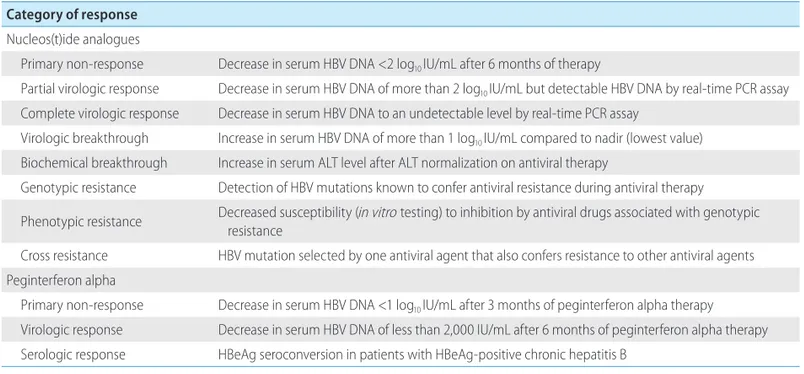
Monitoring during antiviral treatment
1. NAs
In a compliant patient with a primary non-response (decrease in serum HBV DNA of <2 log10 IU/mL after 6 months or more of NA treatment), changing to or adding a more-potent drug should be considered. Serum HBV DNA should be measured every 1 to 3 months for the first few months to ascertain the virologic re-sponse, and then every 3 to 6 months. Serum HBV DNA reduction to an undetectable level by real-time PCR (i.e., <10–15 IU/mL) should ideally be achieved to avoid resistance. Serum HBV DNA monitoring is thus critical to detect treatment failure.