P2-8 / D. S. JO
IMID 2009 DIGEST •
Abstract
The synthesis of nano-size (Y0.955Eu0.1)(P0.7V0.45)O4
red phosphors were conducted by using a Liquid Phase Precursor (LPP) method. In this method, cellulose pulp was used as a template showing the micro fibre structures to obtain the nano size YPV red phosphor. Aqueous solutions of raw materials were impregnated into cellulose pulp and subsequently impregnated pulp was dried and fired at 800-1200 ºC for 1h. The effect of luminescence properties on compositions and temperatures was evaluated with photoluminescence spectrum, X-ray diffraction and FE-SEM, and TEM. High efficiency (~110%) of phosphor of size of ~500nm fired at 1150℃ was obtained compared with the micro size of commer cial product. High efficiency behaviors of nano size phosphors were discussed in this paper.
1. Introduction
Current advances in the information display technology have lead to an increase of demands for the design of luminescent materials with better charact eristics [1].
Interest for high efficiency of nano size phosphors has been increased to use the three-primary color embodiment and energy-saving lights efficiently. In addition, nano or submicron size phosphors have attracted a great interest in the nano scientists and display producing companies.
Eu+3 doped (Y0.955Eu0.1)(P0.7V0.45)O4 phosphor is
one of the most important phosphors having wide
applications in plasma display panels (PDPs) and cold cathode fluorescence lamps (CCFLs).
Recently, various synthes is methods have been performed for t h e development of luminescent materials concerning to the grain size, morphology, agglomeration and defect free surface. Among them, the hydrothermal method has attracted a considerable attention since non agglomerated fine particles with narrow size distribution. However, still there are some drawbacks such as low efficiency, crystallinity and surface defects that must be solved to get better performance [2].
Cho et al. reported in their work the experimental and theoretical investigation of particle size dependent display panel efficiency. Many attempts have been made to obtain a high luminescent phosphor with a grain size of nanometer range [3]. Ac c or di ng t o Ha a s e et a l. , t he ma x i mu m luminescence efficiency does not exceed over 20% for 20-30 nm particle size. Therefore, new orientations in synthesis of nano size phosphor are expected to increase the luminescence efficiency with enhancing the crystallinity of phosphor particles with a new particle synthesis technology [4].
In this paper, we present a facile and new approach called as a Liquid Phase Precursor (LPP) method to synthesize t h e crystalline nano s i z e d (Y0.955Eu0.1)(P0.7V0.45)O4 phosphors.Cellulose pulp is
used as a template of micro fibre structure. Nano si ze phosphors were obtained at low temperature co mpared with conventional solid state reaction meth od. Furthermore, Eu3+ ions are well dispersed since
homogenously mixed hydro chloride and nitric
High Luminescence Properties of YPV nano size
phosphors by a Liquid Phase Precursor Method
D.S. Jo
1, A. Dulda, T
2. Masaki
1, and D. H. Yoon
1, 21School of Advanced Material Science and Engineering, Sungkyunkwan University, Suwan 440-746, Korea
2SKKU Advanced Institute of Nanotechnology (SAINT), Sungkyunkwan University, Suwan 440-746, Korea
Tel: +82 31 290 7361, E-mail: [email protected]
P2-8 / D. S. JO
• IMID 2009 DIGEST
solutions containing composition prepared Eu, Y, V, and P ions was impregnated into each cellulose matrix.
2. Experimental
Synthesis of (Y0.955Eu0.1)(P0.7V0.5)O4 phosphor was
conducted by a liquid phase precursor (LPP) method. In this method, cellulose pulp was used as a template. Raw materials of VO(SO3)·nH2O (99.9%), H3PO4
(99.9%),YCl3·6H2O(99.9%) and EuCl3·6H2O (99.9%)
were prepared with the concentration of 50, 50, 80, and 30% in de-ionized water, respectively. Aqueous solutions of each metal salts corresponding to the mol ratio of (Y0.955Eu0.1)(P0.7V0.5)O4 phosphor were
homogenously mixed by magnetic stirrer.
A mixed solution was impregnated into the cellulose pulp by weight percent of 1:1. The impregnated pulp was dried at 100℃ in a dry oven and then it was calcined at 800°C for 2 hours to remove the cellulose template.
The calcination conditions were as follows: inserting the specimens soon into a preheated furnace at 800 °C, maintaining at this temperature for 2h and taking out from furnace. After then, the calcined powder was fired from room temperature to 1000, 1100, 1150, and 1200 °C at a heating rate of 200 °C/h in air atmosphere.
The phase of fired YPV specimens and commerc ial product were det ermined by using X-ray diffractometer with Cu Ka (λ=1.54178 Å) radiation.
The morphology and crystallinity of the resulting particles were observed by field emission scanning electron microscopy (FESEM, XL-30, Philips) and high resolution transmission electron microscopy (HR-TEM, JEOL 300kV).
The emission and excitation spectra of f i r e d specimens and commercial product were measured at room temperature using a PL spectrophotometer in which xenon lamp (500 W) was used as a source (PLE/PL Drasa PRO 5300, Korea).
Emission at 620 nm was monitored over the range of 200–400 nm and by using 254 nm excitation wave length, emissions were detected over the range of 550-750 nm.
3. Results and discussion
Figure 1 shows the XRD pattern s of (Y0.955
Eu0.1)(P0.7V0.5)O4 s fired at 800, 1000, 1100, 1150 and
1200℃. Compared with the YPV commercial product,
our YPV specimens show the same pattern in tetragonal phase. 20 25 30 35 40 45 50 55 60 65 70 75 80 Commercial 1200o C 1150o C 1100o C 1000o C Intensity 2-Theta 800o C
Fig.1. XRD patterns of the (Y0.955, Eu0.1)(P0.7, V0.45)O4 specimen fired at 800, 1000, 1100, 1150 and 1200℃ and commercial YPV product.
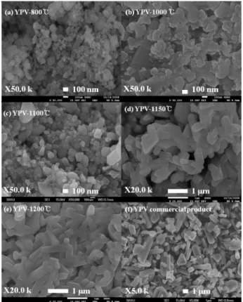
Fig.2. Field emission scanning electron micrograph of red (Y0.955Eu0.1)(P0.7V0.45)O4 phosphors fired at (a) 800℃, (b) 1000℃ (c) 1100℃, (d) 1150℃ and , (e) 1200℃ and (f) commercial YPV product.
Figure 2 shows the FE-SEM image s of our specimens and commercial product. The specimens
P2-8 / D. S. JO
IMID 2009 DIGEST •
are nano sized and cubic shaped. Their sizes are 50-80, 100-150 and 100-250nm fired at 800, 1000 and 1100℃, respectively. O n t h e ot h er ha n d, the specimens fired at 1150 and 1200℃ exhibit the mixed structures of cubic and rod grains. The diffraction peaks increased with the increase of temperatures due to the higher crystallinity of larger size particles.
Figure 3 shows the HRTEM images of YPV specimen fired at 1150℃. They exhibit the perfect crystalline structure without lattice defects (Figure 3a) and smooth surface (Figure 3b). Crystalinity is confirmed with selected area electron diffraction pattern (inset Figure 3b).
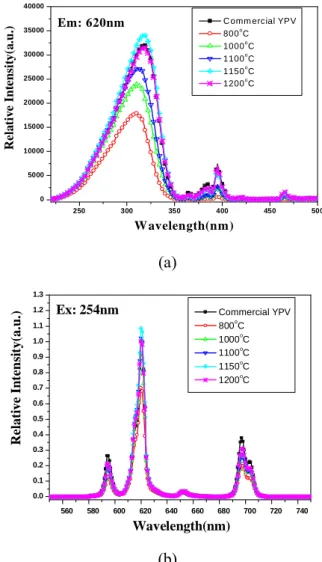
Figure 4a and b show the broad excitation (monitored at 621 nm) and emission (excited at 254nm) spectra of YPV phosphors fired at 800-1200℃ compared with commercial product. The emission efficiency of nano size YPV phosphor of ~ 500nm fired at 1150℃ is found as high as ~10%, compared with that of micro size commercial product. However, the emission efficiency of specimen fired at 1200℃ is decreased as shown in Figure 4b. Upon UV excitation, europium doped YPV phosphors exhibit strong red luminescence (Figure 4b) caused by transitions within the f-electron shell of the europium ions6). The emission spectra are consisted of the tree
of peaks. They show the magnetic dipole transition (595nm) of 5D
0–7F1 and the forced electric dipole
transitions (620, 697nm) of 5D 0–7F2,4.
Fig.3. (a) HR-TEM of the (Y0.955Eu0.1)(P0.7V0.45)O4 fired at 1150℃ for 1h and (b) surface of the specimen. 250 300 350 400 450 500 0 5000 10000 15000 20000 25000 30000 35000 40000
C omm ercial YPV
800o C 1000o C 1100o C 1150o C 1200o C R e la tiv e Int e nsit y(a .u.) W avelength(nm) Em: 620nm (a) 560 580 600 620 640 660 680 700 720 740 0.0 0.1 0.2 0.3 0.4 0.5 0.6 0.7 0.8 0.9 1.0 1.1 1.2 1.3 Commercial YPV 800oC 1000o C 1100oC 1150o C 1200o C R el a ti ve I n te n si ty( a. u. ) Wavelength(nm) Ex: 254nm (b)
Fig. 4 (a) Photoluminescence excitation (PLE) spectrum monitored at 621 nm of (Y0.955Eu0.1) (P0.7V0.45)O4 red phosphor and (b) Normalized Emission spectrum under 254nm excitation fired at
800, 1000, 1100, 1150℃ and 1200℃ and
Commercial YPV product.
4. Summary
The red nano (Y0.955, Eu0.1)(P0.7, V0.5)O4 phosphors
were synthesized at 800-1200℃ by using a liquid phase precursor (LPP) method. The emis s ion efficiency of nano size phosphor under excitation at 254nm was obtained as high as ~10% compared with that of commercial micro size YPV product. The perfect crystalline structures without lattice defects were observed by HRTEM. The emission spectra of Eu3+ energy transfer for red phosphor were observed
at 595nm (5D
P2-8 / D. S. JO
• IMID 2009 DIGEST
5. Acknowledgement
The authors are grateful to Daejoo electronic materials for helping us with phosphor synthesis.
6. References
(1 line spacing)
1. S. M. Hong and K. Y. Kim, JJAP, Vol. 44, No. 3, pp. 1356–1360 (2005).
2. T. Justel, H. Nikol and C. Ronda, Angew. Chem., Int. Ed7., vol. 37, p.3084 (1998).
3. C.W. Cho and D.S. Zang, Appl. Phys. Lett. Vol. 93, No. 3, 031505 (2008)
4. A. Huignard, T. Gacoin and J.-P. Boilot, J. Phys.
Chem. B, Vol. 107, p.6754 (2003).
5. K. D. Hong, IMID’06 Technical Digest, p.877 (2006).