7
X-ray Structure Analysis Online 2009, VOL. 25 7
2009 © The Japan Society for Analytical Chemistry
A number of polypodal ligands derived from 1,n-diaminoalcohols and acyclic amines, such as pyridyl, phenolic, or immidazolyl moieties, have been used in modeling studies related to the active sites of metalloenzymes including dioxygen binding.1 Such examples of macrocyclic polyamine derivatives
are relatively rare. Recently, an endogenously bridged Cu(II) complex incorporating a deprotonated form of the dinucleting ligand, 1,3-di(1,4,7-triazonan-1-yl)propan-2-ol (T2PrOH), has
been reported.2 The two Cu centers were in slightly different
distorted square pyramidal Cu(II) geometries and 3.582(1)Å apart. In order to investigate the effect of changes in the ligand structure, we prepared a new dinucleating ligand, 1,4-di(1,4,7-triazonan-1-yl)butan-2-ol (L-OH). The bridging unit in L-OH is 2-hydroxybutane, while it was 2-hydroxypropane in T2PrOH.
The asymmetrical ligand, L-OH was synthesized according to modified literature procedures.3,4 The Cu
2(L-O)(H2O)2(ClO4)3
complex was prepared by the reaction of L-OH (0.3 mmol, 100 mg), Cu(ClO4)2·6H2O (0.64 mmol, 237 mg) and triethylamine
(0.3 mmol, 42.4 ml) in absolute ethanol (10 ml). The solution turned to dark blue with precipitation, and was stirred for overnight. The reaction mixture was evaporated to dryness and washed with cold ethanol. The solid was dissolved in methanol (60˚C), and was slowly evaporated to give blue plate crystals for X-ray structure analysis. The crystal and structure-refinement data are summarized in Table 1. H atoms, except for water hydrogen atoms, were placed at the calculated positions, with
C–H distances of 0.97 Å and N–H distances of 0.91 Å, and treated as riding atoms, with Uiso(H) = 1.2 Ueq(C). The selected
bond distances and angles are listed in Table 2. A chemical diagram is shown in Fig. 1, and the structure is shown in Fig. 2.
The title compound was obtained as a rod crystal with the monoclinic space group P21. A symmetric unit contains a
Cu2(L–O–) cation, two water molecules, and three ClO4– anion.
X-ray Structure Analysis Online
Crystal Structure of Dinuclear Cu(II) Complex of 1,4-Di(1,4,7-triazonan-1-
yl)butan-2-ol
Youngmee K
IM* and Junghee K
IM**
*Department of Chemistry and Nano Science, Ewha Womans University, Seoul 120-750, Korea
**Department of Chemistry, Sunmoon University, Asan si, Chung Nam 336-840, Korea
A new dinucleating ligand, 1,4-di(1,4,7-triazonan-1-yl)butan-2-ol (L-OH), was synthesized, and a dinuclear Cu(II) complex was prepared and characterized by X-ray crystallography. The compound crystallized in a monoclinic system, and was characterized thus: P21, a = 9.808(9), b = 14.308(13), c = 10.951(10)Å, b = 103.075(15)˚, Z = 2, V = 1497(2)Å3.
The crystal structure was solved by direct methods and refined by full-matrix least-squares on F2 to final values of R1 =
0.0823 and wR2 = 0.2051.
(Received September 12, 2008; Accepted December 26, 2008; Published on web February 10, 2009)
† To whom correspondence should be addressed.
E-mail: [email protected]
Fig. 1 Structural chemical diagram.
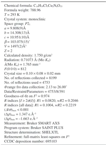
Table 1 Crystal data and experimental data Chemical formula: C16H39Cl3Cu2N6O15
Formula weight: 788.96
T = 293 K
Crystal system: monoclinic Space group: P21 a = 9.808(9)Å b = 14.308(13)Å c = 10.951(10)Å b = 103.075(15)˚ V = 1497(2)Å3 Z = 2 Calculated density: 1.750 g/cm3 Radiation: 0.71073 Å (Mo Ka) l(Mo Ka) = 1.765 mm–1 F(0 0 0) = 812 Crystal size = 0.10 ¥ 0.08 ¥ 0.02 mm No. of reflections collected = 8199 No. of reflections used = 4753
q range fro data collection: 2.13 to 26.00˚
Data/Restraint/Parameters = 4753/8/391 Goodness-of-fit on F2 = 0.974
R indices [I > 2s(I)]: R1 = 0.0820, wR2 = 0.2046 R indices [all data]: R1 = 0.1004, wR2 = 0.2219 (D/s)max < 0.001
(Dr)max = 1.347 e.Å–3
(Dr)min = –1.663 e.Å–3
Measurement: Bruker SMART AXS Program system: Bruker SAINT PLUS Structure determination: SHELXTL Refinement: full-matrix least-squares on F2
8 X-ray Structure Analysis Online 2009, VOL. 25
Figure 2 shows a [{Cu(H2O)2}(L–O–))]3+ cation containing
coordinated water molecules on each Cu ion. The asymmetric ligand L–O– contains two [9]aneN3 units linked by a
2-hydroxylbutyl group. Three nitrogen atoms of each [9]aneN3
moiety are coordinated to each Cu ion, and an oxygen atom of the 2-hydroxylbutyl linker bridges two Cu ions. Each water molecule coordinates each Cu ion, and one of the oxygen atoms bridges two Cu ions asymmetrically with a Cu1–O3 distance of 2.88(1)Å. The coordination geometry of the Cu1 ion is a distorted octahedron with a tertiary amine (N13) of the L–O– and
a water oxygen atom (O3) in trans to each other, and that of the Cu2 ion is a trigonal pyramid (tbp) with a tertiary amine of the L–O– and a water occupying the axial positions. The Cu1–O1(L–
O–) and Cu2–O1(L–O–) bond distances are 1.948(7) and
1.950(7)Å, respectively. The Cu1–O2(water) and Cu2– O3(water) bond distances are 2.032(12) and 2.001(7)Å,
respectively. The Cu1–O1–Cu2 angle is 120.0(3)˚. Two Cu ions are 3.38(3)Å apart.
Acknowledgements
This work was supported by the Korea Science and Engineering Foundation (RO4-2003-000-10097-0).
References
1. K. Selmeczi, M. Reglier, M. Giorgi, and G. Speier, Coord. Chem. Rev., 2003, 245, 191.
2. F. H. Fry, B. Moubaraki, K. S. Murray, L. Spiccia, M. Warren, B. W. Skelton, and A. H. White, Dalton Trans., 2003, 866.
3. S. B. Mhaske and N. P. Argade, J. Org. Chem., 2001, 66, 9038.
4. M. A. Calter and R. K. Orr, Tetrahedron Lett., 2003, 5699.
Fig. 2 Structure of [{Cu(H2O)2}(L–O))]3+ with the labeling scheme
at 30% probability. Hydrogen atoms, except for water hydrogen atoms, were omitted, and all counter anions (ClO4–) are not shown for
clarity. Table 2 Selected bond distances [Å] and angles [˚]
![Fig. 2 Structure of [{Cu(H 2 O) 2 }(L–O))] 3+ with the labeling scheme](https://thumb-ap.123doks.com/thumbv2/123dokinfo/5080761.74378/2.892.81.435.145.416/fig-structure-cu-h-o-l-labeling-scheme.webp)
