RESEARCH ARTICLE
Impact of idiopathic pulmonary fibrosis on
recurrence after surgical treatment for stage
I–III non-small cell lung cancer
Myung Jin Song1, Dae Jun Kim2, Hyo Chae PaikID2, Sukki Cho3, Kwhanmien Kim3, Sanghoon Jheon3, Sang Hoon Lee4☯*, Jong Sun Park1☯*
1 Division of Pulmonary and Critical Care Medicine, Department of Internal Medicine, Seoul National
University College of Medicine, Seoul National University Bundang Hospital, Seongnam, South Korea,
2 Department of Thoracic and Cardiovascular Surgery, Yonsei University College of Medicine, Seoul,
Republic of Korea, 3 Department of Thoracic and Cardiovascular Surgery, Seoul National University College of Medicine, Seoul National University Bundang Hospital, Seongnam, South Korea, 4 Division of Pulmonology, Department of Internal Medicine, Institute of Chest Diseases, Severance Hospital, Yonsei University College of Medicine, Seoul, Republic of Korea
☯These authors contributed equally to this work.
*[email protected](JSP);[email protected](SHL)
Abstract
Background
Idiopathic pulmonary fibrosis (IPF) is an independent risk factor for lung cancer (LC) devel-opment; however, its effect on recurrence after curative surgery remains unclear.
Objectives
This study aimed to determine the impact of IPF on recurrence-free survival following cura-tive surgical resection of stage I–III non-small cell lung cancer (NSCLC) and investigate the effects of patient and surgical factors on the risk of recurrence.
Methods
We reviewed retrospectively collected data of patients with surgically resected stage I–III NSCLC from two tertiary care hospitals in South Korea. By propensity score matching, patients with IPF (LC with IPF) were matched to those without IPF (LC without IPF).
Results
In total, 3416 patients underwent surgical resection, and 96 were diagnosed with underlying IPF. In the LC with IPF group, 89.6% patients were men, and the average age was 69.7 years. Sublobar resection was performed more frequently in the LC with IPF group than in the LC without IPF group, while the rate of mediastinal lymph node dissection and dis-sected node number were lower in the former group. The 5-year recurrence-free survival rate was significantly lower in the LC with IPF group (49.2%) than in the LC without IPF group (69.1%; P<0.001). Multivariable Cox regression analysis revealed that IPF and post-operative stage III were independent risk factors for recurrence.
a1111111111 a1111111111 a1111111111 a1111111111 a1111111111 OPEN ACCESS
Citation: Song MJ, Kim DJ, Paik HC, Cho S, Kim K,
Jheon S, et al. (2020) Impact of idiopathic pulmonary fibrosis on recurrence after surgical treatment for stage I–III non-small cell lung cancer. PLoS ONE 15(6): e0235126.https://doi.org/ 10.1371/journal.pone.0235126
Editor: Hyun-Sung Lee, Baylor College of Medicine,
UNITED STATES
Received: March 12, 2020 Accepted: June 9, 2020 Published: June 29, 2020
Copyright:© 2020 Song et al. This is an open access article distributed under the terms of the Creative Commons Attribution License, which permits unrestricted use, distribution, and reproduction in any medium, provided the original author and source are credited.
Data Availability Statement: All relevant data are
within the paper and its Supporting Information files.
Funding: The authors received no specific funding
for this work.
Competing interests: The authors have declared
that no competing interests exist.
Abbreviations: AE-IPF, acute exacerbation of IPF;
ARDS, acute respiratory distress syndrome; CI, confidence interval; DLCO, diffusing capacity of the
Conclusions
IPF may increase the risk of recurrence after curative surgical treatment for NSCLC. Close surveillance for recurrence is mandatory for patients with underlying IPF.
Introduction
Idiopathic pulmonary fibrosis (IPF) is a devastating lung disease characterized by progressive lung scarring and a histopathological pattern of usual interstitial pneumonia (UIP). The median survival period of IPF ranges from 2.5 to 3.5 years [1,2]. Patients with IPF exhibit a high prevalence of various comorbidities, including chronic obstructive lung disease, pulmo-nary hypertension, coropulmo-nary artery disease, and lung cancer (LC) [3].
The reported prevalence of LC in patients with IPF is 2.7%–45.7% [4–8]. Moreover, the rel-ative risk of LC development is approximately eight times higher in patients with IPF than in the general population [9]. Although adenocarcinoma is the most common histopathological type of LC in the general population, patients with IPF are most commonly affected by squa-mous cell carcinoma, followed by adenocarcinoma [8,10,11].
Patients with stage I and stage II non-small cell lung cancer (NSCLC), as well as those with certain stage III NSCLCs, are considered eligible for curative resection. Complete resection, when possible, remains the mainstay of therapy and is associated with the highest probability of long-term survival. However, if patients with LC who were eligible for surgical resection have underlying IPF, several aspects must be considered. Previous studies have shown that the postoperative morbidity and mortality rates for patients with NSCLC are significantly higher if IPF is present, with the main cause of postoperative mortality being acute exacerbation of IPF (AE-IPF) [12–16]. A few studies have reported the long-term survival of patients with NSCLC and IPF, although overall survival was significantly poorer for these patients than for those without IPF [14,17–20]. However, the effect of IPF on recurrence after curative surgical treat-ment for NSCLC remains unclear. Therefore, the present study aimed to determine the impact of IPF on recurrence-free survival and overall survival following curative surgical resection of stage I–III NSCLC, and investigate the effects of patient and surgical factors on the recurrence and mortality.
Methods
Patients
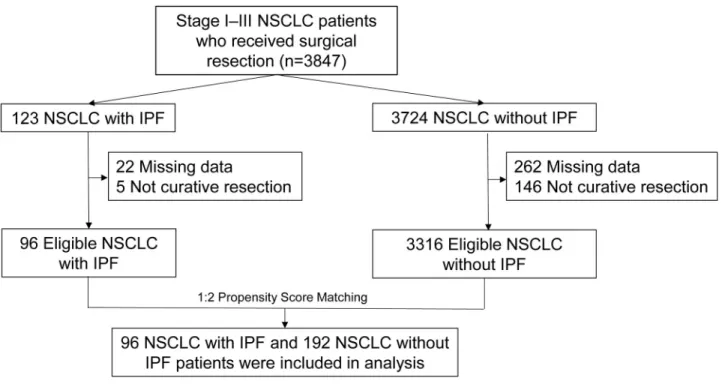
We retrospectively reviewed the medical records of patients with stage I–III NSCLC who underwent curative resection at Seoul National University Bundang Hospital and Severance Hospital between January 2003 and September 2016. Among these patients, we identified those who were diagnosed with underlying IPF. By propensity score matching of confounding variables, including age, sex, the histopathological subtype of NSCLC, the postoperative stage of NSCLC, and the year of NSCLC diagnosis, patients with IPF (LC with IPF group) were matched to those without IPF (LC without IPF group) in a 1:2 ratio. Flow chart of patient recruitment is shown inFig 1. This study was conducted in accordance with the amended
Declaration of Helsinki. Local institutional review boards and independent ethics committees approved the study protocol (Seoul National University Bundang Hospital, IRB number: B-1707/411-402; Severance Hospital, IRB number: 4-2019-0292).
lungs for carbon monoxide; FVC, forced vital capacity; HR, hazard ratio; IPF, idiopathic pulmonary fibrosis; LC, lung cancer; NSCLC, non-small cell lung cancer.
Ethics approval and consent to participate
This study was approved by the Institutional Review Board and Ethics Committee of Seoul National University Bundang Hospital and Severance Hospital (Seoul National University Bundang Hospital, IRB number: B-1707/411-402; Severance Hospital, IRB number: 4-2019-0292). Written informed consent was waived as the nature of retrospective study by IRB. The patient’s medical records between January 2003 and September 2016 were accessed. All data were fully anonymized before accessement.
Definitions
In accordance with diagnostic criteria defined by the International Consensus Statement of the American Thoracic Society and European Respiratory Society in 2018 [1], IPF was diag-nosed via a multidisciplinary approach involving pulmonologists as well as radiologists and pathologists specializing in chest diseases.
The LC stage was defined according to the 8th edition of the American Joint Committee on Cancer staging system. Recurrence-free survival was defined as the time from surgery to the first diagnosis of disease recurrence or the last follow-up visit. Death was considered a censoring event for recurrence. Overall survival was defined as the time from initial LC treatment to death or the last follow-up visit. Local recurrence was defined as evidence of tumor cells within the same lobe of the lung or ipsilateral pulmonary hilum, and regional recurrence was defined as evidence of tumor cells within another lobe of the ipsilateral lung or the ipsilateral or subcarinal lymph nodes (LNs), while distant metastasis was defined as evidence of tumor cells in the con-tralateral lung, concon-tralateral mediastinal LNs, the pleural space, or areas outside the hemithorax.
AE-IPF was defined as acute, clinically significant respiratory deterioration characterized by evidence of new widespread alveolar abnormality, in accordance with the diagnostic criteria proposed by Collard et al. in 2016 [21].
Fig 1. Patient recruitment flow chart. https://doi.org/10.1371/journal.pone.0235126.g001
The gender–age–physiology (GAP) index was applied for the assessment of the severity and prognosis of IPF. The index was calculated as follows: gender (0–1 point), age (0–2 points), forced vital capacity (FVC; 0–2 points), and diffusing capacity of the lungs for carbon monox-ide (DLCO; 0–3 points). Based on the score, IPF was categorized into three stages: I (0–3
points), II (4–5 points), and III (6–8 points). The predicted 1-year mortality rates for GAP stages I, II, and III were 6%, 16%, and 39%, respectively [22].
Statistical analysis
Propensity scores were estimated through multiple logistic regression analysis with sex, age, the postoperative cancer stage, the histopathological subtype of cancer, and the year of cancer diagnosis as independent variables.
Using the matched set, we examined the effect of IPF on recurrence after curative surgical resection of stage I–III NSCLC. Generalized estimating equations were used to compare con-tinuous and categorical variables of propensity score matched population. Cumulative time-to-event distributions (survival, recurrence) were estimated using the stratified Kaplan–Meier method. Independent predictors for recurrence and overall survival were determined by strati-fied Cox proportional hazards models. A P-value <0.05 was considered statistically significant. All statistical analyses were performed using SAS software (version 9.4; SAS Inc., Cary, NC, USA).
Results
Patient characteristics and surgical methods
In total, 3416 patients with NSCLC underwent surgical resection during the study period, and 96 were diagnosed with underlying IPF. Baseline characteristics of the overall cohort before propensity score matching are shown inTable 1. The LC with IPF group and LC without IPF group showed significant differences in potential confounding factors for recurrence, includ-ing age, sex, the histopathological subtype, and the postoperative stage. The average age (69.7 years vs 63.4 years; P<0.001) and proportion of men (89.6% vs 61.8%; P<0.001) were higher Table 1. Baseline characteristics of patients with surgically resected non-small cell lung cancer.
LC with IPF (n = 96) LC without IPF (n = 3316) P-value
Sex (male) 86 (89.6%) 2049 (61.8%) <0.001
Age (year) 69.7± 7.4 63.4± 10.0 <0.001
Histology <0.001
Adenocarcinoma 44 (45.8%) 2262 (68.2%)
Squamous cell carcinoma 48 (50.0%) 922 (27.8%)
Adenosquamous 2 (2.1%) 42 (1.3%) Large cell 0 (0.0%) 64 (1.9%) Sarcomatoid 2 (2.1%) 26 (0.8%) Postoperative Stage <0.001 0 – 74 (2.2%) I 43 (44.8%) 2123 (64.0%) II 35 (36.5%) 608 (18.3%) III 18 (18.8%) 511 (15.4%)
Values are expressed as the mean± standard deviation or number (%). IPF, idiopathic pulmonary fibrosis; LC, lung cancer
in the LC with IPF group than in the LC without IPF group. The most frequent histopatholog-ical subtype was squamous cell carcinoma in the LC with IPF group and adenocarcinoma in the LC without IPF group. Patients with IPF showed more advanced postoperative stages (stages II and III) than did those without IPF.
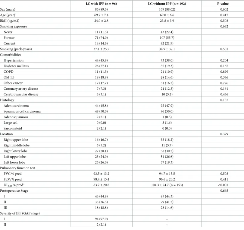
Baseline characteristics of the matched groups are shown inTable 2. The diffusing capacity of the lungs for carbon monoxide (DLCO) was significantly lower in the LC with IPF group
than in the LC without IPF group (83.7±20.8 vs. 104.3±24.7; P<0.001). Other variables, includ-ing the location of the primary tumor, forced vital capacity (FVC), and forced expiratory vol-ume in one second, were comparable between groups. In the LC with IPF group, stages I and II as per the GAP index were documented for 94 (97.9%) and two (2.1%) patients, respectively. There was no patient with GAP stage III.
The treatment-related characteristics of the matched groups are shown inTable 3. There was no significant difference in the proportion of patients receiving induction therapy and adjuvant therapy between the two groups. Sublobar resection, either wedge resection or seg-mentectomy, was more frequently performed in the LC with IPF group than in the LC without IPF group, while the rate of mediastinal lymph node dissection was lower in the LC with IPF group (82.3% vs. 99.0%; P<0.001). The mean number of dissected LNs was also smaller in the LC with IPF group than in the LC without IPF group (16.0 vs. 23.2; P<0.001). The two groups showed no significant difference in the resection margin status (R0, complete resection; R1, microscopic residual tumor).
Treatment-related complications
To assess the incidence of treatment-related complications, we defined the duration between the last treatment and the onset of complications as �4 weeks (Table 4). The incidence of sur-gery-related AE-IPF or acute respiratory distress syndrome (ARDS) was significantly higher in the LC with IPF group than in the LC without IPF group. Adjuvant treatment-related compli-cation rates were comparable between groups. Surgery and adjuvant treatment resulted in AE-IPF in 5.2% and 6.7% patients, respectively.
Recurrence and mortality
During a median follow-up period of 49.2 months, 92 recurrences were documented for the entire matched study population. Recurrences were significantly more frequent in the LC with IPF group than in the LC without IPF group (44.8% vs. 25.5%; P = 0.002). Among the patients with recurrence, the type of recurrence (local, regional recurrence or distant metastasis) did not show significant difference between groups. The 5-year recurrence-free survival rate (49.2%; 95% CI, 39.4–61.4 vs. 69.1%; 95% CI, 61.9–77.0; P<0.001;Fig 2,Table 5) and 5-year overall survival rate (45.9%; 95% CI, 36.5–57.7 vs. 64.8%; 95% CI, 57.9–72.6; P<0.001;Fig 2,
Table 5) were significantly lower in the LC with IPF group than in the LC without IPF group. Cancer-specific survival was also significantly lower in the LC with IPF group than in the LC without IPF group (P = 0.008,S1A Fig,Table 5).
Predictors of recurrence
Univariable Cox proportional hazards regression analysis revealed that IPF, the postoperative stage, surgical extent, and rate of mediastinal lymph node dissection were major predictors of recurrence after surgical treatment. These variables and additional potential confounding vari-able were combined in multivarivari-able regression analysis, which revealed that IPF (HR, 2.68; 95% CI, 1.53–5.76; P = 0.001) and postoperative stage III (vs. stage I; HR, 5.46; 95% CI, 1.72– 17.37; P = 0.004) were independent predictors of recurrence (Table 6).
Table 2. Baseline characteristics of patients with surgically resected non-small cell lung cancer with or without idiopathic pulmonary fibrosis (propensity score-matched population).
LC with IPF (n = 96) LC without IPF (n = 192) P-value
Sex (male) 86 (89.6) 169 (88.02) 0.602 Age (year) 69.7± 7.4 69.0± 6.6 0.417 BMI (kg/m2) 24.0± 2.8 23.8± 3.9 0.503 Smoking exposure 0.642 Never 11 (11.5) 43 (22.4) Former 71 (74.0) 107 (55.7) Current 14 (14.6) 42 (21.9) Smoking (pack-years) 37.1± 25.7 34.9± 32.1 0.501 Comorbidities Hypertension 44 (45.8) 73 (38.0) 0.204 Diabetes mellitus 26 (27.1) 37 (19.3) 0.167 COPD 11 (11.5) 21 (10.9) 0.899 Old TB 18 (18.8) 28 (14.6) 0.346 Other cancer 17 (17.7) 31 (16.2) 0.726
Coronary artery disease 7 (7.3) 24 (12.5) 0.161
Cerebrovascular disease 3 (3.1) 10 (5.2) 0.436
Histology 0.157
Adenocarcinoma 44 (45.8) 92 (47.9)
Squamous cell carcinoma 48 (50.0) 96 (50.0)
Adenosquamous 2 (2.1) 1 (0.5)
Large cell 0 (0.0) 3 (1.6)
Sarcomatoid 2 (2.1) 0 (0.0)
Location 0.379
Right upper lobe 16 (16.7) 35 (18.2)
Right middle lobe 5 (5.2) 11 (5.7)
Right lower lobe 27 (28.1) 58 (30.2)
Left upper lobe 23 (24.0) 51 (26.6)
Left lower lobe 25 (26.0) 37 (19.3)
Pulmonary function test
FVC % pred 93.5± 13.2 94.7± 15.5 0.503 FEV1% pred 98.4± 15.4 96.6± 20.2 0.411 DLCO% pred� 83.7± 20.8 104.3± 24.7 (n = 153) <0.001 Postoperative Stage 0.665 I 43 (44.8) 85 (44.3) II 35 (36.5) 79 (41.2) III 18 (18.8) 28 (14.6)
Severity of IPF (GAP stage)
I 94 (97.9) –
II 2 (2.1) –
Values are expressed as the mean± standard deviation or number (%).
BMI, body mass index; COPD, chronic obstructive pulmonary disease; DLCO, diffusing capacity of the lungs for carbon monoxide; ECOG, Eastern Cooperative
Oncology Group; FEV1, forced expiratory volume in one second; FVC, forced vital capacity; GAP, gender, age, and physiology; IPF, idiopathic pulmonary fibrosis; LC,
lung cancer
�39 patients with missing data in LC without IPF group
Predictors of overall survival
Univariable Cox proportional hazards regression analysis revealed that IPF, Eastern Coopera-tive Oncology Group score, FVC, postoperaCoopera-tive stage, surgical extent, and surgery-related AE-IPF or ARDS were major covariates for overall survival. These variables and additional potential confounding variable were combined in multivariable analysis, which revealed that IPF (HR, 1.98; 95% CI, 1.12–3.47; P = 0.018), postoperative stage III NSCLC (vs. stage I; HR, 3.39; 95% CI, 1.28–8.97; P = 0.014), and surgery-related AE-IPF or ARDS (HR, 14.02; 95% CI, 1.55–126.89; P = 0.019) were independent predictors of overall survival (Table 6).
Discussion
In the present study, we demonstrated the impact of IPF on recurrence and survival following curative surgical resection of stage I–III NSCLC in a propensity score-matched study popula-tion. Both recurrence-free survival and overall survival after curative surgical resection were significantly compromised in patients with IPF. IPF and postoperative stage III were predic-tors of both recurrence and overall survival. Surgery-related AE-IPF or ARDS was also identi-fied as an independent risk factor for mortality.
Epidemiological studies have revealed that IPF is an independent risk factor for LC devel-opment [4,6,11,23], although the pathogenesis connecting the two diseases remains poorly understood. Common pathways for the development of LC in IPF, yet with distinct roles in the pathogenesis of both conditions, include genetic and epigenetic alterations; abnormal Table 3. Treatment-related variables for patients with surgically resected non-small cell lung cancer with or without idiopathic pulmonary fibrosis (propensity score-matched population).
LC with IPF (n = 96) LC without IPF (n = 192) P-value
Induction treatment 0.273 Chemotherapy 1 (1.0) 5 (2.6) Concurrent chemoradiotherapy 0 (0.0) 1 (0.5) Surgery Surgical extent <0.001 Wedge resection 16 (16.7) 3 (1.6) Segmentectomy 8 (8.3) 8 (4.2) Lobectomy 70 (72.9) 150 (78.1) Bilobectomy 1 (1.0) 18 (9.4) Pneumonectomy 1 (1.0) 13 (6.8) MLND 79 (82.3) 190 (99.0) <0.001 Dissected LN 16.0± 13.4 23.2± 12.8 <0.001 Number of positive LN 1.0± 2.3 1.1± 2.4 0.629 Resection margin 0.481 R0 92 (95.8) 180 (93.8) R1 4 (4.2) 12 (6.3) Adjuvant treatment 0.187 Chemotherapy 24 (25.0) 64 (33.3) Radiotherapy 3 (3.1) 4 (2.1) Concurrent chemoradiotherapy 0 (0.0) 7 (3.7)
Sequential radiotherapy after chemotherapy 3 (3.1) 5 (2.6)
Values are expressed as mean± SD or number (%).
IPF, idiopathic pulmonary fibrosis; MLND, mediastinal lymph node resection; LC, lung cancer; LN, lymph node https://doi.org/10.1371/journal.pone.0235126.t003
expression of microRNAs; cellular and molecular aberrances; and activation of specific signal-ing transduction pathways [24–26]. Several pieces of evidence related to therapeutic agents also suggest a pathogenic link between IPF and LC. Nintedanib, which was first approved for use in combination with docetaxel for the second-line treatment of advanced NSCLC [27], has also been approved as an antifibrotic agent for the treatment of IPF. A previous retrospective study reported that the incidence of LC was lower in patients with IPF receiving pirfenidone, another antifibrotic agent approved for IPF treatment, than in those without pirfenidone treat-ment [28], while a preclinical experiment showed that the combination of pirfenidone and cis-platin led to increased apoptosis and synergistic death of both cancer-associated fibroblasts and NSCLC cells [29].
Patients with LC and underlying IPF are commonly encountered in clinical practice; how-ever, for these patients, it is difficult to predict the efficacy of treatment for LC due to the increased incidence of treatment-related complications and poor prognosis of IPF itself [12–
14,30]. Reports on the long-term prognosis of patients with NSCLC accompanied by IPF who underwent curative surgical resection have consistently documented lower 5-year survival and recurrence-free survival for patients with IPF than for those without [14,17–20]. In a study performed by the Japanese Association for Chest Surgery, which analyzed predictors of overall survival after the surgical treatment of LC in patients with interstitial lung disease [31], it was found that wedge resection, a predicted FVC of <80%, and tumors located in the inferior lobe were poor prognostic factors for overall survival. Furthermore, subgroup analysis of patients with stage I cancer showed that the survival curves for those treated with wedge resection and those treated with lobectomy crossed at approximately 1 year after surgery, with 5-year sur-vival rates of 29.2% and 68.6%, respectively. From this result, the authors inferred that wedge Table 4. Treatment-related complications in patients with surgically resected non-small cell lung cancer with or without idiopathic pulmonary fibrosis.
(A) Surgery-related complications
LC with IPF (n = 96) LC without IPF (n = 192) P-value
Pneumonia 11 (11.5) 25 (13.0) 0.706 Pneumothorax 20 (20.8) 24 (12.0) 0.509 BPF 1 (1.0) 2 (1.0) 1.000 Prolonged MV 2 (2.1) 10 (5.2) 0.224 Arrythmia 1 (1.0) 2 (1.0) 1.000 Bleeding 2 (2.1) 3 (1.6) 0.751 ARDS� 2 (2.1) 2 (1.0) 0.255 AE–IPF 5 (5.2) – ARDS or AE–IPF 7 (7.3) 2 (1.0) 0.012
(B) Adjuvant treatment-related complication (n = 110)
Postoperative hospital day 10.2± 10.0 9.8± 11.9 0.780
LC with IPF (n = 30) LC without IPF (n = 80) P-value
Cytopenia 2 (6.7) 17 (21.2) 0.091
GI trouble 8 (26.7) 28 (35.0) 0.409
Pneumonia 6 (20.0) 6 (7.5) 0.071
Pneumonitis 1 (3.4) 1 (1.2) 0.469
AE–IPF 2 (6.7) 0 (0.0) 0.126
Values are expressed as number (%).
�ARDS other than AE-IPF
AE-IPF, acute exacerbation of idiopathic pulmonary fibrosis; ARDS, acute respiratory distress syndrome; BPF, bronchopleural fistula; GI, gastrointestinal; LC, lung cancer; MV, mechanical ventilation
Fig 2. Probability of recurrence-free survival and overall survival for patients with surgically resected non-small cell lung cancer with or without IPF. (A) Recurrence-free survival was significantly lower in the LC with IPF group
than in the LC without IPF (P<0.001). (B) Overall survival was significantly lower in the LC with IPF group than in the LC without IPF (P<0.001).
resection is associated with a lower incidence of surgery-related complications and better short-term outcomes than is lobectomy, although the long-term outcomes are worse with wedge resection, probably due to a higher recurrence rate. In the present study, however, mul-tivariable analysis for recurrence-free survival showed that IPF and postoperative stage III were independent risk factors for recurrence after adjustment for other variables, including the surgical extent. In order to confirm that inferior recurrence-free survival and overall survival in the LC with IPF group were not due to the difference in surgical extent between groups, subgroup analysis was performed for patients who underwent lobectomy in the pro-pensity score matched population. Subgroup analysis showed that recurrence-free survival, overall survival and cancer-specific survival were significantly inferior in the LC with IPF group than in the LC without IPF group (S1andS2Figs).
Unlike previous studies on patients with NSCLC and IPF, we assessed detailed surgery-related variables, including whether or not mediastinal lymph node dissection was performed, the number of dissected LNs and resection margin status. However, none were found to be sig-nificant predictors of recurrence-free survival. Therefore, IPF itself appears to increase the risk of lung carcinogenesis and recurrence after curative resection. This may be explained by the aforementioned pathways for carcinogenesis in IPF, along with the possibility that tumors that developed within fibrotic lesions could be obscured by honeycombing or reticular changes in fibrotic lesions and consequently overlooked. Moreover, the probability of undiscovered posi-tive LNs is higher for patients with IPF; this can be explained by the lower number of dissected LNs in our IPF group, which could lead to stage migration and remnant malignancy [32].
Previous studies showed that morbidity and mortality rates after the surgical treatment of NSCLC were significantly higher in patients with IPF than in those without IPF, and inferred Table 5. Recurrence and overall survival in patients with surgically resected non-small cell lung cancer with or without idiopathic pulmonary fibrosis.
LC with IPF (n = 96) LC without IPF (n = 192) P-value
Recurrence 43 (44.8) 49 (25.5) 0.002
Type of recurrence 0.062
Local recurrence 11 (25.6%) 4 (8.2%)
Regional recurrence 7 (16.3%) 13 (26.5%)
Distant metastasis 25 (58.1%) 32 (65.3)
5-year recurrence-free survival rate (%), 95% CI 49.2 (39.4–61.4) 69.1 (61.9–77.0) <0.001 Recurrence-free survival (months), 95% CI 48.8 (23.0–NA) NA
Death 61 (63.5%) 67 (34.9%) <0.001
Cause of death
Lung cancer progression 30 (31.2%) 36 (18.8%)
Surgery-related complication� 5 (5.2%) 9 (4.7%)
IPF progression or acute exacerbation 10 (10.4%) NA
Pneumonia 9 (9.4%) 8 (4.2%)
Other�� 7 (2.4%) 14 (7.3%)
5-year survival rate (%), 95% CI 45.9 (36.5–57.7) 64.8 (57.9–72.6) <0.001 Overall survival (months), 95% CI 49.5 (35.9–68.0) 172.0 (NA–NA)
Cancer specific survival (months), 95% CI 89.3 (68.0–NA) 172.0 (NA–NA) 0.008 Values are expressed as number (%).
IPF, idiopathic pulmonary fibrosis; LC, lung cancer
�The duration between surgery and the onset of complications was �4 weeks ��1 heart failure, 1 stomach perforation, 3 sepsis, 3 other cancer, 13 unknown
Table 6. Stratified Cox regression analyses for recurrence and mortality after surgical treatment of non-small cell lung cancer in patients with or without idiopathic pulmonary fibrosis (propensity score-matched population).
(A) Stratified Cox regression analyses for recurrence
Univariable Multivariable
HR 95%CI p-value HR 95%CI p-value
IPF 2.86 1.72–4.76 <0.001 2.68 1.53–5.76 0.001 Sex 2.34 0.83–6.59 0.108 Age 0.97 0.92–1.02 0.236 Smoking (pack-years) 0.99 0.98–1.00 0.173 ECOG �2 5.16 0.52–50.41 0.158 FVC % pred 1.00 0.98–1.01 0.640 FEV1% pred 1.00 0.99–1.02 0.527 Postoperative Stage I 1.00 II 2.00 0.91–4.39 0.084 2.39 0.78–7.30 0.339 III 5.19 1.98–13.64 <0.001 5.46 1.72–17.37 0.004 Surgery extent Wedge resection 1.00 Segmentectomy 0.30 0.06–1.59 0.158 0.85 0.11–6.85 0.882 Lobectomy 0.26 0.07–0.93 0.038 1.36 0.23–8.07 0.738 Biolobectomy 0.22 0.04–1.17 0.076 1.64 0.17–16.05 0.670 Pneumonectomy 0.77 0.12–4.83 0.780 3.56 0.30–41.80 0.313 MLND 0.17 0.05–0.65 0.009 0.24 0.04–1.70 0.154 Dissected LN (n) 0.99 0.96–1.01 0.257 Resection margin 1.47 0.44–4.86 0.529 Adjuvant chemotherapy� 1.28 0.72–2.27 0.400 0.95 0.42–2.18 0.905
(B) Stratified Cox regression analyses for mortality
Univariable Multivariable
HR 95%CI p-value HR 95%CI p-value
IPF 2.38 1.56–3.64 <0.001 1.98 1.12–3.47 0.018 Sex 0.81 0.33–2.00 0.652 Age 0.97 0.93–1.02 0.227 Smoking (pack-years) 1.00 1.00–1.01 0.722 ECOG �2 5.16 1.03–25.86 0.046 1.73 0.90–3.33 0.101 FVC % pred 0.98 0.97–1.00 0.049 0.98 0.96–1.01 0.219 FEV1% pred 0.99 0.98–1.00 0.113 Postoperative Stage I 1.00 II 1.49 0.81–2.73 0.196 1.83 0.79–4.23 0.158 III 3.56 1.67–7.60 <0.001 3.39 1.28–8.97 0.014 Surgery extent Wedge resection 1.00 Segmentectomy 1.02 0.22–4.64 0.982 1.01 0.17–5.92 0.993 Lobectomy 0.27 0.09–0.80 0.018 0.41 0.10–1.61 0.200 Biolobectomy 0.55 0.16–1.90 0.345 0.86 0.18–4.14 0.848 Pneumonectomy 1.67 0.38–7.25 0.496 2.43 0.39–15.31 0.344 MLND 0.60 0.21–1.68 0.330 Dissected LN (n) 0.99 0.97–1.01 0.259 Resection margin 1.00 0.36–2.78 1.000 (Continued )
that AE-IPF is the main cause of morbidity and mortality [12–14,30]. The present study also found a higher incidence of surgery-related AE-IPF or ARDS in the LC with IPF group. Multi-variable analysis for overall survival reconfirmed that surgery-related AE-IPF or ARDS, along with IPF itself and postoperative stage III, was an independent risk factor for overall survival. Therefore, close monitoring of postoperative respiratory complications in patients with IPF is important. DLCO, which showed a significant difference between the LC with IPF and LC
without IPF groups in the propensity score-matched population, was excluded from the Cox regression model due to 39 patients with missing DLCOdata in the LC without IPF group.
The DLCOtest was mainly missed in patients without underlying lung fibrosis and respiratory
symptoms before surgery. Nevertheless, since DLCOhas been reported as a risk factor for
post-operative respiratory complication, patients with a low DLCOshould be carefully evaluated,
particularly for surgery-related AE-IPF or ARDS [33].
This study had several limitations. First, as the surgical extent was not included in propen-sity score matching due to lack of data, the LC with IPF group received sublobar resection more frequently compared to the LC without IPF group in the propensity matched population, which can be a risk factor for recurrence. To cope with these limitations, stratified Cox regres-sion analysis was conducted including the surgical extent, and it revealed that IPF and cancer stage were independent risk factor for recurrence regardless of surgical extent. Second, more detailed surgical and tumor variables that can affect recurrence, including the distance from the tumor to the resection margin, visceral pleural invasion, angiolymphatic invasion, and the degree of tumor differentiation, were not available for analysis. Lastly, this study had a retro-spective nonrandomized design, which is associated with various biases and confounding factors. However, we obtained the data from two tertiary care hospitals to minimize biases. Further studies with a larger patient sample are required to validate our findings.
Conclusions
The present study revealed that the recurrence-free survival rate after curative surgical resec-tion of stage I–III NSCLC was significantly lower for patients with IPF than for those without IPF, and that IPF and postoperative stage III were predictors of both recurrence and mortality. These findings suggest that close surveillance for recurrence after curative surgical resection of NSCLC is mandatory if the patient has underlying IPF. We suggest that the conventional regimen involving follow-up computed tomography every 6 months after surgery should be replaced with a regimen involving follow-up visits at shorter intervals in patients with underly-ing IPF, regardless of the surgical extent especially in higher cancer stage.
Supporting information
S1 Fig. Probability of cancer specific survival for patients with surgically resected non-small cell lung cancer with or without IPF. (A) Cancer-specific survival was significantly
Table 6. (Continued)
AE+ARDS 16.00 2.00–127.90 0.009 14.02 1.55–126.89 0.019
Adjuvant chemotherapy� 0.94 0.56–1.58 0.830 0.92 0.45–1.88 0.810
AE or ARDS, acute exacerbation of idiopathic pulmonary fibrosis or acute respiratory distress syndrome; DLCO, diffusing capacity of the lungs for carbon monoxide;
ECOG, Eastern Cooperative Oncology Group; FVC, forced vital capacity; FEV1, Forced expiratory volume in one second; MLND, modified lymph node resection; LN,
lymph node; IPF, idiopathic pulmonary fibrosis
�Use of any chemotherapy including chemotherapy, Concurrent chemoradiotherapy, Sequential radiotherapy after chemotherapy
inferior in the LC with IPF group compared to the LC without IPF group in the whole propen-sity score matched population (P = 0.008). (B) Cancer-specific survival was significantly inferior in the LC with IPF group compared to the LC without IPF group in patients who underwent lobectomy among the propensity score-matched population (P = 0.007). (PPTX)
S2 Fig. Probability of recurrence-free survival and overall survival in patients who under-went lobectomy among propensity score-matched population. (A) Recurrence-free
survival was significantly inferior in the LC with IPF group than in the LC without IPF group (P = 0.003). (B) Overall survival was also significantly inferior in the LC with IPF group than in the LC without IPF group (P<0.001).
(PPTX)
S1 Data. Relevant data.
(XLSX)
Author Contributions
Conceptualization: Myung Jin Song, Sang Hoon Lee, Jong Sun Park.
Data curation: Myung Jin Song, Dae Jun Kim, Hyo Chae Paik, Sukki Cho, Kwhanmien Kim,
Sanghoon Jheon, Sang Hoon Lee, Jong Sun Park.
Formal analysis: Myung Jin Song, Dae Jun Kim, Hyo Chae Paik, Sukki Cho, Kwhanmien
Kim, Sanghoon Jheon, Sang Hoon Lee, Jong Sun Park.
Methodology: Myung Jin Song, Sang Hoon Lee, Jong Sun Park. Supervision: Sang Hoon Lee, Jong Sun Park.
Validation: Myung Jin Song, Sang Hoon Lee, Jong Sun Park. Visualization: Myung Jin Song, Sang Hoon Lee, Jong Sun Park.
Writing – original draft: Myung Jin Song, Sang Hoon Lee, Jong Sun Park.
Writing – review & editing: Myung Jin Song, Dae Jun Kim, Hyo Chae Paik, Sukki Cho,
Kwhanmien Kim, Sanghoon Jheon, Sang Hoon Lee, Jong Sun Park.
References
1. Raghu G, Remy-Jardin M, Myers JL, Richeldi L, Ryerson CJ, Lederer DJ, et al. Diagnosis of Idiopathic Pulmonary Fibrosis. An Official ATS/ERS/JRS/ALAT Clinical Practice Guideline. Am J Respir Crit Care Med. 2018; 198(5):e44–e68.https://doi.org/10.1164/rccm.201807-1255STPMID:30168753
2. Ley B, Collard HR, King TE Jr. Clinical course and prediction of survival in idiopathic pulmonary fibrosis. Am J Respir Crit Care Med. 2011; 183(4):431–40.https://doi.org/10.1164/rccm.201006-0894CIPMID: 20935110
3. King CS, Nathan SD. Idiopathic pulmonary fibrosis: effects and optimal management of comorbidities. Lancet Respir Med. 2017; 5(1):72–84.https://doi.org/10.1016/S2213-2600(16)30222-3PMID: 27599614
4. Le Jeune I, Gribbin J, West J, Smith C, Cullinan P, Hubbard R. The incidence of cancer in patients with idiopathic pulmonary fibrosis and sarcoidosis in the UK. Respir Med. 2007; 101(12):2534–40.https:// doi.org/10.1016/j.rmed.2007.07.012PMID:17870458
5. Matsushita H, Tanaka S, Saiki Y, Hara M, Nakata K, Tanimura S, et al. Lung cancer associated with usual interstitial pneumonia. Pathol Int. 1995; 45(12):925–32.https://doi.org/10.1111/j.1440-1827. 1995.tb03417.xPMID:8808297
6. Ozawa Y, Suda T, Naito T, Enomoto N, Hashimoto D, Fujisawa T, et al. Cumulative incidence of and predictive factors for lung cancer in IPF. Respirology. 2009; 14(5):723–8.https://doi.org/10.1111/j. 1440-1843.2009.01547.xPMID:19659650
7. Turner-Warwick M, Lebowitz M, Burrows B, Johnson A. Cryptogenic fibrosing alveolitis and lung can-cer. Thorax. 1980; 35(7):496–9.https://doi.org/10.1136/thx.35.7.496PMID:7434310
8. Kato E, Takayanagi N, Takaku Y, Kagiyama N, Kanauchi T, Ishiguro T, et al. Incidence and predictive factors of lung cancer in patients with idiopathic pulmonary fibrosis. ERJ Open Res. 2018; 4(1).
9. Hubbard R, Venn A, Lewis S, Britton J. Lung cancer and cryptogenic fibrosing alveolitis. A population-based cohort study. Am J Respir Crit Care Med. 2000; 161(1):5–8.https://doi.org/10.1164/ajrccm.161. 1.9906062PMID:10619790
10. Tomassetti S, Gurioli C, Ryu JH, Decker PA, Ravaglia C, Tantalocco P, et al. The impact of lung cancer on survival of idiopathic pulmonary fibrosis. Chest. 2015; 147(1):157–64.https://doi.org/10.1378/chest. 14-0359PMID:25166895
11. Yoon JH, Nouraie M, Chen X, Zou RH, Sellares J, Veraldi KL, et al. Characteristics of lung cancer among patients with idiopathic pulmonary fibrosis and interstitial lung disease—analysis of institutional and population data. Respir Res. 2018; 19(1):195.https://doi.org/10.1186/s12931-018-0899-4PMID: 30285867
12. Sato T, Teramukai S, Kondo H, Watanabe A, Ebina M, Kishi K, et al. Impact and predictors of acute exacerbation of interstitial lung diseases after pulmonary resection for lung cancer. J Thorac Cardiovasc Surg. 2014; 147(5):1604–11.e3.https://doi.org/10.1016/j.jtcvs.2013.09.050PMID:24267779
13. Chida M, Kobayashi S, Karube Y, Hayama M, Tamura M, Ishihama H, et al. Incidence of acute exacer-bation of interstitial pneumonia in operated lung cancer: institutional report and review. Ann Thorac Car-diovasc Surg. 2012; 18(4):314–7.https://doi.org/10.5761/atcs.oa.11.01839PMID:22446955
14. Watanabe A, Higami T, Ohori S, Koyanagi T, Nakashima S, Mawatari T. Is lung cancer resection indi-cated in patients with idiopathic pulmonary fibrosis? J Thorac Cardiovasc Surg. 2008; 136(5):1357–63, 63.e1–2.https://doi.org/10.1016/j.jtcvs.2008.07.016PMID:19026828
15. Kushibe K, Kawaguchi T, Takahama M, Kimura M, Tojo T, Taniguchi S. Operative indications for lung cancer with idiopathic pulmonary fibrosis. Thorac Cardiovasc Surg. 2007; 55(8):505–8.https://doi.org/ 10.1055/s-2007-965645PMID:18027337
16. Kumar P, Goldstraw P, Yamada K, Nicholson AG, Wells AU, Hansell DM, et al. Pulmonary fibrosis and lung cancer: risk and benefit analysis of pulmonary resection. J Thorac Cardiovasc Surg. 2003; 125 (6):1321–7.https://doi.org/10.1016/s0022-5223(03)00028-xPMID:12830051
17. Saito Y, Kawai Y, Takahashi N, Ikeya T, Murai K, Kawabata Y, et al. Survival after surgery for pathologic stage IA non-small cell lung cancer associated with idiopathic pulmonary fibrosis. Ann Thorac Surg. 2011; 92(5):1812–7.https://doi.org/10.1016/j.athoracsur.2011.06.055PMID:21944440
18. Voltolini L, Bongiolatti S, Luzzi L, Bargagli E, Fossi A, Ghiribelli C, et al. Impact of interstitial lung disease on short-term and long-term survival of patients undergoing surgery for non-small-cell lung cancer: anal-ysis of risk factors. Eur J Cardiothorac Surg. 2013; 43(1):e17–23.https://doi.org/10.1093/ejcts/ezs560 PMID:23129356
19. Chiyo M, Sekine Y, Iwata T, Tatsumi K, Yasufuku K, Iyoda A, et al. Impact of interstitial lung disease on surgical morbidity and mortality for lung cancer: analyses of short-term and long-term outcomes. J Thorac Cardiovasc Surg. 2003; 126(4):1141–6.https://doi.org/10.1016/s0022-5223(03)00791-8PMID: 14566260
20. Lee T, Park JY, Lee HY, Cho YJ, Yoon HI, Lee JH, et al. Lung cancer in patients with idiopathic pulmo-nary fibrosis: clinical characteristics and impact on survival. Respir Med. 2014; 108(10):1549–55. https://doi.org/10.1016/j.rmed.2014.07.020PMID:25175479
21. Collard HR, Ryerson CJ, Corte TJ, Jenkins G, Kondoh Y, Lederer DJ, et al. Acute Exacerbation of Idio-pathic Pulmonary Fibrosis. An International Working Group Report. Am J Respir Crit Care Med. 2016; 194(3):265–75.https://doi.org/10.1164/rccm.201604-0801CIPMID:27299520
22. Ley B, Ryerson CJ, Vittinghoff E, Ryu JH, Tomassetti S, Lee JS, et al. A multidimensional index and staging system for idiopathic pulmonary fibrosis. Annals of internal medicine. 2012; 156(10):684–91. https://doi.org/10.7326/0003-4819-156-10-201205150-00004PMID:22586007
23. Antoniou KM, Tomassetti S, Tsitoura E, Vancheri C. Idiopathic pulmonary fibrosis and lung cancer: a clinical and pathogenesis update. Curr Opin Pulm Med. 2015; 21(6):626–33.https://doi.org/10.1097/ MCP.0000000000000217PMID:26390339
24. Karampitsakos T, Tzilas V, Tringidou R, Steiropoulos P, Aidinis V, Papiris SA, et al. Lung cancer in patients with idiopathic pulmonary fibrosis. Pulm Pharmacol Ther. 2017; 45:1–10.https://doi.org/10. 1016/j.pupt.2017.03.016PMID:28377145
25. Vancheri C. Common pathways in idiopathic pulmonary fibrosis and cancer. Eur Respir Rev. 2013; 22 (129):265–72.https://doi.org/10.1183/09059180.00003613PMID:23997054
26. Vancheri C. Idiopathic pulmonary fibrosis and cancer: do they really look similar? BMC Med. 2015; 13:220.https://doi.org/10.1186/s12916-015-0478-1PMID:26399408
27. Reck M, Kaiser R, Mellemgaard A, Douillard JY, Orlov S, Krzakowski M, et al. Docetaxel plus nintedanib versus docetaxel plus placebo in patients with previously treated non-small-cell lung cancer (LUME-Lung 1): a phase 3, double-blind, randomised controlled trial. Lancet Oncol. 2014; 15(2):143–55. https://doi.org/10.1016/S1470-2045(13)70586-2PMID:24411639
28. Miura Y, Saito T, Tanaka T, Takoi H, Yatagai Y, Inomata M, et al. Reduced incidence of lung cancer in patients with idiopathic pulmonary fibrosis treated with pirfenidone. Respir Investig. 2018; 56(1):72–9. https://doi.org/10.1016/j.resinv.2017.09.007PMID:29325685
29. Mediavilla-Varela M, Boateng K, Noyes D, Antonia SJ. The anti-fibrotic agent pirfenidone synergizes with cisplatin in killing tumor cells and cancer-associated fibroblasts. BMC Cancer. 2016; 16:176. https://doi.org/10.1186/s12885-016-2162-zPMID:26935219
30. Shintani Y, Ohta M, Iwasaki T, Ikeda N, Tomita E, Kawahara K, et al. Predictive factors for postoperative acute exacerbation of interstitial pneumonia combined with lung cancer. Gen Thorac Cardiovasc Surg. 2010; 58(4):182–5.https://doi.org/10.1007/s11748-009-0569-zPMID:20401711
31. Sato T, Watanabe A, Kondo H, Kanzaki M, Okubo K, Yokoi K, et al. Long-term results and predictors of survival after surgical resection of patients with lung cancer and interstitial lung diseases. J Thorac Car-diovasc Surg. 2015; 149(1):64–9, 70.e1–2.https://doi.org/10.1016/j.jtcvs.2014.08.086PMID: 25439777
32. Liang W, He J, Shen Y, Shen J, He Q, Zhang J, et al. Impact of Examined Lymph Node Count on Pre-cise Staging and Long-Term Survival of Resected Non-Small-Cell Lung Cancer: A Population Study of the US SEER Database and a Chinese Multi-Institutional Registry. J Clin Oncol. 2017; 35(11):1162–70. https://doi.org/10.1200/JCO.2016.67.5140PMID:28029318
33. Goto T. Measuring Surgery Outcomes of Lung Cancer Patients with Concomitant Pulmonary Fibrosis: A Review of the Literature. Cancers (Basel). 2018; 10(7).