Sang-Ho Cho
Jong-Won Ha, Jong-Doo Lee, Young-Gook Ko, Mijin Yun, Se-Joong Rim, Namsik Chung and
F-2-Deoxyglucose Positron Emission Tomography
18
Constrictive Pericarditis With
Assessment of Pericardial Inflammation in a Patient With Tuberculous Effusive
Print ISSN: 0009-7322. Online ISSN: 1524-4539
Copyright © 2006 American Heart Association, Inc. All rights reserved.
is published by the American Heart Association, 7272 Greenville Avenue, Dallas, TX 75231
Circulation
doi: 10.1161/CIRCULATIONAHA.105.554139
2006;113:e4-e5
Circulation.
http://circ.ahajournals.org/content/113/1/e4
World Wide Web at:
The online version of this article, along with updated information and services, is located on the
http://circ.ahajournals.org//subscriptions/
is online at:
Circulation
Information about subscribing to
Subscriptions:
http://www.lww.com/reprints
Information about reprints can be found online at:
Reprints:
document. Permissions and Rights Question and Answer
this process is available in the
click Request Permissions in the middle column of the Web page under Services. Further information about Office. Once the online version of the published article for which permission is being requested is located,
can be obtained via RightsLink, a service of the Copyright Clearance Center, not the Editorial
Circulation
in
Requests for permissions to reproduce figures, tables, or portions of articles originally published
Permissions:
at CONS KESLI on June 17, 2014
http://circ.ahajournals.org/
Downloaded from http://circ.ahajournals.org/ at CONS KESLI on June 17, 2014 Downloaded from
Assessment of Pericardial Inflammation in a Patient With
Tuberculous Effusive Constrictive Pericarditis With
18
F-2-Deoxyglucose Positron Emission Tomography
Jong-Won Ha, MD, PhD; Jong-Doo Lee, MD, PhD; Young-Gook Ko, MD; Mijin Yun, MD;
Se-Joong Rim, MD, PhD; Namsik Chung, MD, PhD; Sang-Ho Cho, MD, PhD
C
onstrictive pericarditis is an uncommon but treatable cause of heart failure that results from a variety of acute inflammatory processes. Although complete surgical peri-cardiectomy remains the only definitive treatment, complete resection may not be easy in the presence of residual inflammation and friable pericardium. However, no reliable diagnostic test is available to accurately evaluate the inflam-mation of the pericardium. This case illustrates that a nonin-vasive imaging modality,18F-2-deoxyglucose (FDG) positron emission tomography, may be useful for the assessment of pericardial inflammation.A 75-year-old woman was evaluated for progressively worsening exertional dyspnea. On physical examination, her jugular vein was distended, and her liver was enlarged. An ECG showed sinus tachycardia with low-voltage QRS. Echo-cardiography showed a moderate amount of pericardial effu-sion without significant hemodynamic compromise. How-ever, abnormal septal motion (septal bouncing) was noted, suggestive of constrictive physiology. FDG positron emission
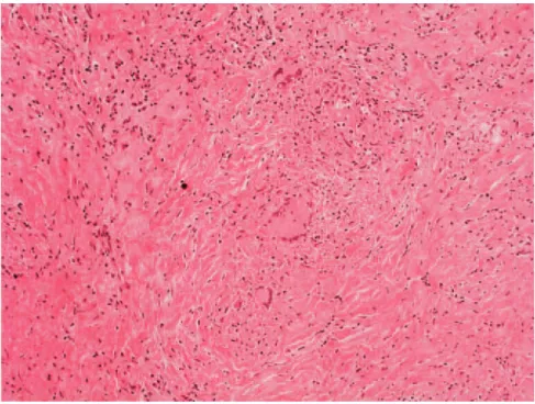
tomography showed prominent uptake in the visceral and parietal pericardium, suggesting active inflammation (Figure 1, left panel). Closed pericardiostomy with biopsy was performed. After pericardiostomy with drainage, echocardio-graphic features of constrictive physiology persisted and thus verified the presence of effusive constrictive pericarditis. Pathology showed chronic granulomatous inflammation with caseous necrosis consistent with tuberculosis (AFB stain: positive; Figure 2). Antituberculosis medication was started (isoniazid 400 mg, ethambutol 800 mg, rifampin 600 mg, pyrazinamide 1500 mg/d). Follow-up FDG positron emission tomography 12 months after administration of antituberculo-sis medication showed no visible uptake in the visceral and parietal pericardium, suggesting resolved inflammation (Fig-ure 1, right panel).
Disclosures
None.
From the Departments of Internal Medicine, Radiology, and Pathology, Yonsei University College of Medicine, Seoul, South Korea.
Correspondence to Jong-Won Ha, MD, PhD, Cardiology Division, Department of Internal Medicine, Yonsei University College of Medicine, Seoul, South Korea. E-mail [email protected]
(Circulation. 2006;113:e4-e5.) © 2006 American Heart Association, Inc.
Circulation is available at http://www.circulationaha.org DOI: 10.1161/CIRCULATIONAHA.105.554139 Figure 1. Left, PET scan showed homogenous uptake in the
visceral and parietal pericardium, suggesting active inflamma-tion. Right, Follow-up FDG positron emission tomography 12 months after administration of antituberculosis medication showed no visible uptake in the visceral and parietal pericar-dium, suggesting resolved inflammation.
e4
Images in Cardiovascular Medicine
at CONS KESLI on June 17, 2014
http://circ.ahajournals.org/
Figure 2. Pathology showed chronic granulomatous inflammation with case-ous necrosis consistent with
tuberculosis.
Ha et al Pericardial Inflammation in Constriction e5
at CONS KESLI on June 17, 2014
http://circ.ahajournals.org/