Review Article
The Advances of Laparoscopic Gastrectomy for Gastric Cancer
Yeon-Ju Huh and Joo-Ho Lee
Department of Surgery, Ewha Womans University Mokdong Hospital, Yangcheon-gu, Seoul 07985, Republic of Korea Correspondence should be addressed to Joo-Ho Lee; [email protected]
Received 31 March 2017; Accepted 19 July 2017; Published 5 September 2017 Academic Editor: Michel Kahaleh
Copyright © 2017 Yeon-Ju Huh and Joo-Ho Lee. This is an open access article distributed under the Creative Commons Attribution License, which permits unrestricted use, distribution, and reproduction in any medium, provided the original work is properly cited.
Laparoscopic gastrectomy is evolving. With the increasing expertise and experience of oncologic surgeons in the minimally invasive surgery for gastric cancer, the indication for laparoscopic gastrectomy is expanding to advanced cases. Many studies have demonstrated the benefits of minimally invasive surgery, including reduced risk of surgery-related injury, reduced blood loss, less pain, and earlier recovery. In order to establish concrete evidence for the suitability of minimal invasive surgery for gastric cancer, many multicenter RCTs, comparing the short- and long-term outcomes of laparoscopic versus open surgery, are in progress. Advances in laparoscopic gastrectomy are moving toward increasingly minimally invasive approaches that enable the improvement of the quality of life of patients, without compromising on oncologic safety.
1. Introduction
In spite of a decrease in its incidence, gastric cancer is still the fifth most common malignancy and the third leading cause of cancer-related death in the world (723,000 deaths, 8.8% of the total) according to GLOBOCAN, 2012. Half of the total global incidence occurs in Eastern Asia (mainly in China) [1]. Especially in Korea and Japan, gastric cancer is one of the most prevalent malignancies, and the proportion of early gastric cancer (EGC) has increased, partly because of nationwide surveillance [2, 3]. Given the heightened incidence of early-stage gastric cancer, the continuing accumulation of surgical experience, and the concomitant advances in instrumentation, the laparoscopic approach has become more commonly employed for the treatment of gastric cancer. Since the 1990s, laparoscopic surgery has been performed for the treatment of EGC in patients with a relatively low risk of lymph node metastasis. Here, the meaningful surgeries that have been historical turning points were summarized (Table 1). Ohgami et al. [4] reported a laparoscopic wedge resection using a lesion-lifting method
in 1991, which was thefirst case of laparoscopic surgery for
stomach cancer. Intragastric mucosal resection was also reported by Ohashi [5] in the early 1990s. In 1994, successful laparoscopy-assisted distal gastrectomy (LADG) with lymph
node dissection (LND) for EGC was introduced by Kitano et al. [6]. With the development of endoscopic treatments, such as endoscopic mucosal resection and endoscopic sub-mucosal dissection, the need of laparoscopic wedge resection and intragastric mucosal resection has decreased, whereas the application of LADG with LND has increased for treating patients with EGC who are at potential risk of lymph node metastasis. Laparoscopic surgery for EGC has become popu-lar based on several prospective randomized controlled trials (RCTs) that generally reported improved short-term surgical outcomes with comparable oncological safety to that of open surgery [7–10].
2. Advances in the Technical Aspect
2.1. Hand-Assisted Laparoscopic Surgery (HALS). The techni-cal difficulties of laparoscopic surgery include the limited mobility of the instruments and the absence of tactile sensa-tion. In HALS, however, the surgeon’s left hand is inserted into the abdominal cavity through a special pressurized sleeve, approximately 6-7 cm long, which preserves the
operator’s tactile sensation [11]. Incorporating both the
laparoscopy-assisted open surgicalfield and the high
resolu-tion of laparoscopy, HALS combines the advantages of lapa-roscopic surgery and laparotomy. Increased tactile sensation Volume 2017, Article ID 9278469, 12 pages
and depth perception with the inserted hand might solve sev-eral of the technical difficulties of gastrectomy with LND [11]. In the early phase of the development of laparoscopic surgery, it also conferred the advantages of relatively low morbidity and high operative safety. Kim et al. [12] suggested that hand-assisted gastrectomy could be a good learning technique for laparoscopic gastrectomy beginners. However, there are some disadvantages to this method. A hand can encroach upon the intra-abdominal working space, and the sleeve for maintaining the pneumoperitoneum is expensive. Moreover, HALS is ergonomically unfavorable for surgeons, which leads to shoulder and forearm fatigue and strain for the surgeon [13]. As surgical expertise has evolved over time, the number of surgeons who use this method has definitively decreased.
2.2. Laparoscopy-Assisted Gastrectomy. In laparoscopy-assisted gastrectomy (LAG), after laparoscopic full mobiliza-tion of the stomach, resecmobiliza-tion of the stomach and anastomo-sis is performed extracorporeally through a small incision in the epigastrium for the accurate localization of the lesion and secure reconstruction. For the precise localization of the lesion in LAG, the effectiveness of endoscopic clips and lapa-roscopic ultrasonography has been reported [14]. The useful-ness of intraoperative navigation, using three-dimensional (3-D) computed tomographic (CT) angiography during extended LND in LADG, was also emphasized [15], as was
the importance of the preoperative identification of vascular
trees by 3-D CT angiography [16].
Laparoscopy-assisted total gastrectomy (LATG) is tech-nically demanding in terms of reconstruction and LND such as stations 11d, 4sa, and 10 [3, 17]. Therefore, the use of LATG for the treatment of upper gastric cancer has not been generalized. Various reconstruction methods for esophagoje-junostomy have been reported, using circular- and linear-stapled anastomosis.
2.3. Totally Laparoscopic Gastrectomy. In LAG, the surgical process via minilaparotomy is sometimes difficult to per-form, especially in patients with obesity [18]. Totally, laparo-scopic distal gastrectomy (TLDG) that all procedures are carried out laparoscopically has been made possible pursuing minimal invasiveness. TLDG received attention when the first delta-shaped anastomosis was introduced [19]. Several
intracorporeal anastomosis techniques have been reported, including Billroth II anastomosis using linear staplers, beta-shaped Roux-en-Y reconstruction, overlap method, and semi-loop after total gastrectomy and inverted T-shaped anastomosis using linear staplers [20–24]. Nowa-days, the totally laparoscopic procedure is performed for
advanced cancer or remnant cases. The first case of totally
laparoscopic total gastrectomy for completion was reported by Shinohara et al. [25].
2.4. Reconstruction Methods. For the standardization of lapa-roscopic gastrectomy, the procedure must achieve reproduc-ibility, safety, and simplicity in terms of not only the LND but also the reconstruction component. Billroth I anastomosis has been known to offer the advantages of greater simplicity of performance, fewer postanastomosis anatomical changes, increased physiological pathways, and lower incidences of adhesion and internal herniation. Consequently, it has been the most commonly performed anastomosis after distal gas-trectomy in Korea and Japan [26]. During laparoscopic sur-gery, intracorporeal gastroduodenostomy has been typically considered a challenging technique because of the difficulty of operating in the narrow working space around the duode-nal stump. Therefore, the laparoscopy-assisted reconstruc-tion procedure has been popularly performed for a long time. 2.4.1. Delta Anastomosis. Recently, delta-shaped anastomo-sis, one of the well-established intracorporeal gastroduode-nostomy methods, is popularly performed; however, it still demands delicate and precise laparoscopic techniques of the operators and assistants. The technique proceeds in the following way. First, the duodenal bulb is transected using
an endoscopic linear stapler at a 90°angle from the usual line.
The stomach is then divided in the customary fashion. Small entry holes are created along the edge of the stomach and the duodenum. The posterior walls of both the stomach and the duodenum are approximated using a 45 mm linear stapler. Then, the staple line is meticulously inspected for any defects to ascertain the color of the anastomosis, after which the common entry hole is closed with one or two linear staplers [27]. The most serious problem regarding anastomosis is leakage. Poor blood supply and excessive tension at the anas-tomotic site may cause the leakage of anastomosis. Regarding blood supply, the delta technique, in particular, carries a Table 1: Previous historical report of laparoscopic gastrectomy for gastric cancer.
Author Year Operation Indication Country Analysis
Ohgami et al. 1992 Laparoscopic wedge resection EGC Japan n = 6
Ohashi et al. 1995 Laparoscopic intragastric mucosal resection EGC Japan n = 8
Kitano et al. 1994 Laparoscopic-assisted Billroth I gastrectomy EGC Japan Case report
Azagra et al. 1999 Laparoscopic-assisted total gastrectomy EGC, AGC Belgium n = 13
Uyama et al. 1999 LADG with D2 LND AGC Japan n = 12
Ohki et al. 1999 Hand-assisted laparoscopic distal gastrectomy EGC Japan Case report
Kanaya et al. 2002 Totally laparoscopic Billroth I gastrecotmy EGC Japan n = 9
Giulianotti et al. 2003 Distal and total robotic gastrectomy EGC, AGC Italy n = 18
potential risk of anastomosis ischemia since the tissue around the duodenal stump has to be dissected to prepare a sufficient space for the anastomosis. In Billroth I recon-struction, the small size of the remnant stomach might lead to anastomotic-site tension and leakage [28]. There-fore, in case of small remnant stomach or short duodenal bulb, a Billroth II or Roux-en-Y reconstruction might achieve a safer anastomosis.
2.4.2. Roux-en-Y Anastomosis. The Roux-en-Y method is known to be favorable to postoperative quality of life (QOL) with lower incidence of bile reflux and anastomotic leakage [10, 11]. However, it is more laborious because it requires the creation of two anastomoses and one duodenal stump closure. The jejunum is divided at a point approxi-mately 15 cm distal to the ligament of Treitz and
jejunojeju-nostomy which is performed 30–40 cm distal from the
jejunal division using a linear stapler. After the creation of the jejunojejunostomy, side-to-side anastomosis between the distal part of the divided jejunum and remnant stomach is carried out by using a linear stapler. The common entry hole is closed by hand-sewn running suture or using linear stapler [29]. Anastomotic leakage hardly occurs after Roux-Y gastrojejunostomy because of the good blood supply and minimal tension at the anastomosis site. Meanwhile, there are some reports that an excessively long Roux-limb can cause internal herniation or Roux stasis syndrome [29, 30].
In addition, even though reflux is known to be rare in the
RY group, there is debate remaining as to whether the inci-dence of reflux-related gastritis is different or not. There-fore, apparent superiority of any particular method was not confirmed. Extracorporeal jejunojejunostomy via peri-umbilical minilaparotomy is generally accepted. The intra-corporeal anastomosis is time-consuming and technically demanding in the hands of unskilled and inexperienced surgeons, and for the extraction of the resected stomach, an incision of at least 3 cm is necessary anyway. Therefore, some surgeons prefer making extracorporeal jejunojeju-nostomy through periumbilical small incisions in order to reduce operating time.
2.4.3. Esophagojejunostomy Using Circular-Stapled Anastomosis. Owing to the difficulty of achieving a secure purse-string suture and the technical challenge of performing an
esopha-gojejunostomy in a narrow and deep surgicalfield, LATG or
laparoscopic total gastrectomy (LTG) is performed relatively less often compared with LADG.
Esophagojejunostomy using a circular stapler is the most famous technique in the open surgery. For extracorporeal approaches in LATG, a 4 to 6 cm-sized epigastric incision is made to access the esophagogastric junction. Many reports pertaining to the extracorporeal method describe the
proce-dures in simple terms, such as“the insertion of an anvil with
the purse-string suture.” However, the procedure is often very difficult because the purse-string device is too bulky to handle in a small minilaparotomy space, and suturing through a small incision in the deep and narrow operative field is an exacting process [31, 32]. However, a purse-string instrument with a smaller head has been developed,
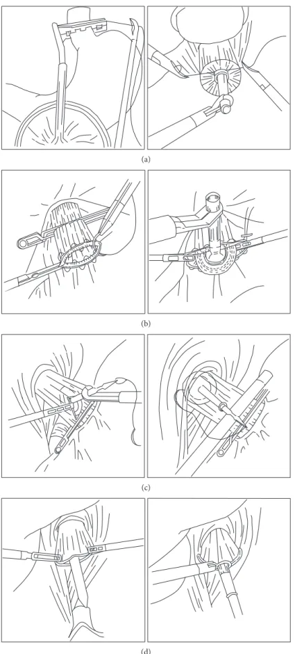
which can be inserted through a 12 mm port into the abdom-inal cavity. Accordingly, a purse-string suture can be per-formed under laparoscopic vision [33] (Figure 1(a)). Some surgeons prefer intracorporeal hand-sewn purse-string sutures because of their relative simplicity (Figure 1(b)). Secured anvil insertion into the esophageal stump is the next challenging step. After the purse-string suture, the esopha-geal stump is opened by two forceps, at which point, an anvil can be inserted easily [34, 35]. Another method for laparo-scopic anvil insertion is to use an anvil attached with a thread through a small esophagotomy at the anterior wall of the esophagus. The thread is then pulled outside to place the anvil at the esophageal edge, and the esophagus is transected using a linear stapler [36] (Figure 1(c)). Oral insertion of an anvil (Orvil, OrVil™, Covidien, Tokyo, Japan) was introduced as an alternative to reduce procedure complexity [37, 38] (Figure 1(d)). Despite concerns, the esophageal mucosal injury, a specific complication of a transoral anvil passage, would occur; such mucosal injury has been rarely reported [39]. The location of the camera port and the small size of the incision for circular stapler insertion are also very important to enable a proper view during this procedure. Most surgeons prefer an upper midline incision for anvil insertion in cases of extracorporeal anastomosis. The same incision is used for the insertion of a circular stapler with a laparoscope from the left lower port [31] or the left upper port [40]. Some surgeons use an extended umbilical port wound [41] or a left lower port wound [37].
2.4.4. Esophagojejunostomy Using Linear-Stapled Anastomosis.
The handling of the linear stapler is easier in LTG field
because linear staplers are thinner and have superior mobility of the tip compared to circular staplers. Moreover, surgeons can perform the linear-stapled anastomosis regardless of the esophageal diameter and can achieve a larger anastomo-sis than in the case of circular-stapled anastomoanastomo-sis. In addi-tion, linear-stapled anastomosis helps the surgeon reduce the torsion of the jejunal limb, which is a severe complication of circular-stapled anastomosis.
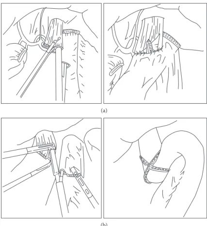
For the overlap method (Figure 2(a)), a small entry hole is made about 5 cm distal to the stapler line on the jejunal limb, while another enterotomy is made on the left wall of the esophageal stump. An anastomosis is performed by inserting the stapler between the esophageal enterotomy and the entry hole of the jejunal limb toward the cephalic side of the lumen. The jejunal limb is positioned at the left side of the esopha-geal stump. Then, the common entry hole is closed using an intracorporeal hand-sewn suture. For the functional end-to-end anastomosis (FEEA) (Figure 2(b)), the esophagus is transected intracorporeally in the horizontal direction. Two small entry holes are made at the edges of the tip of the jejunal end and the transected esophagus. The linear sta-pler is inserted into the holes, and the anastomosis is created. The common entry hole is closed with a linear stapler.
There are two differences between the overlap method
and FEEA: the peristaltic direction of the esophagojeju-nostomy and the methods of closing the common entry hole. Esophagojejunostomy in FEEA is performed in the antiperistaltic direction; therefore, the jejunal limb needs
(a)
(b)
(c)
(d)
Figure 1: The several methods of anvil insertion for esophagojejunostomy using a circular stapler after total gastrectomy. (a) Anvil insertion using a small-head purse-string instrument. (b) Anvil insertion after laparoscopic hand-sewn purse-string sutures. (c) Insertion of an anvil attached with a thread through an esophagotomy at the anterior wall of the esophagus. (d) An oral anvil insertion method (OrVil).
to be lifted further up in FEEA than in the overlap method. In addition, the closure of the common entry hole in FEEA is performed using a linear stapler, whereas the closure is per-formed using an intracorporeal suture in the overlap method. Thus, since a larger working space is needed in FEEA than in the overlap method, the dissection around the crura of the diaphragm has to be performed in FEEA, which can increase the risk of a diaphragmatic hernia. In contrast, advanced suturing skills are needed in the overlap method, and intracorporeal suturing frequently results in prolonged operative time [42].
2.5. Laparoscopic Function-Preserving Surgery
2.5.1. Pylorus-Preserving Gastrectomy (PPG). PPG was first
introduced for the treatment of peptic ulcer, and since then, it has been used as a surgical treatment for EGC to preserve function and maintain a better QOL [43, 44]. According to Japanese gastric cancer treatment guidelines, PPG is the treatment option for clinically T1N0M0 gastric cancers in the middle third of the stomach at least 4.0 cm away from the pylorus. The distance from the lesion to the pylorus needs
to be carefully considered because a short antral cuff may lead
to postoperative gastric stasis, which is one of the typical
complications of PPG. Considering the need for a sufficient
distal resection margin of>1 cm for EGC, in addition to the
length of the antral cuff, the distance from the lesion to the
pylorus should be greater than 4.0 cm. The standard tech-nique for PPG includes preservation of the hepatic branch of the vagus nerve and the infrapyloric vessels to allow for the structural and functional preservation of the pylorus. The LND of station 5 is usually omitted during PPG to preserve the hepatic branch of the vagus nerve. Finally, gas-trogastrostomy is performed using either the hand-sewn method or the linear stapler. By preserving pyloric function, it confers potential nutritional advantages and carries a lower incidence of disturbed bowel habits, as well as fewer postgas-trectomy disorders such as dumping syndrome and alkaline reflux [45]. Recently, laparoscopy-assisted PPG (LAPPG) has been reported to be beneficial compared to conventional PPG in terms of the preservation of functionality and minimal invasiveness [46].
2.5.2. Vagus Nerve-Preserving Gastrectomy (VPG). VPG was designed to decrease postgastrectomy symptoms caused by injury to the vagus nerve. It preserves not only the celiac branches of the posterior vagal trunk that innervate the small intestine but also the hepatic branches of the anterior vagal trunk that innervate the liver and biliary tract [47]. However, there is a specific concern regarding the incomplete LND for nerve preservation. During the LND, nonvisible nerve injury can occur because of energy devices and traction, making it
difficult to achieve the complete removal of lymph nodes
without nerve injury. Even if the nerve is grossly preserved, (a)
(b)
Figure 2: Two representative types of anastomosis using linear stapler after total gastrectomy. (a) The overlap method. (b) The functional end-to-end anastomosis method.
it is hard to confirm whether the function of the nerve has been preserved. Furthermore, a longer operative time is required to dissect around the nerve compared to that of
con-ventional gastrectomy. In spite of these technical difficulties,
VPG continues to be performed using auxiliary methods such as nerve monitoring according to an ongoing study that has not been published.
2.5.3. Proximal Gastrectomy (PG). In laparoscopic PG (LPG), the type of reconstruction may be the most important issue. Esophagogastrostomy has served as the most simple recon-struction method, though most patients end up suffering from postoperative reflux esophagitis and/or anastomotic
stricture. Antireflux procedures including fundoplication,
gastric tube formation, pyloroplasty, and esophagopexy with crural repair have been attempted in order to reduce the
inci-dence of anastomotic stricture and reflux esophagitis.
How-ever, the results of these efforts have been far from
satisfactory [48–50], and two effective alternatives to esopha-gogastrostomy after PG have been introduced, namely, jeju-nal interposition and double-tract reconstruction (DTR). Jejunal interposition was introduced as a strategy to reduce severe reflux; however, laparoscopic jejunal interposition has not been frequently performed because of the inherent
technical complexity of the creation of a jejunalflap and three
anastomoses, with the consequently prolonged operative time [51, 52]. Alternatively, DTR consists of three anastomo-ses: Roux-en Y esophagojejunostomy, gastrojejunostomy 15 cm distant from the esophagojejunostomy, and jejunojeju-nostomy 20 cm distant from the gastrojejujejunojeju-nostomy. Ahn et al. reported that LPG with DTR for proximal EGC showed excellent postoperative outcomes, particularly with respect to decreased reflux symptoms. This procedure also showed the tendency for improvements in nutritional status, accept-able oncologic outcome rates, surgical time, and complica-tion rates [53]. This procedure involves the creacomplica-tion of an additional anastomosis, a gastrojejunostomy by stapling,
which adds only 5–10 min to the conventional LTG
anasto-mosis procedure. Lastly, delayed gastric emptying becomes less of a concern since even if delayed gastric emptying occurs, there is an alternative passage route for food, in con-trast to jejunal interposition.
2.6. Single-Incision Distal Gastrectomy (SIDG). Reduced port or single-port laparoscopic surgery was developed to reduce scars and surgical stress, and indeed, single-port laparoscopic surgery through the umbilicus does not leave any visible scar. A vertical 2.5 cm-sized transumbilical incision is made, and a commercial single-port device is placed in the umbilical inci-sion. An additional assistant trocar is not needed. Then, a flexible scope is used to secure a clear view of the operative field. For effective dissection, curved instruments are used when operating in the lesser curvature side and the supra-pancreatic area. Regarding suprasupra-pancreatic LND, the neck and body of the pancreas are sometimes overly protruded, which makes it difficult to dissect the lymph nodes behind the pancreas using a straight instrument from the umbilicus. SIDG is not yet generalized because of the continuing need for more advanced techniques and scarring evidences of
oncological safety. In addition, although previous reports of SIDG showed comparable morbidity and an absence of open conversion, the risk of unexpected intraoperative accidents in pure, single-port surgery cannot be ignored. A large-scale study is necessary to confirm the safety and oncologic outcomes of SIDG.
2.7. Robot Gastrectomy. Robotic systems for surgery were implemented in early 2000 to overcome the drawbacks of laparoscopic surgery. The features of the robotic systems, such as 3-D vision, the elimination of physiologic tremor, and the articulated arms, assist the surgeon by facilitating
manipulation in the surgicalfield [54]. An articulated
endo-scopic wrist allows the operator seven degrees of freedom,
via 180°articulation and 540°rotation. Moreover, the
magni-fied 3-D high-definition imaging system and very stable cam-era platform, which is controlled by the opcam-erator, are
significant characteristics. Another advantage of robotic
sur-gery involves its ability to enable more precise intracorporeal suturing, such that reconstruction after total gastrectomy is greatly facilitated [55]. With the use of wristed instruments, the meticulous dissection of cardiac LND, mobilization of the distal esophagus, and insertion of the anvil head into the esophageal stump are more easily performed. In addition, the learning curve of robotic surgery is quite steep because of its simplicity and the potential for early adaptation [56, 57]. Recently, the robotic surgery with image-guided assistance has been reported. Kim et al. [58] demonstrated that the operators could employ a vascular map and avoid vascular injury or damage to other organs through the intraoperative
image-guidance feature. During robotic gastrectomy, thefine
movements and magnified view allow surgeons to perform clear LND without any vascular injury and with minimal intraoperative bleeding and to preserve the small branches of the splenic vessels.
3. Clinical Outcomes
3.1. Morbidity and Mortality. In LDG, the incidence of oper-ative complications is reportedly lower overall than that in conventional ODG. In a previous prospective RCT, we reported that LADG confers the clear advantage of fewer pul-monary complications as compared to open gastrectomy [59]. Furthermore, a meta-analysis of 5 RCTs and 17 non-RCTs with 3411 patients reported that LADG might result in less blood loss, less consumption of analgesics, and shorter hospital stays, without an increase in total hospitalization costs [60]. While Kim et al. (COACT 0301) [61] reported sig-nificant reductions in mild complications in LADG (LADG
versus ODG; 23.2% versus 41.5%,p = 0 012), the Korean
lap-aroscopic gastrointestinal surgery study (KLASS) group per-formed a phase 3 multicenter RCT (KLASS-01) to establish even stronger evidence, and their morbidity and mortality data was recently reported [62]. The overall complication
rate was significantly lower in the LADG group (LADG
versus ODG; 13.0% versus 19.9%, p = 0 001); in particular,
the wound complication rate in the LADG group was signif-icantly lower than that in the ODG group (3.1% versus 7.7%,
terms of either major intra-abdominal complications (7.6%
versus 10.3%, p = 0 095) or mortality rates (0.6% versus
0.3%, p = 0 687) between the two groups. For cases of
advanced cancer, a Chinese group recently reported the sur-gical safety of LDG with D2 LND when compared with con-ventional ODG [63]. In their multicenter prospective RCT,
no significant difference in postoperative morbidity was
shown (LDG versus ODG; 15.2% versus 12.9%,p = 0 285).
The mortality rate was also similar between the two groups
(LDG versus ODG; 0.4% versus 0%,p = 0 249).
In contrast, only a few studies have reported on the feasi-bility and safety of LTG and the results remain controversial [40, 64–66]. Some authors reported that LATG offers techni-cal feasibility and safety, comparable to open total gastrec-tomy. LATG was also shown to be associated with fewer postoperative complications, less pain, and rapid recovery. However, the heterogeneity of the results may have been caused by the following limitations: the mostly retrospective nature of the studies, small sample sizes, mostly EGC cases, and short follow-up periods.
Recently, several retrospective studies reported that TLDG was technically feasible, less invasive, and safer than LADG [67–70]. In a meta-analysis including five studies with 652 patients, TLDG was associated with less intraoperative
blood loss, earlierfirst flatus, and lower postoperative
mor-bidity than LADG. Our group performed a prospective RCT to evaluate the overall feasibility of TLDG [71] in which it was shown that TLDG is as safe and feasible as LADG, with a comparable rate of complications. Indeed, short of our expectation, no significant differences were shown between the TLDG and LADG groups in terms of postoperative course such as recovery, postoperative pulmonary function, and inflammatory parameters. It could be the reason that
the parameters used in the clinicalfield to evaluate the early
surgical outcomes could not accurately reflect the subtle
dif-ference in surgical invasiveness between TLDG and LADG. Recently, the Korean Robot Gastrectomy Study Group reported the results of a multicenter prospective, clinical trial comparing robotic gastrectomy with laparoscopic gastrec-tomy in EGC [72]. Patients were enrolled for treatment with
either robotic (n = 185) or laparoscopic (n = 185)
gastrec-tomy. The study showed similar overall complication rates (11.9% versus 10.3%; robotic versus laparoscopic) and major complication rates (1.1% versus 1.1%; robotic versus laparo-scopic) with no operative mortality in either groups. In the
robotic surgery group, the operative time was significantly
longer (221 minutes versus 178 minutes; robotic versus
lapa-roscopic,p < 0 001) compared with that in the laparoscopic
group. There were no significant differences between the two groups regarding the rates of open conversion, operative blood loss, length of stay, or diet build-up. The authors concluded that robotic gastrectomy was not superior to laparoscopic gastrectomy. It is true that the robot group paid higher costs [South Korean won 13,748,422.5 (US$13,470)
(robotic) versus 9,165,862 (US$8980) (laparoscopic);
P < 0 001], but most of the high cost will not be a big
problem in the future due to depreciation and mainte-nance costs. Rather than simply calculating the cost per operation, we should study the indications that require
robotic gastrectomy, apply robotic surgery where necessary, and reduce the social burden by reducing complications. 3.2. Survival and Recurrence. Regarding oncological safety, our group reported the long-term safety of LADG for EGC [73]. Tumor recurrence occurred in 0.9% and the rate of cancer-related death was 0.5% (only one patient) during the median 55-month follow-up period, and the overall 5-year survival rate was not significantly different between the LADG and ODG groups (95.9% versus 94.9%; LADG versus ODG). Zeng et al. [60] also reported comparable long-term survival rates between both groups in a meta-analysis. The long-term results of a prospective RCT (COACT 0301) with 164 patients of the median 74.3-month follow-up period showed that the survival rates of the LADG and ODG groups were comparable (5-year overall survival: 97.6% versus 96.3,
p = 0 721; 5-year disease-free survival: 98.8% versus 97.6%
in LADG versus ODG,p = 0 514) [61].
There have been several studies on the oncologic out-comes of laparoscopic gastrectomy with extended
lymphade-nectomy for advanced gastric cancer (AGC). Thefindings of
these studies suggested that the 3-year overall survival (75.3%) and disease-free survival (69.0%) for laparoscopic surgery were comparable to those of published studies on
conventional open surgery [74–76].
Two multicenter RCTs, the KLASS-01 study and the Japanese Clinical Oncology Group (JCOG) 0912 trials, are currently ongoing on large-scale studies and seek to elu-cidate the oncological outcomes of laparoscopic gastrectomy
for EGC. The KLASS-01 trial is thefirst multicenter RCT to
compare laparoscopic surgery with open surgery in patients with clinical stage I gastric cancer. Overall survival is the pri-mary endpoint, and the secondary endpoints include mor-bidity and mortality, disease-free survival, quality of life,
inflammatory responses, and cost effectiveness. A total of
1416 patients (705 and 711 patients in the LADG and ODG
groups, resp.) were enrolled from 2006 to 2010, and thefinal
results will be reported in the near future [77]. The JCOG started a multicenter RCT in 2010 to compare LADG with ODG in 920 patients with stage I gastric cancer from 33 insti-tutions. In the JCOG 0912 study, the overall survival rate is the primary endpoint, and the secondary endpoints include short-term clinical outcomes, adverse events, proportion of LADG completion and conversion to open surgery, relapse-free survival, and postoperative QOL [78].
The long-term results of laparoscopic surgery for AGC remain controversial because of the paucity of reliable stud-ies. While D2 LND is the standard for AGC treatment in open, laparoscopic, and robotic surgeries, it is very challeng-ing to perform. Even in East Asia, where there are many more cases and experiences of gastric cancer than in Western countries, only a few qualified surgeons are capable of per-forming a complete D2 LND using a laparoscopic approach. Significant improvements in laparoscopic surgical instru-ments and techniques have been progressing for decades [79]. This accumulation of laparoscopic expertise has led to the use of extended LND and to attempts by some experi-enced surgeons to extend the indication of laparoscopic gas-trectomy to locally advanced cases. Recently, it was reported
that no significant difference in overall survival and disease-free survival between open gastrectomy and laparoscopic gastrectomy in AGC was shown in a meta-analysis using 10 studies with a total of 1819 patients [80]. There are several
difficulties or limitations in applying laparoscopic surgery
to AGC, including total omentectomy, splenic hilar dissec-tion for proximal gastric cancer, bulky positive nodes or large primary tumor, esophageal invasion, and peritoneal lavage [81]. Several data reported that laparoscopic gastrectomy with D2 LND was technically feasible and safe for patients with AGC, with acceptable rates of morbidity and mortality and satisfactory long-term outcomes [74–76, 82, 83]. Achiev-ing excellent long-term results in laparoscopic gastrectomy depends on the standardization of the D2 LND. To that end, the KLASS team sought to achieve a consensus as to the D2 LND procedure [84]. All surgeons participating in the KLASS study were each asked to submit three laparo-scopic and three open D2 gastrectomy videos. Each unedited video was allocated to several peer reviewers and reviewed blindly. Based on the results of experts’ reviews, the review evaluation committee decided whether the surgeon could be included in the KLASS-02 trial. This systematic approach will serve as a crucial example for surgical standardization.
Three multicenter trials, the KLASS-02, the Japanese Laparoscopic Surgery Study Group (JLSSG) 0901, and the CLASS (Chinese laparoscopic gastrointestinal surgery study)-01, are current large-scale studies seeking to elucidate the oncological outcomes of laparoscopic gastrectomy for AGC. The KLASS-02 trial is a phase III study evaluating
the efficacy of LDG with D2 LND for AGC. The primary
endpoint is 3-year disease-free survival, and the estimated population size is 1050. The JLSSG 0901 trial is a phase II/III study comparing LADG and ODG in patients with clinical T2 to T4aM0 gastric cancer. After the recruitment of 180 patients, the rate of major complications will be analyzed. The study will continue the recruitment until 500 patients are enrolled unless there is early termination due to a high complication rate [85]. Lastly, the CLASS-01 trial performed by the Chinese group is a phase III study, and the study design is similar to that of the KLASS-02 trial. In the near future, these well-designed studies may help to establish concrete, reliable evidence for the expansion of the indication for laparoscopic gastrectomy to advanced cases.
3.3. Quality of Life. Laparoscopic gastrectomy has many merits over open gastrectomy, such as less pain, shorter postoperative hospital stays, earlier recovery, and superior cosmetic outcomes. In several meta-analyses, the superior-ity of laparoscopic gastrectomy in terms of postoperative recovery was shown [86, 87]. In another report, LADG showed improved short-term symptoms and functional outcomes [61].
Function-preserving surgery was introduced to improve the QOL of patients. Recently, the nutritional and functional benefits of LAPPG were compared to those of LADG [46]. It was reported that the incidence of delayed gastric emptying was lower, though other complications occurred more fre-quently in LADG than in LAPPG. Decreased serum albumin and protein levels at one to six months postoperatively and
greater abdominal fat volumes at postoperative one year were observed in LADG compared with LAPPG. The authors concluded that LAPPG could be considered a superior treatment option for middle-third EGC over LADG in terms of its lower incidence of gallstone and nutritional advantages. In addition, an RCT reported that the patients who underwent VPG showed significantly less diarrhea and less appetite loss at 12 months. They concluded that VPG could improve postoperative QOL compared with conven-tional gastrectomy [47].
In proximal EGC, the application of PG has been limited until now. In a systematic meta-analysis comparing TG with PG, PG with esophagogastrostomy showed a higher inci-dence of reflux esophagitis and anastomotic stenosis [88]. However, several positive results of LPG with modified reconstruction methods have been reported [48, 89]. Accord-ingly, LPG can be considered an attractive treatment option for proximal EGC as a minimally invasive surgery to preserve functionality, including reduction of postoperative com-plaints, prevention of anemia, improved nutrition, and
improved production of gut hormones [90–93].
4. Conclusions
Laparoscopic gastrectomy has advanced to look for mini-mally invasive approaches as well as to maintain the onco-logic safety. In accordance with the evolution of surgical instrumentation and increased laparoscopic surgical experi-ence, its indication has been extended to advanced cases. Recent studies show that the oncologic outcomes of laparo-scopic gastrectomy for EGC are comparable to those of open gastrectomy. The demonstration of a similarly optimal result regarding the safety of laparoscopic gastrectomy in AGC is awaited. The results of several ongoing multicenter RCTs are expected to establish concrete evidence of the widespread suitability of laparoscopic gastrectomy in the treatment of gastric cancer.
Additional Points
Core Tip. Laparoscopic gastrectomy will continue to advance toward increasingly minimally invasive approaches to improve the quality of life of patients with gastric cancer, without compromising oncologic safety.
Conflicts of Interest
All authors have no conflict of interests.
Authors
’ Contributions
Yeon-Ju Huh and Joo-Ho Lee contributed equally to this work including the designing of research, performing the research, and writing the paper.
Acknowledgments
This study was supported by the Basic Science Research Program through the National Research Foundation of
Korea (NRF) funded by the Ministry of Education, Science and Technology (no. 2016R1D1A1B03932360).
References
[1] L. A. Torre, F. Bray, R. L. Siegel, J. Ferlay, J. Lortet-Tieulent,
and A. Jemal,“Global cancer statistics, 2012,” CA: A Cancer
Journal for Clinicians, vol. 65, no. 2, pp. 87–108, 2015.
[2] M. Inoue and S. Tsugane,“Epidemiology of gastric cancer in
Japan,” Postgraduate Medical Journal, vol. 81, no. 957,
pp. 419–424, 2005.
[3] O. Jeong and Y. K. Park, “Clinicopathological features and
surgical treatment of gastric cancer in South Korea: the results of 2009 nationwide survey on surgically treated gastric
cancer patients,” Journal of Gastric Cancer, vol. 11, no. 2,
pp. 69–77, 2011.
[4] M. Ohgami, K. Kumai, Y. Otani, G. Wakabayashi, T. Kubota,
and M. Kitajima,“Laparoscopic Wedge Resection of the
Stom-ach for Early Gastric Cancer Using a Lesion-Lifting Method,”
Digestive Surgery, vol. 11, no. 5, pp. 64–67, 1994.
[5] S. Ohashi, “Laparoscopic intraluminal (intragastric) surgery
for early gastric cancer. A new concept in laparoscopic sur-gery,” Surgical Endoscopy, vol. 9, no. 2, pp. 169–171, 1995.
[6] S. Kitano, Y. Iso, M. Moriyama, and K. Sugimachi,
“Laparos-copy-assisted Billroth I gastrectomy,” Surgical Laparoscopy &
Endoscopy, vol. 4, no. 2, pp. 146–148, 1994.
[7] H. Hayashi, T. Ochiai, H. Shimada, and Y. Gunji,“Prospective
randomized study of open versus laparoscopy-assisted distal gastrectomy with extraperigastric lymph node dissection for
early gastric cancer,” Surgical Endoscopy, vol. 19, no. 9,
pp. 1172–1176, 2005.
[8] C. G. Huscher, A. Mingoli, G. Sgarzini et al.,“Laparoscopic
versus open subtotal gastrectomy for distal gastric cancer: five-year results of a randomized prospective trial,” Annals of
Surgery, vol. 241, no. 2, pp. 232–237, 2005.
[9] S. Kitano, N. Shiraishi, K. Fujii, K. Yasuda, M. Inomata, and
Y. Adachi, “A randomized controlled trial comparing open
vs laparoscopy-assisted distal gastrectomy for the treatment of early gastric cancer: an interim report,” Surgery, vol. 131,
Supplement 1, pp. S306–S311, 2002.
[10] Y. W. Kim, Y. H. Baik, Y. H. Yun et al.,“Improved quality of
life outcomes after laparoscopy-assisted distal gastrectomy for early gastric cancer: results of a prospective randomized clinical trial,” Annals of Surgery, vol. 248, pp. 721–727, 2008. [11] S. Tanimura, M. Higashino, Y. Fukunaga, and H. Osugi,
“Hand-assisted laparoscopic distal gastrectomy with regional
lymph node dissection for gastric cancer,” Surgical
Laparos-copy EndosLaparos-copy & Percutaneous Techniques, vol. 11, no. 3, pp. 155–160, 2001.
[12] Y. W. Kim, J. M. Bae, J. H. Lee et al.,“The role of hand-assisted
laparoscopic distal gastrectomy for distal gastric cancer,”
Surgical Endoscopy, vol. 19, no. 1, pp. 29–33, 2005.
[13] D. E. M. Litwin, A. Darzi, J. Jakimowicz et al.,“Hand-assisted
laparoscopic surgery (HALS) with the HandPort System
-initial experience with 68 patients,” Annals of Surgery,
vol. 231, no. 5, pp. 715–721, 2000.
[14] W. J. Hyung, J. S. Lim, J. H. Cheong et al., “Intraoperative
tumor localization using laparoscopic ultrasonography in
laparoscopic-assisted gastrectomy,” Surgical Endoscopy,
vol. 19, no. 10, pp. 1353–1357, 2005.
[15] S. Takiguchi, M. Sekimoto, Y. Fujiwara et al., “Laparoscopic
lymph node dissection for gastric cancer with intraoperative navigation using three-dimensional angio computed
tomogra-phy images reconstructed as laparoscopic view,” Surgical
Endoscopy, vol. 18, no. 1, pp. 106–110, 2004.
[16] S. Usui, S. Hiranuma, T. Ichikawa, M. Maeda, S. E. Kudo, and
T. Iwai, “Preoperative imaging of surrounding arteries by
three-dimensional CT: is it useful for laparoscopic
gastrec-tomy?,” Surgical Laparoscopy, Endoscopy & Percutaneous
Techniques, vol. 15, no. 2, pp. 61–65, 2005.
[17] S. Kitano and N. Shiraishi, “Current status of laparoscopic
gastrectomy for cancer in Japan,” Surgical Endoscopy, vol. 18,
no. 2, pp. 182–185, 2004.
[18] M. G. Kim, K. C. Kim, B. S. Kim et al., “A totally
laparo-scopic distal gastrectomy can be an effective way of
per-forming laparoscopic gastrectomy in obese patients (body
mass index > 30),” World Journal of Surgery, vol. 35, no. 6,
pp. 1327–1332, 2011.
[19] S. Kanaya, T. Gomi, H. Momoi et al.,“Delta-shaped
anastomo-sis in totally laparoscopic Billroth I gastrectomy: new
tech-nique of intraabdominal gastroduodenostomy,” Journal of
the American College of Surgeons, vol. 195, no. 2, pp. 284–
287, 2002.
[20] J. Du, J. Shuang, J. Li et al.,“Totally laparoscopic Billroth II
gastrectomy with a novel, safe, simple, and time-saving anasto-mosis by only stapling devices,” Journal of Gastrointestinal
Surgery : Official Journal of the Society for Surgery of the
Alimentary Tract, vol. 16, no. 4, pp. 738–743, 2012.
[21] K. Motoyama, K. Kojima, M. Hayashi, K. Kato, M. Inokuchi,
and K. Sugihara, “Beta-shaped intracorporeal Roux-en-Y
reconstruction after totally laparoscopic distal gastrectomy,”
Gastric Cancer: Official Journal of the International Gastric Cancer Association and the Japanese Gastric Cancer
Associa-tion, vol. 17, no. 3, pp. 588–593, 2014.
[22] H. Okabe, K. Obama, E. Tanaka et al.,“Intracorporeal
esopha-gojejunal anastomosis after laparoscopic total gastrectomy for
patients with gastric cancer,” Surgical Endoscopy, vol. 23, no. 9,
pp. 2167–2171, 2009.
[23] K. Inaba, S. Satoh, Y. Ishida et al., “Overlap method: novel
intracorporeal esophagojejunostomy after laparoscopic total gastrectomy,” Journal of the American College of Surgeons,
vol. 211, no. 6, pp. e25–e29, 2010.
[24] E. Nagai, K. Ohuchida, K. Nakata et al.,“Feasibility and safety
of intracorporeal esophagojejunostomy after laparoscopic total gastrectomy: inverted T-shaped anastomosis using linear
staplers,” Surgery, vol. 153, no. 5, pp. 732–738, 2013.
[25] T. Shinohara, N. Hanyu, Y. Tanaka, K. Murakami, A.
Watanabe, and K. Yanaga, “Totally laparoscopic complete
resection of the remnant stomach for gastric cancer,” Langen-beck's Archives of Surgery / Deutsche Gesellschaft fur Chirurgie,
vol. 398, no. 2, pp. 341–345, 2013.
[26] H. J. Lee, N. Shiraishi, H. H. Kim et al.,“Standard of practice
on laparoscopic gastric cancer surgery in Korea and Japan:
experts' survey,” Asian Journal of Endoscopic Surgery, vol. 5,
no. 1, pp. 5–11, 2012.
[27] S. Kanaya, Y. Kawamura, H. Kawada et al., “The
delta-shaped anastomosis in laparoscopic distal gastrectomy: analysis of the initial 100 consecutive procedures of
intra-corporeal gastroduodenostomy,” Gastric Cancer : Official
Journal of the International Gastric Cancer Association and the Japanese Gastric Cancer Association, vol. 14, no. 4, pp. 365–371, 2011.
[28] H. Kitagami, M. Morimoto, M. Nozawa et al., “Evaluation of the delta-shaped anastomosis in laparoscopic distal gas-trectomy: midterm results of a comparison with roux-en-Y
anastomosis,” Surgical Endoscopy, vol. 28, no. 7, pp. 2137–
2144, 2014.
[29] K. Kojima, H. Yamada, M. Inokuchi, T. Kawano, and K.
Sugihara, “A comparison of Roux-en-Y and Billroth-I
reconstruction after laparoscopy-assisted distal gastrectomy,”
Annals of Surgery, vol. 247, no. 6, pp. 962–967, 2008.
[30] M. Hirao, K. Fujitani, and T. Tsujinaka, “Delayed gastric
emptying after distal gastrectomy for gastric cancer,”
Hepato-Gastroenterology, vol. 52, no. 61, pp. 305–309, 2005.
[31] O. Jeong, S. Y. Ryu, X. F. Zhao, M. R. Jung, K. Y. Kim, and
Y. K. Park, “Short-term surgical outcomes and operative
risks of laparoscopic total gastrectomy (LTG) for gastric
carcinoma: experience at a large-volume center,” Surgical
Endoscopy and Other Interventional Techniques, vol. 26,
no. 12, pp. 3418–3425, 2012.
[32] T. Bo, Y. Peiwu, Q. Feng et al., “Laparoscopy-assisted vs.
open total gastrectomy for advanced gastric cancer: long-term outcomes and technical aspects of a case-control
study,” Journal of Gastrointestinal Surgery, vol. 17, no. 7,
pp. 1202–1208, 2013.
[33] S. Usui, K. Nagai, S. Hiranuma, N. Takiguchi, A. Matsumoto,
and K. Sanada,“Laparoscopy-assisted esophagoenteral
anasto-mosis using endoscopic purse-string suture instrument “Endo-PSI (II)” and circular stapler,” Gastric Cancer : Official Journal of the International Gastric Cancer Association and the Japanese Gastric Cancer Association, vol. 11, no. 4, pp. 233–237, 2008.
[34] H. I. Kim, I. Cho, D. S. Jang, and W. J. Hyung,“Intracorporeal
esophagojejunostomy using a circular stapler with a new purse-string suture technique during laparoscopic total
gas-trectomy,” Journal of the American College of Surgeons,
vol. 216, no. 2, pp. E11–EE6, 2013.
[35] T. Kinoshita, T. Oshiro, K. Ito, H. Shibasaki, S. Okazumi, and
R. Katoh, “Intracorporeal circular-stapled
esophagojejunost-omy using hand-sewn purse-string suture after laparoscopic
total gastrectomy,” Surgical Endoscopy and Other
Interven-tional Techniques, vol. 24, no. 11, pp. 2908–2912, 2010.
[36] S. Nunobe, N. Hiki, S. Tanimura et al.,“Three-step
esophago-jejunal anastomosis with atraumatic anvil insertion technique
after laparoscopic total gastrectomy,” Journal of
Gastrointesti-nal Surgery, vol. 15, no. 9, pp. 1520–1525, 2011.
[37] S. Sakuramoto, S. Kikuchi, N. Futawatari et al.,“Technique
of esophagojejunostomy using transoral placement of the pretilted anvil head after laparoscopic gastrectomy for gastric
cancer,” Surgery, vol. 147, no. 5, pp. 742–747, 2010.
[38] C. Kunisaki, H. Makino, T. Oshima et al.,“Application of the
transorally inserted anvil (OrVil (TM)) after
laparoscopy-assisted total gastrectomy,” Surgical Endoscopy and Other
Interventional Techniques, vol. 25, pp. 1300–1305, 2011.
[39] G. M. Campos, D. Jablons, L. M. Brown, R. M. Ramirez, C.
Rabl, and P. Theodore,“A safe and reproducible anastomotic
technique for minimally invasive Ivor Lewis oesophagectomy: the circular-stapled anastomosis with the trans-oral anvil,” European Journal of Cardio-thoracic Surgery, vol. 37, no. 6,
pp. 1421–1426, 2010.
[40] G. A. Jeong, G. S. Cho, H. H. Kim, H. J. Lee, S. W. Ryu,
and K. Y. Song,“Laparoscopy-assisted total gastrectomy for
gastric cancer: a multicenter retrospective analysis,” Surgery,
vol. 146, no. 3, pp. 469–474, 2009.
[41] S. Nunobe, N. Hiki, S. Tanimura et al.,“Three-step
esophago-jejunal anastomosis with atraumatic anvil insertion technique
after laparoscopic total gastrectomy,” Journal of
Gastrointesti-nal Surgery : Official Journal of the Society for Surgery of the
Alimentary Tract, vol. 15, no. 9, pp. 1520–1525, 2011.
[42] M. Morimoto, H. Kitagami, T. Hayakawa, M. Tanaka, Y.
Matsuo, and H. Takeyama,“The overlap method is a safe
and feasible for esophagojejunostomy after
laparoscopic-assisted total gastrectomy,” World Journal of Surgical
Oncol-ogy, vol. 12, no. 1, p. 392, 2014.
[43] K. Nishikawa, H. Kawahara, T. Yumiba et al.,
“Func-tional characteristics of the pylorus in patients undergoing
pylorus—preserving gastrectomy for early gastric cancer,”
Surgery, vol. 131, no. 6, pp. 613–624, 2002.
[44] N. Hiki, S. Shimoyama, H. Yamaguchi, K. Kubota, and M.
Kaminishi, “Laparoscopy-assisted pylorus-preserving
gas-trectomy with quality controlled lymph node dissection in
gastric cancer operation,” Journal of the American College
of Surgeons, vol. 203, no. 2, pp. 162–169, 2006.
[45] S. Nunobe, M. Sasako, M. Saka, T. Fukagawa, H. Katai, and
T. Sano, “Symptom evaluation of long-term postoperative
outcomes after pylorus-preserving gastrectomy for early gas-tric cancer,” Gasgas-tric Cancer : Official Journal of the Interna-tional Gastric Cancer Association and the Japanese Gastric
Cancer Association, vol. 10, no. 3, pp. 167–172, 2007.
[46] Y. S. Suh, D. S. Han, S. H. Kong et al.,“Laparoscopy-assisted
pylorus-preserving gastrectomy is better than laparoscopy-assisted distal gastrectomy for middle-third early gastric
can-cer,” Annals of Surgery, vol. 259, no. 3, pp. 485–493, 2014.
[47] S. M. Kim, J. Cho, D. Kang et al.,“A randomized controlled
trial of vagus nerve-preserving distal gastrectomy versus con-ventional distal gastrectomy for postoperative quality of life
in early stage gastric cancer patients,” Annals of Surgery,
vol. 263, no. 6, pp. 1079–1084, 2016.
[48] S. H. Ahn, J. H. Lee, D. J. Park, and H. H. Kim,“Comparative
study of clinical outcomes between laparoscopy-assisted prox-imal gastrectomy (LAPG) and laparoscopy-assisted total
gas-trectomy (LATG) for proximal gastric cancer,” Gastric
Cancer : Official Journal of the International Gastric Cancer Association and the Japanese Gastric Cancer Association,
vol. 16, no. 3, pp. 282–289, 2013.
[49] J. Y. An, H. G. Youn, M. G. Choi, J. H. Noh, T. S. Sohn, and S.
Kim,“The difficult choice between total and proximal
gastrec-tomy in proximal early gastric cancer,” American Journal of
Surgery, vol. 196, no. 4, pp. 587–591, 2008.
[50] C. H. Yoo, B. H. Sohn, W. K. Han, and W. K. Pae,“Long-term
results of proximal and total gastrectomy for adenocarcinoma
of the upper third of the stomach,” Cancer Research and
Treat-ment : Official Journal of Korean Cancer Association, vol. 36,
no. 1, pp. 50–55, 2004.
[51] I. Uyama, A. Sugioka, J. Fujita, Y. Komori, H. Matsui, and
A. Hasumi, “Completely laparoscopic proximal gastrectomy
with jejunal interposition and lymphadenectomy,” Journal
of the American College of Surgeons, vol. 191, no. 1, pp. 114–
119, 2000.
[52] T. Kinoshita, N. Gotohda, Y. Kato, S. Takahashi, M. Konishi,
and T. Kinoshita, “Laparoscopic proximal gastrectomy with
jejunal interposition for gastric cancer in the proximal third of the stomach: a retrospective comparison with open sur-gery,” Surgical Endoscopy, vol. 27, no. 1, pp. 146–153, 2013. [53] S. H. Ahn, D. H. Jung, S. Y. Son, C. M. Lee, D. J. Park, and
for proximal early gastric cancer,” Gastric Cancer : Official Journal of the International Gastric Cancer Association and the Japanese Gastric Cancer Association, vol. 17, no. 3,
pp. 562–570, 2014.
[54] H. Hur, J. Y. Kim, Y. K. Cho, and S. U. Han,“Technical
feasi-bility of robot-sewn anastomosis in robotic surgery for gastric
cancer,” Journal of Laparoendoscopic & Advanced Surgical
Techniques Part A, vol. 20, no. 8, pp. 693–697, 2010.
[55] A. Coratti, M. Annecchiarico, M. Di Marino, E. Gentile, F.
Coratti, and P. C. Giulianotti, “Robot-assisted gastrectomy
for gastric cancer: current status and technical considerations,”
World Journal of Surgery, vol. 37, no. 12, pp. 2771–2781,
2013.
[56] H. I. Kim, M. S. Park, K. J. Song, Y. Woo, and W. J. Hyung, “Rapid and safe learning of robotic gastrectomy for gastric cancer: multidimensional analysis in a comparison with laparoscopic gastrectomy,” European Journal of Surgical Oncology : The Journal of the European Society of Surgical Oncology and the British Association of Surgical Oncology,
vol. 40, no. 10, pp. 1346–1354, 2014.
[57] J. Y. Park, M. J. Jo, B. H. Nam et al.,“Surgical stress after
robot-assisted distal gastrectomy and its economic implications,”
The British Journal of Surgery, vol. 99, no. 11, pp. 1554–1561, 2012.
[58] Y. M. Kim, S. E. Baek, J. S. Lim, and W. J. Hyung,“Clinical
application of image-enhanced minimally invasive robotic
surgery for gastric cancer: a prospective observational study,”
Journal of Gastrointestinal Surgery, vol. 17, no. 2, pp. 304–
312, 2013.
[59] J. H. Lee, H. S. Han, and J. H. Lee,“A prospective randomized
study comparing open vs laparoscopy-assisted distal gastrec-tomy in early gastric cancer: early results,” Surgical Endoscopy,
vol. 19, no. 2, pp. 168–173, 2005.
[60] Y. K. Zeng, Z. L. Yang, J. S. Peng, H. S. Lin, and L. Cai, “Laparoscopy-assisted versus open distal gastrectomy for early gastric cancer: evidence from randomized and nonrandomized
clinical trials,” Annals of Surgery, vol. 256, no. 1, pp. 39–52,
2012.
[61] Y. W. Kim, H. M. Yoon, Y. H. Yun et al.,“Long-term outcomes
of laparoscopy-assisted distal gastrectomy for early gastric cancer: result of a randomized controlled trial (COACT
0301),” Surgical Endoscopy, vol. 27, no. 11, pp. 4267–4276,
2013.
[62] W. Kim, H. H. Kim, S. U. Han et al.,“Decreased morbidity of
laparoscopic distal gastrectomy compared with open distal gastrectomy for stage I gastric cancer: short-term outcomes from a multicenter randomized controlled trial (KLASS-01),”
Annals of Surgery, vol. 263, no. 1, pp. 28–35, 2016.
[63] Y. Hu, C. Huang, Y. Sun et al.,“Morbidity and mortality of
laparoscopic versus open D2 distal gastrectomy for advanced
gastric cancer: a randomized controlled trial,” Journal of
Clinical Oncology : Official Journal of the American Society
of Clinical Oncology, vol. 34, no. 12, pp. 1350–1357, 2016.
[64] S. E. Lee, K. W. Ryu, B. H. Nam et al.,“Technical feasibility
and safety of laparoscopy-assisted total gastrectomy in gas-tric cancer: a comparative study with laparoscopy-assisted
distal gastrectomy,” Journal of Surgical Oncology, vol. 100,
no. 5, pp. 392–395, 2009.
[65] T. Shinohara, S. Kanaya, K. Taniguchi, T. Fujita, K. Yanaga,
and I. Uyama,“Laparoscopic total gastrectomy with D2 lymph
node dissection for gastric cancer,” Archives of Surgery,
vol. 144, no. 12, pp. 1138–1142, 2009.
[66] N. Wada, Y. Kurokawa, S. Takiguchi et al., “Feasibility of
laparoscopy-assisted total gastrectomy in patients with
clinical stage I gastric cancer,” Gastric Cancer : Official
Journal of the International Gastric Cancer Association and the Japanese Gastric Cancer Association, vol. 17, no. 1, pp. 137–140, 2014.
[67] O. Ikeda, Y. Sakaguchi, Y. Aoki et al.,“Advantages of totally
laparoscopic distal gastrectomy over laparoscopically assisted
distal gastrectomy for gastric cancer,” Surgical Endoscopy and
Other Interventional Techniques, vol. 23, no. 10, pp. 2374–
2379, 2009.
[68] K. Y. Song, C. H. Park, H. C. Kang et al., “Is totally
laparoscopic gastrectomy less invasive than
laparoscopy-assisted gastrectomy? Prospective, multicenter study,”
Journal of Gastrointestinal Surgery, vol. 12, no. 6, pp. 1015– 1021, 2008.
[69] B. S. Kim, J. H. Yook, Y. B. Choi et al.,“Comparison of early
outcomes of intracorporeal and extracorporeal gastroduode-nostomy after laparoscopic distal gastrectomy for gastric
cancer,” Journal of Laparoendoscopic & Advanced Surgical
Techniques Part A, vol. 21, no. 5, pp. 387–391, 2011.
[70] M. Sugimoto, T. Kinoshita, H. Shibasaki et al., “Short-term
outcome of total laparoscopic distal gastrectomy for
overweight and obese patients with gastric cancer,” Surgical
Endoscopy and Other Interventional Techniques, vol. 27,
no. 11, pp. 4291–4296, 2013.
[71] J. Woo, J. H. Lee, K. N. Shim, H. K. Jung, H. M. Lee, and
H. K. Lee,“Does the difference of invasiveness between totally
laparoscopic distal gastrectomy and laparoscopy-assisted dis-tal gastrectomy lead to a difference in early surgical outcomes?
A prospective randomized trial,” Annals of Surgical Oncology,
vol. 22, no. 6, pp. 1836–1843, 2015.
[72] H. I. Kim, S. U. Han, H. K. Yang et al.,“Multicenter
prospec-tive comparaprospec-tive study of robotic versus laparoscopic
gastrec-tomy for gastric adenocarcinoma,” Annals of Surgery,
vol. 263, no. 1, pp. 103–109, 2016.
[73] J. H. Lee, C. K. Yom, and H. S. Han,“Comparison of long-term
outcomes of laparoscopy-assisted and open distal gastrectomy for early gastric cancer,” Surgical Endoscopy, vol. 23, no. 8,
pp. 1759–1763, 2009.
[74] J. Park do, S. U. Han, W. J. Hyung et al.,“Long-term outcomes
after laparoscopy-assisted gastrectomy for advanced gastric
cancer: a large-scale multicenter retrospective study,” Surgical
Endoscopy, vol. 26, no. 6, pp. 1548–1553, 2012.
[75] T. Shinohara, S. Satoh, S. Kanaya et al.,“Laparoscopic versus
open D2 gastrectomy for advanced gastric cancer: a retrospec-tive cohort study,” Surgical Endoscopy, vol. 27, no. 1, pp. 286– 294, 2013.
[76] J. H. Lee, S. H. Ahn, J. Park do, H. H. Kim, H. J. Lee, and
H. K. Yang,“Laparoscopic total gastrectomy with D2
lymph-adenectomy for advanced gastric cancer,” World Journal of
Surgery, vol. 36, no. 10, pp. 2394–2399, 2012.
[77] H. H. Kim, S. U. Han, M. C. Kim et al.,“Prospective
random-ized controlled trial (phase III) to comparing laparoscopic dis-tal gastrectomy with open disdis-tal gastrectomy for gastric adenocarcinoma (KLASS 01),” Journal of the Korean Surgical
Society, vol. 84, no. 2, pp. 123–130, 2013.
[78] K. Nakamura, H. Katai, J. Mizusawa et al.,“A phase III study of
laparoscopy-assisted versus open distal gastrectomy with nodal dissection for clinical stage IA/IB gastric cancer
(JCOG0912),” Japanese Journal of Clinical Oncology, vol. 43,
[79] L. L. Swanstrom and J. L. Pennings,“Laparoscopic control of short gastric vessels,” Journal of the American College of
Surgeons, vol. 181, no. 4, pp. 347–351, 1995.
[80] Y. Y. Choi, J. M. Bae, J. Y. An, W. J. Hyung, and S. H. Noh, “Laparoscopic gastrectomy for advanced gastric cancer: are the long-term results comparable with conventional open
gastrectomy? A systematic review and meta-analysis,” Journal
of Surgical Oncology, vol. 108, no. 8, pp. 550–556, 2013.
[81] T. Kinoshita and A. Kaito,“Current status and future
perspec-tives of laparoscopic radical surgery for advanced gastric
cancer,” Translational Gastroenterology and Hepatology,
vol. 2, p. 43, 2017.
[82] J. X. Lin, C. M. Huang, C. H. Zheng et al.,“Is all advanced
gastric cancer suitable for laparoscopy-assisted gastrectomy with extended Lymphadenectomy? A case-control study using
a propensity score method,” Annals of Surgical Oncology,
vol. 23, no. 4, pp. 1252–1260, 2016.
[83] X. F. Zhao, O. Jeong, M. R. Jung, S. Y. Ryu, and Y. K. Park, “A propensity score-matched case-control comparative study of laparoscopic and open extended (D2) lymph node
dissection for distal gastric carcinoma,” Surgical Endoscopy,
vol. 27, no. 8, pp. 2792–2800, 2013.
[84] H. I. Kim, H. Hur, Y. N. Kim et al.,“Standardization of D2
lymphadenectomy and surgical quality control
(KLASS-02-QC): a prospective, observational, multicenter study
[NCT01283893],” BMC Cancer, vol. 14, p. 209, 2014.
[85] Y. Kodera, M. Fujiwara, N. Ohashi et al., “Laparoscopic
surgery for gastric cancer: a collective review with
meta-analysis of randomized trials,” Journal of the American College
of Surgeons, vol. 211, no. 5, pp. 677–686, 2010.
[86] E. F. Vinuela, M. Gonen, M. F. Brennan, D. G. Coit, and
V. E. Strong, “Laparoscopic versus open distal gastrectomy
for gastric cancer: a meta-analysis of randomized controlled
trials and high-quality nonrandomized studies,” Annals of
Surgery, vol. 255, no. 3, pp. 446–456, 2012.
[87] L. Jiang, K. H. Yang, Q. L. Guan et al.,“Laparoscopy-assisted
gastrectomy versus open gastrectomy for resectable gastric cancer: an update meta-analysis based on randomized
con-trolled trials,” Surgical Endoscopy, vol. 27, no. 7, pp. 2466–
2480, 2013.
[88] L. Wen, X. Z. Chen, B. Wu et al.,“Total vs. proximal
gastrec-tomy for proximal gastric cancer: a systematic review and
meta-analysis,” Hepato-Gastroenterology, vol. 59, no. 114,
pp. 633–640, 2012.
[89] Y. J. Huh, H. J. Lee, S. Y. Oh et al., “Clinical outcome of
modified laparoscopy-assisted proximal gastrectomy
com-pared to conventional proximal gastrectomy or Total gastrec-tomy for upper-third early gastric cancer with special
references to postoperative reflux esophagitis,” Journal of
Gastric Cancer, vol. 15, no. 3, pp. 191–200, 2015.
[90] Y. Adachi, T. Inoue, Y. Hagino, N. Shiraishi, K. Shimoda, and
S. Kitano,“Surgical results of proximal gastrectomy for
early-stage gastric cancer: jejunal interposition and gastric tube
reconstruction,” Gastric Cancer : Official Journal of the
Inter-national Gastric Cancer Association and the Japanese Gastric
Cancer Association, vol. 2, no. 1, pp. 40–45, 1999.
[91] K. Takeshita, N. Saito, I. Saeki et al.,“Proximal gastrectomy
and jejunal pouch interposition for the treatment of early cancer in the upper third of the stomach: surgical techniques
and evaluation of postoperative function,” Surgery, vol. 121,
no. 3, pp. 278–286, 1997.
[92] H. Furukawa, M. Hiratsuka, S. Imaoka et al.,“Limited surgery
for early gastric cancer in cardia,” Annals of Surgical Oncology,
vol. 5, no. 4, pp. 338–341, 1998.
[93] J. Kameyama, H. Ishida, Y. Yasaku, A. Suzuki, H. Kuzu, and
M. Tsukamoto, “Proximal gastrectomy reconstructed by
interposition of a jejunal pouch. Surgical technique,” The
European Journal of Surgery = Acta Chirurgica, vol. 159,
Submit your manuscripts at
https://www.hindawi.com
Stem Cells
International
Hindawi Publishing Corporationhttp://www.hindawi.com Volume 2014
Hindawi Publishing Corporation
http://www.hindawi.com Volume 2014
INFLAMMATION
Hindawi Publishing Corporation
http://www.hindawi.com Volume 2014
Behavioural
Neurology
Endocrinology
International Journal ofHindawi Publishing Corporation
http://www.hindawi.com Volume 2014 Hindawi Publishing Corporation
http://www.hindawi.com Volume 2014
Disease Markers
Hindawi Publishing Corporation
http://www.hindawi.com Volume 2014
BioMed
Research International
Oncology
Journal ofHindawi Publishing Corporation
http://www.hindawi.com Volume 2014
Hindawi Publishing Corporation
http://www.hindawi.com Volume 2014 Oxidative Medicine and Cellular Longevity Hindawi Publishing Corporation
http://www.hindawi.com Volume 2014
PPAR Research
The Scientific
World Journal
Hindawi Publishing Corporation
http://www.hindawi.com Volume 2014
Immunology Research
Hindawi Publishing Corporation
http://www.hindawi.com Volume 2014
Journal of
Obesity
Journal ofHindawi Publishing Corporation
http://www.hindawi.com Volume 2014
Hindawi Publishing Corporation
http://www.hindawi.com Volume 2014 Computational and Mathematical Methods in Medicine
Ophthalmology
Journal ofHindawi Publishing Corporation
http://www.hindawi.com Volume 2014
Diabetes Research
Journal ofHindawi Publishing Corporation
http://www.hindawi.com Volume 2014
Hindawi Publishing Corporation
http://www.hindawi.com Volume 2014 Research and Treatment
AIDS
Hindawi Publishing Corporation
http://www.hindawi.com Volume 2014
Gastroenterology Research and Practice
Hindawi Publishing Corporation
http://www.hindawi.com Volume 2014