INTRODUCTION
Early prenatal diagnosis of congenital anomalies often app-roaches the limits of ultrasonography in the first trimester of pregnancy. Further evaluation can be performed by embryo-fetoscopy which provides direct visualization of the embryo and fetus.
With the development of small fiberoptic endoscopes, trans-abdominal embryofetoscopy has been introduced in the first trimester of continuing pregnancies for early prenatal diag-nosis of genetic syndromes with recognizable external fetal abnormalities that are beyond the resolution of current ultra-sound in the first trimester (Table 1) (1-7).
This report of a case demonstrates the potential of first tri-mester embryofetoscopy, used in a patient whose fetus was at high risk for short rib-polydactyly syndrome (SRPS), type II (Majewski).
CASE REPORT
A 30-yr-old married woman, gravida 4, termination of pregnancy 3, presented for prenatal diagnosis at 9 weeks of gestation in July, 2003 because she had had three babies affect-ed by SRPS resulting in termination of each pregnancy
con-secutively since 1997.
Her first pregnancy, at the age of 24, was terminated at 23 weeks of gestation at local private clinic due to oligohy-dramnios and short limbs of the fetus. She was also told that the baby had cleft lip and polydactyly on gross examination without an autopsy. In the following year, she was referred to our hospital at 26 weeks of gestation with suspicious short limbs by ultrasound. SRPS, type II (Majewski type) was diag-nosed after her second termination by the presence of narrow constricted thorax and short ribs, median cleft lip and palate, pre- and postaxial polysyndactyly in both hands and feet, renal tubular and glomerular cysts and short long bones including tibia. She was informed that recurrence risk of having another affected baby is 25% since SRPS is an autosomal recessive disorder. She became pregnant again at the age of 26 and pre-natal sonographic evaluation was begun in our unit at 11+5 weeks of gestation. The short limbs suggested by conventional ultrasound at 18+2weeks and preaxial polydactyly of hand and foot by 3D ultrasound at 21 weeks of gestation made her third pregnancy terminated. A cleft lip was also noticed.
From the first beginning of prenatal care for her current pregnancy, early embryofetoscopic evaluation was suggested for the prenatal diagnosis of the SRPS because an embryofe-toscope has been available in our unit since 2001. A transvagi-nal scan revealed that a crown-rump length (2.55 cm) of the
Kook Lee, Jin Woo Lee*, Doo Byung Chay, Sang Hee Lee, Si Hyun Cho, Bo Wook Kim, Ju Youn Hwang, Min Soo Park�
Perinatal Center, Department of Obstetrics and Gynecology, Yongdong Severance Hospital, Department of Obstetrics and Gynecology*, Severance Hospital and Perinatal Center�
, Department of Pediatrics, Yongdong Severance Hospital, Yonsei University, College of Medicine, Seoul, Korea
Address for correspondence
Kook Lee, M.D.
Perinatal Center, Department of Obstetrics and Gynecology, Yongdong Severance Hospital Yonsei University, College of Medicine, Yongdong P.O.Box 1217, Seoul, Korea
Tel : +82.2-2019-3431, Fax : +82.2-3462-8209 E-mail : dr3431@yumc.yonsei.ac.kr
165 J Korean Med Sci 2006; 21: 165-8
ISSN 1011-8934
Copyright � The Korean Academy of Medical Sciences
Transabdominal Embryofetoscopy for the Detection of Short
Rib-polydactyly Syndrome, Type Ⅱ(Majewski), in the First Trimester
Our aim was to demonstrate the potential of first-trimester embryofetoscopy for prenatal diagnosis in a continuing pregnancy. A patient at risk for giving birth to an infant with short rib-polydactyly syndrome, type II (Majewski), presented for prena-tal diagnosis at 9 weeks of gestation. A 1 mm semirigid fiberoptic endoscope with an 18 gauge examination sheath and a single-chip digital camera were used for trans-abdominal embryofetoscopy. Transtrans-abdominal embryofetoscopy was performed at 13 weeks of gestation. Direct visualization of the fetus was achieved and no gross limb or facial abnormalities were seen. This case shows that embryofetoscopy is a useful tool for early diagnosis in high-risk patients in the first trimester for continu-ing pregnancies.
Key Words : Fetoscopy; Prenatal Diagnosis, Pregnancy Trimester, First; Short Rib-Polydactyly Syndrome, Type II (Majewski)
Received : 1 December 2004
166 K. Lee, J.W. Lee, D.B. Chay, et al.
fetus was compatible with clinical dates (9+3weeks). Com-bined screening test for chromosomal abnormalities with fetal nuchal translucency (1.2 mm) and maternal serum bio-chemistry of free -hCG (1.02 MoM) and PAPP-A (0.53 MoM) was negative (<1:300 ) at 11 weeks of gestation. Fetal digits and toes could not be delineated. At 12+4weeks of ges-tation, a transabdominal embryofetoscopy was offered and she was informed about the procedure, the risks and even the possibility of impaired visualization. The following week, the couple elected to undergo an embryofetoscopy. Informed consent was obtained. An ultrasonography was performed
for localization of the fetus and the placenta: a live fetus was in a supine position with the breech presentation and nor-mal biometry of 6.99 cm CRL (13+2weeks), 2.64 cm BPD (14+5weeks), 8.96 cm HC (14+1weeks), 7.43 cm AC (14+0 weeks), and 0.73 cm FL (12+2weeks). The placenta was locat-ed anteriorly (Fig. 1).
Embryofetoscopy was performed in the following manner. The abdomen was cleansed with a betadine solution. Under local anesthesia with 1% lidocaine hydrochloride solution given into the subcutaneous tissues down to the myometri-um, an 18-gauge needle (1.3 mm diameter) was inserted transabdominally with ultrasound guidance through the uterine wall and the lower margin of anterior placenta into the amniotic cavity. The 1 mm endoscope (Karl Storz, Tut-tlingen, Germany), connected to a xenon light source, was placed through the lumen of a 18-gauge needle after removal of the stylet and systematic visualization of the fetus was begun with gentle movements of the needle.
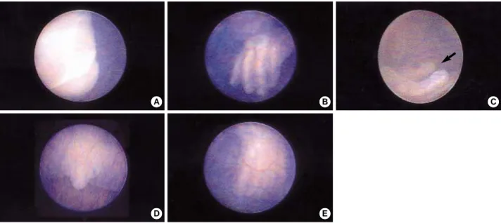
The face, eyes, nose and lips (Fig. 2A) appeared to be nor-mal. The hands were clearly seen and there was no polydactyly or syndactyly of the fingers (Fig. 2B, C). The fetal cord inser-tion site was clearly seen. The external genitalia was seen and still somewhat indeterminate but suggestive of a male (Fig. 2D). Both feet were seen and again, there was no poly- or syn-dactyly of the toes (Fig. 2E). Direct visualization of the fetus was achieved and no gross limb or facial abnormalities were seen. The duration of the procedure was 15 min. She was discharged 2 days after procedure without any complications of fluid leakage, bleeding or uterine contraction. Follow-up
Investigator Case
No. Indication
Needle
(Endoscope diameter) Findings
Duration of procedure (min) Gestation age
(weeks)
Quintero et al. 1993 (1) 11 1 R/O Meckel-Gruber 18 G (0.7 mm) Polydactyly
-syndrome encephalocele
Ginsberg et al. 1994 (2) 11 1 R/O Carpenter 20 G (0.5 mm) Normal 23
syndrome
Hobbins et al. 1994 (3) 11+5 1 R/O Smith-Lemli- 18 G (0.75 mm) Polydactyly 15
10+4 1 Opitz syndrome Normal 5
Reece et al. 1995 (4) 12 1 Suspicious NTD 16 G (0.8 mm) Normal spine
-1996 (5) 12 1 R/O Robert’s Normal
-syndrome
Ville et al. 1996 (6) 12 1 Suspicious 18 G (1.0 mm) Confirmed
-club hands +NTD+short forearm
11 1 Encephalocele confirmed+facial cleft
12 1 Facial cleft Confirmed
-Ville et al. 1997 (7) 12-14 1 R/O Pierre Robin 18 G (1.0 mm) Confirmed
-sequence
1 Increased NT & Cutaneous angioma &
-generalized edema clinodactyly
1 Increased NT & Facial dysmorphia
-generalized edema
Lee et al. 2004 13+5 1 R/O short rib-poly- 18 G (1.0 mm) Normal 15
(The present report) dactyly syndrome
Table 1.Reported cases of prenatal diagnosis in continuing pregnancies by first-trimester transabdominal embryofetoscopy
NTD, neural tube defect; NT, nuchal translucency; -, not recorded; R/O, Rule out.
Fig. 1.Transabdominal sonogram of the fetus in supine position with the breech presentation and a crown-rump length of 6.99 cm and the anterior placenta before embryofetoscopy
Embryofetoscopy for the Detection of Short Rib-polydactyly Syndrome 167 ultrasound in the second trimester was advised and was
per-formed so that normal biometry could be obtained. At 20+4 weeks of gestation, normal fetal anatomy and adequate femur length were confirmed. Serial ultrasound examinations were performed throughout the remainder of pregnancy and con-tinued to reveal normal fetal growth and development with-out evidence of fetal malformations.
A normal full-term female infant was born at 39+3 weeks of gestation via a normal vaginal delivery. The infant weighed 3,250 g and was 20 inches in length. On physical examina-tion, there was no evidence of facial or limb abnormalities. At our last follow-up at 2-months of age, the infant weighed 6,427 g and continued to do well with a normal ophthalmo-logic examination.
DISCUSSION
Short rib-polydactyly syndrome is a lethal skeletal dyspla-sia with marked limb reduction, narrow constricted thorax, short horizontal ribs, pre- and postaxial polydactyly and fre-quent cardiac defects and cystic renal dysplasia. The Majew-ski type (type II) has additional cleft lip/plalate and dispro-portionately shortened tibia (8-10).
SRPS is an autosomal recessive disorder with 25% recur-rence risk (11). Considering the limitations of therapy, pre-natal diagnosis with selective termination of pregnancy is an important option for couples at risk. At the present time, the responsible gene for disorder has not been identified and therefore, the only method of prenatal diagnosis has to be made on the detection of its phenotypic manifestations.
The prenatal diagnosis of Majewski type has been made in fetuses at risk by identification of short tibia, polydactyly,
and cleft lip at fetoscopy using 1.7 mm diameter of endoscope at 16 weeks of gestation (12), or severe micromelia, short ribs with narrow thorax, and polydactyly at ultrasound (13, 14). The earliest sonographic diagnosis has been made at the 16th week of gestation (14). Despite the use of vaginal ultrasound, fetal cleft lip has not been diagnosed until 14 weeks of gestation (15, 16) and is usually recognized during the second trimester.
The fingers and toes of the fetus could always be clearly identified with embryoscopy as early as 9 weeks of gestation (17). Prenatal diagnosis of cleft lip was made at 11 weeks of gestation using embryoscopy (18). Therefore, embryofetoscopy offers a distinct advantage over current diagnostic techniques in that it can assess limb while also ruling out facial anoma-lies early in gestation.
Recently first-trimester embryofetoscopy has been utilized for early prenatal diagnosis of external developmental defects to either confirm or rule out. Our use of thin gauge needle embrofetoscopy in a pregnancy at risk for SRPS highlights the potential of this technique for investigating early in ges-tation the anatomical features of the fetus and embryo in utero. Care should be taken in determining a fetal sex by external genitalia in the first trimester. Until 11 weeks of gestation the external genitalia of the two sexes are similar in appear-ance; the phallus is as big in the female as in the male and develops into penis or clitoris at 11 weeks of gestation. Labium minora and majora are formed from 11 to 14 weeks of ges-tation (19). Our embryofetoscopy at 13 weeks of gesges-tation demonstrated the fetus looked male. We would not see the genitalia during the follow up fetal ultrasound examination after embryofetoscopy. But female external genitalia was observed after birth. During first-trimester embryofetoscopy it is pointed out again that distinguishing features of the
Fig. 2.Embryofetoscopic view of the fetus at 13+5weeks gestation demonstrating normal anatomical structures: (A) lips, (B) four fingers of left hand, (C) thumb of left hand, (D) external genitalia and (E) toes.
A B C
168 K. Lee, J.W. Lee, D.B. Chay, et al. external genitalia appear in 11 weeks of gestation but the
external genitalial organs are not fully differentiated into male or female until 14 weeks of gestation (20). Occasional sex reversal (46, XY with female phenotype) can occur in type I and ambiguous genitalia can be seen in type II (10). Chromo-somal study of the peripheral blood after birth confirmed a normal female, 46, XX although SRPS had been excluded.
Published data do not exist regarding morbidity specifically related to transabdominal embryofetoscopy in continuing pregnancies. The risks of fetal loss, infection, and amniotic membrane rupture are expected to be similar to those associ-ated with early amniocentesis (21, 22). This procedure has a fetal loss rate of 2-2.5% (23, 24). The post procedure cours-es of the patient in this report were completely uneventful. There had been no evidence of damage to the retina in infants born after first trimester transcervical embryoscopy (25).
This report demonstrates the confirmatory potential of transabdominal embryofetoscopy in prenatal diagnosis. It is emphasized that the risks of first-trimester embryofetoscopy still remain to be established.
ACKNOWLEDGEMENTS
We would like to thank Professor E. Albert Reece, Temple University School of Medicine, Philadelphia, U.S.A. for his valuable advice on the setting up the embryofetoscopy unit.
REFERENCES
1. Quintero RA, Abuhamad A, Hobbins JC, Mahoney MJ.
Transab-dominal thin gauge embryo-fetoscopy: a technique for early prena-tal diagnosis and its use in the diagnosis of a case of Meckel Gruber syndrome. Am J Obstet Gynecol 1993; 168: 1552-7.
2. Ginsberg NA, Zbarz D, Strom C. Transabdominal embryoscopy for
the detection of Carpenter syndrome during the first trimester. J Assist Reprod Genet 1994; 11: 373-5.
3. Hobbins JC, Jones OW, Gottesfeld S, Persutte W. Transvaginal
ultra-sonography and transabdominal embryoscopy in the first-trimester diagnosis of Smith-Lemli-Opitz syndrome, type II. Am J Obstet Gynecol 1994; 171: 546-9.
4. Reece EA, Homko CJ, Wiznitzer A, Goldstein I. Needle embryo
-fetoscopy and early prenatal diagnosis. Fetal Diagn Ther 1995; 10: 81-2.
5. Reece EA, Homko CJ, Koch S, Chan L. First-trimester needle embryo
-fetoscopy and prenatal diagnosis. Fetal Diagn Ther 1997; 12: 136-9.
6. Ville Y, Berbard JP, Doumerc S, Multon O, Fernandez H, Frydman R, Barki G. Transabdominal fetoscopy in fetal anomalies diagnosed
by ultrasound in the first trimester of pregnancy. Ultrasound Obstet Gynecol 1996; 8: 11-5.
7. Ville Y, Khalil A, Homphray T, Moscoso G. Diagnostic embryoscopy
and fetoscopy in the first trimester of pregnancy. Prenat Diagn 1997; 17: 1237-46.
8. Majewski F, Pfeiffer RA, Lenz W, Muller R, Feil G, Seiler R. Poly
-syndaktylie, verkurzte Gliedmassen und Genitalfehlbildungen: Kenn
-zeichen eines selbstandigen Syndroms? Z Kinderheilkd 1971; 111: 118-38.
9. Spranger J, Grimm B, Weller M, Weissenbacher G, Herrmann J, Gilbert E, Krepler R. Short Rib-Polydactyly (SRP) syndromes, Types
Majewski and Saldino-Noonan. Z Kinderheilkd 1974; 116: 73-94.
10. Jones KL. Short rib polydactyly syndrome. In Smith’s recognizable
patterns of human malformation, 5th ed. WB Saunders: Philadelphia; 1997; 334-7.
11. Wu MH, Kuo PL, Lin SJ. Prenatal diagnosis of recurrence of short
rib poly- dactyly syndrome. Am J Med Genet 1995; 55: 279-84.
12. Toftager-Larsen K, Benzie RJ. Fetoscopy in prenatal diagnosis of
the Majewski and the Saldino-Noonan types of the short rib-poly-dactyly syndromes. Clin Genet 1984; 26: 56-60.
13. Thomson GS, Reynolds CP, Cruickshank J. Antenatal detection of
recurrence of Majewski dwarf (short rib-polydactyly syndrome type II Majewski). Clin Radiol 1982; 33: 509-17.
14. Gembruch U, Hansmann M, Fodisch HJ. Early prenatal diagnosis
of short rib-polydactyly (SRP) syndrome type I (Majewski) by ultra-sound in a case at risk. Prenat Diagn 1985; 5: 357-62.
15. Bronshtein M, Mashiah N, Blumenfeld I, Blumenfeld Z.
Pseudo-prognathism: an auxiliary ultrasonographic sign for transvaginal ultrasonographic diagnosis of cleft lip and palate in the early sec-ond trimester. Am J Obstet Gynecol 1991; 165: 1314-6.
16. Bronshtein M, Gershoni-Baruch R. Prenatal transvaginal diagnosis
of the ectro-dactyly, ectodermal dysplasia, cleft palate (EEC) syn-drome. Prenat Diagn 1993; 13: 519-22.
17. Dumez Y, Mandelbrot L, Dommergues M. Diagnostic embryoscopy,
50 cases of early first trimester prenatal diagnosis. Prenat Diagn 1992; 12: S10.
18. Dommergues M, Lemerrer M, Couly G, Delezoide AL, Dumez Y.
Prenatal diagnosis of cleft lip at 11 menstrual weeks using embryo
-scopy in the van der Woude syndrome. Prenat Diagn 1995; 15: 378-81.
19. England MA. Genitals: external. In: A colour Atlas of Life before
Birth, Normal fetal development, Wolfe Medical Publications Ltd: London; 1990; 157-62.
20. Moore KL. Development of the external genitalia. In: The
Develop-ing Human. Clinically Oriented Embryology, 4th ed. Saunders: Phi
-ladelphia; 1988; 271-3.
21. Henry GP, Miller WA. Early amniocentesis. J Reprod Med 1992;
37: 396-402.
22. Hanson FW, Zorn EM, Tennant FR, Marianos S, Samuels S.
Amnio-centesis before 15 weeks gestation: Outcome, risks and technical problems. Am J Obstet Gynecol 1987; 156: 1524-31.
23. Penso CA, Sandstrom MM, Garber MF, Ladoulis M, Stryker JM, Benacerraf BB. Early amniocentesis: report of 407 cases with
neona-tal follow-up. Obstet Gynecol 1990; 76: 1032-6.
24. Nicolaides K, Brizot M, Patel F, Snijders R. Comparison of
chori-onic villous sampling and amniocentesis for fetal karyotyping at 10-13 weeks’ gestation. Lancet 1994; 334: 435-9.
25. Dumez Y, Daffos F. L’echographie interventionelle en medecine
foetale. In: Echo-graphie des malformations foetales, Guillet, P (ed). Vigot: Paris; 1990; 407-8.
. . . .