Dendritic cells as targets of
corticotropin-releasing hormone
Hee Jung Lee
Department of Medicine
Dendritic cells as targets of
corticotropin-releasing hormone
Directed by Professor Kwang Hoon Lee
The Master’s Thesis
submitted to the Department of Medicine
and the Graduate School of Yonsei University
in partial fulfillment of the
requirements for the degree of
Master of Medical science
Hee Jung Lee
This certifies that the Master’s Thesis
of Hee Jung Lee is approved.
Thesis Supervisor: Prof. Kwang Hoon Lee
Thesis Committee Member: Prof. Min-Geol Lee
Thesis Committee Member: Prof. Jongsun Kim
The Graduate School
Yonsei University
Acknowledgements
I very much appreciate my thesis supervisor, professor
Kwang Hoon Lee, for his supervision and encouragement to
study this subject.
I appreciate professors Min-Geol Lee and Jongsun Kim who
gave me experienced advice and warm support. I also thank Dr.
Ju Hee Lee, Dr. Chang Ook Park, Nam Soo Chang and Wen
Hao Wu for great support.
I am truly grateful to my family members, especially my
parents and my husband, who have been by my side with love
during the years of my study. I give my love and admiration to
them.
i
Table of Contents
ABSTRACT…….……….……….……….………...1
I. INTRODUCTION……….……….…..3
II. PATIENTS AND METHODS
1. Patients………7
2. Culture of human dendritic cells ……….7
3. RT-PCR analysis……….………8
4. Cytokine and chemokine production………10
5. Detection of CRH-R1 and CRH-R2 by Western blot analysis
………11
III. RESULTS
1. Expression of mRNA for CRH receptor isoforms in DC………..12
2. Western blot anaysis of CRH receptor……….…14
3. The effect of CRH on phenotyping of DC during in vitro generation
………15
4. Effect of CRH on IL-18 mRNA production in DC of atopic
dermatitis patients and nonatopic healthy controls……….…17
5. Effect of CRH on cytokine and chemokine production in DC of
atopic dermatitis patients ……….…19
ii
IV. DISCUSSION………...23
V. CONCLUSION………...…….27
Ⅵ. REFERENCES………..28
iii
List of Figures
Figure 1A. RT-PCR analysis of CRH-R1 isoforms mRNA in DCs of two healthy controls. mRNA for CRH-R1α ……….12 Figure 1B. RT-PCR analysis of CRH-R1 isoforms mRNA in DCs of two healthy controls. mRNA for CRH-R1 β ………. ..12 Figure 2. RT-PCR analysis of CRH-R2 isoforms (α, β, γ) mRNA in DCs of two healthy controls.……….……….…… 13 Figure 3A. Western blot analysis of CRH-R1 in DC of healthy control……. . 14 Figure 3B. Western blot analysis of CRH-R2 in DC of healthy control……. . 14 Figure 4A. CD1a expression of in-vitro generated DCs performed by using flow cytometry…….……. …………. …………. …………. ………….….… .. .... 16 Figure 4B. CD83 expression of in-vitro generated DCs performed by using flow cytometry…….……. …………. …………. …………. ………….….… .. .... 16 Figure 5A, B. RT-PCR analysis of IL-18 mRNA in DCs of atopic dermatitis patients and nonatopic healthy control...……...……...……. ...……...……..18 Figure 6A, B. Flow cytometric analysis of IL-18 protein in DCs of atopic dermatitis patients. ...….…………...……...……...……...……...…….. ...……20 Figure 6C. Flow cytometric analysis of IL-6, CCL17, CCL18, CCL22 protein in DCs of atopic dermatitis patients………..…….. ...……21
iv
List of Tables
- 1 -
ABSTRACT
Dendritic cell as targets of corticotropin-releasing hormone
Hee Jung Lee
Department of Medicine
The Graduate School, Yonsei University
(Directed by Professor Kwang Hoon Lee)
Dendritic cell (DC) plays an important role in the generation and regulation of immune responses, but also is considered to represent the link between allergen uptake and the clinical manifestation of allergic disease, such as atopic dermatitis. Recent evidence suggests that crosstalk between mast cells, nerves and keratinocytes might be involved in exacerbation of the inflammatory conditions by stress, but the mechanism still remains unclear. Corticotropin releasing hormone (CRH), which activates the hypothalamic-pituitary-adrenal axis under stress, also has proinflammatory peripheral effects. However, there have been no reports about CRH receptor expression and functional role of CRH in DC. The purpose of this study was to investigate the expression of CRH receptors and the functional role of CRH in the monocyte-derived DC (MoDC) of atopic dermatitis patients and nonatopic healthy control. In this study, mRNAs for CRH-R1α, 1β, as well as CRH-R1 protein was detected in MoDC. CRH-R2α (but not R2β or R2γ) mRNA and CRH-R2 protein were present in MoDC. Exposure of DC to lipopolysaccharide (LPS) or tumor necrosis factor α (TNF-α), which activate DC, didn’t alter the expression of CRH receptors. Exposure of DC to CRH resulted in decrease of IL-18 in both atopic dermatitis patients and nonatopic healthy control. This effect was more prominent in atopic
- 2 -
dermatitis patients. However, CRH didn’t alter the expressions of IL-6, CCL17, CCL18 and CCL22. Therefore, our results demonstrate that CRH could modulate immune responses by acting directly on DC .
3
Dendritic cell as targets of
corticotropin-releasing hormone
Hee Jung Lee
Department of Medicine
The Graduate School, Yonsei University
(Directed by Professor Kwang Hoon Lee)
I. INTRODUCTION
Atopic dermatitis, psoriasis and other inflammatory skin diseases are
known to be exacerbated by stress1. Recent evidence suggests that crosstalk
between mast cells, neurons and keratinocytes might be involved in such
4
Corticotropin releasing hormone (CRH), a 41-amino acid long peptide is a
main trigger of the hypothalamo-pituitary-adrenal (HPA) axis2-4. Stress
signals in the hypothalamus stimulate expression and release of CRH,
resulting in propiomelanocortin (POMC) expression and adrenocorticotropin
(ACTH) release by the pituitary3. ACTH elicits cortisol release by adrenal
cortex that is responsible for attenuation of the stress response at the central
and peripheral level3.
The skin has its own neuroendocrine system, which is tightly linked into
systemic neuroendocrine axes, probably in order to coordinate peripheral
responses to stress and to maintain cutaneous and global homeostasis5. CRH
is also produced in the skin, where it can act as a regulatory element of local
neuroendocrine interactions5. CRH has been described to act as a growth
factor6,7, apoptosis regulator8 and differentiation factor9. CRH also acts as
pro-inflammatory factor, since it stimulated degranulation of mast cells and
5
growth factor release by mast cells11, IL-6 release by keratinocytes12 and
IL-1β release by monocytes13. CRH also has an anti-inflammatory activity since it diminishes NF-kB activation in epidermal melanocytes14, IL-18 expression
in human HaCaT keratinocytes15. Thus, effects of CRH are dependent on the
cell type and on experimental conditions.
CRH exerts its effects by binding to specific cell surface receptors, of
which two receptor subtypes, CRH-R1 and CRH-R216. Mapping of the
cutaneous CRH signaling system in humans revealed that the CRH receptor
type 1 (CRH-R1) is expressed in all major cellular populations of epidermis,
dermis, and subcutis16. CRH-R1 appears to be the most prevalent isoform of
CRH-R, and the CRH-R2 gene is expressed solely in the dermis and adnexal
structures16. The pathophysiological relevance of CRH-R1 may be reflected
by the observation that CRH-R1 is involved in stress-induced exacerbation of
chronic contact dermatitis in rats17. Furthermore, afftected skin areas from
6
Nevertheless, the functional significance of CRH-R expression in peripheral
tissues is still unresolved. Moreover, there have been few studies regarding
the signals which regulate the expression of CRH-R.
Dendritic cell (DC) is a highly specialized professional antigen-presenting
cell usually located at surveillance interfaces of the human body such as the
skin or mucosa, and is thought to play an important role in the generation and
regulation of immune responses. In particular, DC is considered to represent
the link between allergen uptake and the clinical manifestation of allergic
disease, such as atopic dermatitis19. There have been no reports about CRH-R
expression and functional role of CRH in DC.
The purpose of this study is to investigate the expression of CRH-R, the
signals which regulate the expression of CRH-R, and the functional role of
7
II. PATIENTS AND METHODS
1. Patients
Blood samples are obtained with informed consent from AD patients
according to the criteria of Hanifin and Rajka20. Six AD and six nonatopic
healthy controls are included. The patients, who have not received any
systemic or topical treatment with immunosuppressive drugs for at least four
weeks before collection of blood sample are included. The institutional review
board approved this study.
2. Culture of human dendritic cell
The culture media containes RPMI 1640 (Gibco laboratories, Grand Island,
NY), 2 μM L-glutamine (Gibco), 100 IU/ml penicillin (Gibco), 100 μg/ml
streptomycin (Gibco), and 10% fetal bovine serum (Hyclone, Logan, UT).
8
mononuclear cells (PBMCs) via the cell attachment method21. PBMCs are
attached to six-well plates for 40 minutes. The supernatant and the floating
cells are discarded, and the attached cells are used for culture. Monocytes are
plated in six-well plates at a final concentration of 3×10⁶ cells in 3 ml of
culture medium. DCs are generated by culturing monocytes for six days in
medium supplemented with 500 U/ml GM-CSF, 1000 U/ml IL-4 at days 0, 2,
4, 622. At days 2, 4, and 6, one-third of the medium is removed, and an
equivalent volume of fresh medium supplements with the above mentioned
cytokines. For activation of DC, cells are treated with either
lipopolysaccharide (LPS, Sigma, St Louis, Mo) or tumor necrosis factor α (TNF-α, R&D systems, Minneapolis, MN) for 24h during the last day of culture.
3. RT-PCR analysis
9
the RNeasy mini kit (Qiagen, Hilden, Germany) following the manufacturer's
protocol. Reverse transcription reactions were performed using 1µg of total RNA. Oligonucleotide primers of previously published sequences for
CRH-R1 and CRH-R2 isoforms (Table 1) were used. IL-18 primers were: forward,
5’-AGGAATAAAGATGGCTGCTGAAC-3’; reverse, 5’-GCTCACCAC
AACCTCTACCTCC-3’.
Table 1. PCR primers used for human CRH-R1 and CRH-R2
Amplification is performed on a GeneAmp PCR system 2700 (Applied
Biosystems, Mountain View, CA). PCR is conducted under the following
10
extension at 72℃ for 1 min for 40 cycles. Specific PCR fragments are
separated on a 1% agarose gel and visualized using ethidium bromide staining.
The amounts of PCR products were determined by densitometry using TINA
2.10e software (Raytest, Straubehardt, Germany) and evaluated
semi-quantitatively by grading the ratio between the specific products and the
β-actin band.
4. Cytokine and chemokine production
For stimulation of cytokine production, DCs are distributed to six-well
plates and stimulated with CRH (Sigma) in complete culture medium. CRH
was 96% purity and endotoxin was removed from it. DC was stained for 30
min on ice with human anti IL-6, IL-18, mouse anti-CCL17, anti-CCL22, and
anti-CCL18 monoclonal antibody (R&D systems) in phosphate-buffered
saline containing 0.4% bovine serum albumin. After washing, cells were
11
followed by fixing and permeabilizing the cells with a formaldehyde/saponin
solution (Sigma). Analysis was performed on a FACSCalibur flow cytometer
(Becton Dickinson, Mountain View, CA). Results were expressed as the
relative fluorescence intensity (RFI) calculated from the mean fluorescence
intensity (MFI) as follows: RFI = (MFIparameter evaluated - MFIcontrol)/ MFIcontrol.
5. Detection of CRH-R1 and CRH-R2 by Western blot analysis
DCs were lysed, and prepared for Western blot analysis. Samples were
resolved on 10% SDS-PAGE and then transferred to nitrocellulose (NC)
membranes. The membranes were incubated with goat anti-human CRH-R1
that specifically recognizes CRH-R1, and goat anti-human CRH-R2 that
specifically recognizes CRH-R2 (Santa Cruz Biotechnology, Santa Cruz, CA).
Following stripping and washing, the membranes were incubated with bovine
anti-goat HRP-conjugated serum (Santa Cruz Biotechnology). The protein
12
III. RESULTS
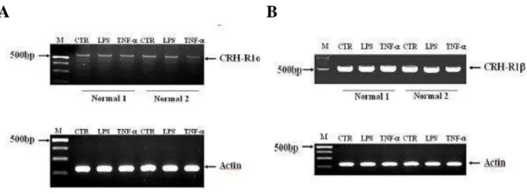
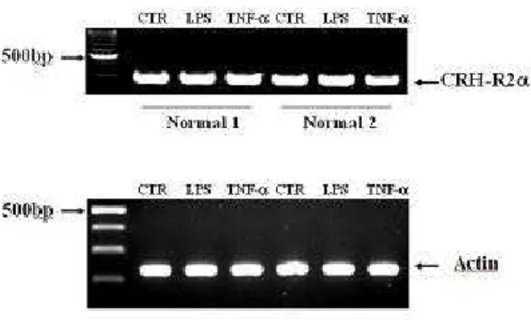
1.
Expression of mRNA for CRH receptor isoforms in DCRT-PCR analysis showed that both immature and mature DCs expressed
specific mRNAs for CRH-R1α, 1β (Figure 1A, B). CRH-R1β was more abundant than CRH-R1α. RT-PCR for CRH-R2 isoforms showed that DC expressed only CRH-R2α (Figure 2). LPS and TNF-α, which were known to activate DCs, didn’t alter the expression of mRNA for CRH-R.
A B
Figure 1. RT-PCR analysis of CRH-R1 isoforms mRNA in DCs of two healthy controls. mRNA for CRH-R1α (A), 1β (B) were detected. For activation, DCs were stimulated for 24h in
13
complete culture medium with LPS or TNF-α during the last day of DC culture. The control lane (CTR) corresponds to untreated cell population. Both LPS and TNF-α didn’t alter the
expression level of CRH-R1α, 1β. Results are representative of three independent experiments
Figure 2. RT-PCR analysis of CRH-R2 isoforms (α, β, γ) mRNA in DCs of two healthy controls. Only mRNA for CRH-R2α was detected. For activation, DCs were stimulated for 24h
in complete culture medium with LPS or TNF-α during the last day of DC culture. The control lane corresponds to untreated cell population. Both LPS and TNF-α didn’t alter the expression
14
2. Western blot analysis of CRH receptor
Western blot analysis was used to detect CRH receptor protein expression in
DC of healthy control. SDS-PAGE of whole cell lysates from DC was
performed using Abs that specifically recognize CRH-R1 or CRH-R2. The Ab
to CRH-R1 yieled a single band of ~50 kDa (Figure 3A). Western blot
analysis for CRH-R2 identified a strong band of 49 kDa and a minor band of
51 kDa (Figure 3B). Lipopolysaccharide (LPS) and TNF-α, which were known to activate DCs, didn’t alter the expression of CRH receptor protein
(Figure 3A, B).
A B
Figure 3. Western blot analysis of CRH-R1 (A) and CRH-R2 (B) in DC of healthy control. DC was collected, lysed and samples of the whole cell lysates were run on 10% SDS-PAGE, using
15
Abs that specifically recognize CRH-R1 or CRH-R2. The Ab to CRH-R1 yieled a single band of ~50 kDa (A). Western blot analysis for CRH-R2 identified a strong band of 49 kDa and a
minor band of 51 kDa (B). For activation, DC was stimulated for 24h in complete culture medium with LPS or TNF-α during the last day of DC culture. The control lane (CTR)
corresponds to untreated cell population. LPS and TNF-α didn’t alter the expression of CRH receptor protein. Results are representative of three independent experiments MW, molecular
weight marker.
3. The effect of CRH on phenotyping of DC during in vitro
generation
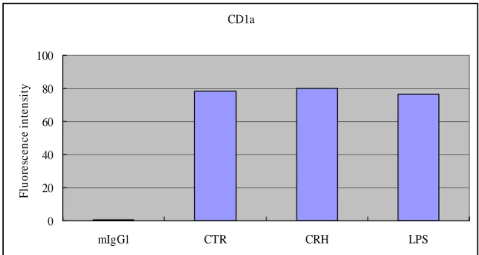
Phenotypic analysis of MoDC was performed by using flow cytometry. To
investigate the effect of CRH on phenotyping of DC, cells were exposed to
CRH at 50nM for 24h during the last day of culture. Expression of CD1a
wasn’t affected by both CRH and LPS (Figure 4A). Although CRH didn’t
16
CD1a 0 20 40 60 80 100 mIgG1 CTR CRH LPS F lu o re sc e n c e i n te n si ty CD83 0 20 40 60 80 100 mIgG1 CTR CRH LPS F lu o re sc e n c e i n te n si ty A BFigure 4. Phenotypic analysis of MoDC was performed by using flow cytometry. To investigate the effect of CRH on phenotyping of DC, cells were exposed to CRH at 50nM for 24h during
the last day of culture. LPS was used for positive control. The control (CTR) corresponds to untreated cell population. Expression of CD1a wasn’t affected by both CRH and LPS (Figure
17
4A). Although CRH didn’t alter the expression of CD83, LPS increased CD83 markedly (Figure 4B).
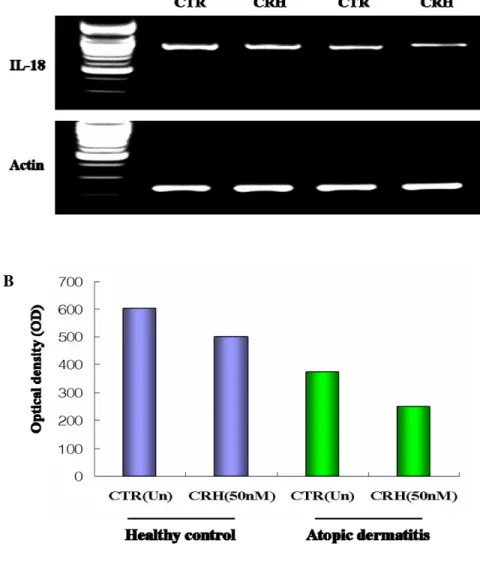
4. Effect of CRH on IL-18 mRNA production in DC of atopic
dermatitis patients and nonatopic healthy controls
To study the effect of CRH on IL-18 mRNA expression in DC, the cells
were exposed to CRH at 50nM for 6h. Total RNA was extracted and RT-PCR
for IL-18 were performed. IL-18 mRNA expression was reduced by CRH in
both atopic dermatitis patients and nonatopic healthy control (Figure 5A).
However, the effect was more prominent in atopic dermatitis patients than
18
A
B
Figure 5. RT-PCR analysis of IL-18 mRNA in DC of atopic dermatitis patients and nonatopic healthy control. For studying effect of CRH, DCs were treated for 6h in complete culture
medium with CRH at 50nM during the last day of DC culture. The control lane corresponds to untreated cell population. IL-18 mRNA expression was reduced by CRH in both atopic
19
atopic dermatitis patients than nonatopic healthy control (Figure 5B). Results are representative of three independent experiments.
5. Effect of CRH on cytokine and chemokine production in DC of
atopic dermatitis patients.
To investigate the effect of CRH on cytokine and chemokine protein
expression in DC of atopic dermatitis patients, the cells were subjected to
flow cytometric analyses using human anti IL-6, IL-18, mouse anti-CCL17,
anti-CCL22, and anti-CCL18 monoclonal antibody (R&D systems,
Minneapolis, MN). Cells were exposed to CRH at 50nM for 24h during the
last day of culture. IL-18 expression was reduced by CRH in atopic dermatitis
patients (Figure 6A, B). However, there was no change in expression of IL-6,
20
A BIL-18 0 2 4 6 8 10 CT R CR H R F I
21
C22
Figure 6. Flow cytometric analysis of IL-18 protein in DCs of atopic dermatitis patients. For studying effect of CRH, DCs were treated for 24h in complete culture medium with CRH at
50nM during the last day of DC culture. The control (CTR) corresponds to untreated cell population. IL-18 expression was reduced by CRH in atopic dermatitis patients (Figure 5A, B).
There was no change in expression of IL-6, CCL17, CCL18 and CCL22 (Figure 6C). Results are representative of three independent experiments.
23
IV. DISCUSSION
To our knowledge, this is the first report that mRNA of CRH receptor
isoforms is expressed in human DCs. We detected mRNA for CRH-R1α, 1β,
as well as CRH-R1 protein. CRH-R2α (but not R2β or R2γ) mRNA and
CRH-R2 protein were present in MoDC. CRH-R1 mRNA is widely expressed
in mammalian brain and pituitary and is responsible for activation of the
POMC gene and ACTH and β-endorphin release from the anterior pituitary23. In human peripheral tissues, CRH-R1 is expressed in a wide range of tissues
such as the testis, ovary, endometrium, myometrium, placenta, adrenal gland,
adipose tissue, skin, spleen, heart, and specific cells of the immune system4.
The gene for human CRH-R1 contains 14 exons, and the complete gene
product is a 444 amino acid protein, termed CRH-R1β, that exhibits impaired agonist binding and signaling properties24. Excision of exon 6, which encodes
24
of CRH-R1α mRNA. This appears to be the main functional CRH-R1 receptor variant containing 415 amino acids, which primarily mediates CRH
actions4. Concerning CRH-R2 variants, CRH-R2α and 2β are found in both human and rodents, and 2γ has so far been found only in the limbic regions of the human CNS25-27. These three variants differ only in their N-terminal
extracellular domains. The different N termini do not significantly alter
agonist binding and signaling properties of the various CRH-related peptides,
although the CRH-R2β is about 10-fold more potent in second messenger activation compared with CRH-R2α or R2γ27. These variants, however, do exhibit significant differences in their tissue distribution. Slominski et al.28
reported that none of CRH-R2 subtypes was detected in epidermal normal and
malignant keratinocytes, normal and malignant melanocytes, dermal
fibroblasts, adipose tissue. By contrast, CRH-R2α was detectable in cells derived from human scalp adnexal structures (hair follicle keratinocytes and
25
mRNA splicing mechanisms of CRH-R might explain many different
biological actions of CRH in various tissues.
CRH profoundly influences the function of the immune system indirectly,
through the activation of the HPA axis and sympathetic system, and directly,
through the local modulatory actions on inflammatory responses3.
Epidemiological and experimental studies suggest that stress and stress
hormones influence the development, course, and pathology of certain allergic,
autoimmune/inflammatory, infectious, and neoplastic diseases, mainly by
stimulating Th2 instead of Th1-type immune response, which means by
enhancing humoral and suppressing cellular immunity29. Atopic dermatitis,
psoriasis, and other inflammatory skin diseases are known to be exacerbated
by stress. It was recently reported that CRH regulates the expression of IL-18
in HaCaT cell15. However this is the first time that CRH decreases expression
of IL-18 in DCs. Moreover, this effect of CRH on IL-18 production was more
26
Because IL-18 functions primarily as an IFN-γ inducer and a promoter of Th1 responses in T cells, our results not only supports the previous hypothesis that
stress downregulates cellular immunity, but also address that CRH could
27
V. CONCLUSION
In conclusion, DCs express mRNAs for CRH-R1α, 1β and CRH-R2α, but
not those for R2β and R2γ. Both CRH-R1 and CRH-R2 protein were also
present in DCs. Furthermore, it was found that CRH regulates the expression
of IL-18 in DCs via specific CRH-Rs. These findings suggest that CRH could
modulate immune responses by acting directly on DCs and may provide an
insight into the pathophysiology of neuroinflammatory skin disease such as
28
REFERENCES
1. Katsarou-Katsari A, Filippou A, Theoharides TC. Effect of stress and other psychological factors on the pathophysiology and treatment of dermatoses. Int J Immunopathol Pharmacol 1999;12:7-11.
2. Vale W, Spiess J, Rivier C, Rivier J. Characterization of a 41-residue ovine hypothalamic peptide that stimulates secretion of corticotropin and beta-endorphin. Science 1981;213:1394–7.
3. Chrousos GP. The hypothalamic–pituitary–adrenal axis and immune-mediated inflammation. N Engl J Med 1995;332:1351–63.
4. Hillhouse EW, Grammatopoulos DK. The molecular mechanisms
underlying the regulation of the biological activity of corticotropin releasing hormone receptors: implications for physiology and pathophysiology. Endocr Rev 2006;27:260-86
5. Arck PC, Slominski A, Theoharides TC, Peters EM, Paus R.
Neuroimmunology of stress: skin takes center stage. J Invest Dermatol 2006;126:1697-704
6. Korbonits M, Morris DG, Nanzer A, Kola B, Grossman AB. Role of regulatory factors in pituitary tumour formation. Front Horm Res 2004;32:63–95.
29
7. Slominski A, Zbytek B, Pisarchik A, Zmijewski RM, Wortsman J. CRH functions as a growth factor/cytokine in the skin. J Cell Physiol
2006;206:780–91.
8. Dermitzaki E, Tsatsanis C, Gravanis A, Margioris AN. Corticotropin releasing hormone induces Fas ligand production and apoptosis in PC12 cells via activation of p38 mitogen-activated protein kinase. J Biol Chem 2002;277:12280–7.
9. Zbytek B, Slominski AT. Corticotropin releasing hormone induces keratinocyte differentiation in the adult human epidermis. J Cell Physiol 2005;203:118–26.
10. Theoharides TC, Donelan JM, Papadopoulou N, Cao J, Kempuraj D, Conti P. Mast cells as targets of corticotropinreleasing factor and related peptides. Trends Pharmacol Sci 2004;25:563–8
11. Cao J, Papadopoulou N, Kempuraj D, Boucher WS, Sugimoto K, Cetrulo CL, Theoharides TC. Human mast cells express corticotrophin-releasing hormone (CRH) receptors and CRH leads to selective secretion of vascular endothelial growth factor. J Immunol 2005;174:7665-75
12. Zbytek B, Mysliwski A, Slominski A, Wortsman J, Wei ET, Mysliwska J. Corticotropin releasing hormone affects cytokine production in human HaCaT keratinocytes. Life Sci 2002;70:1013–21.
30
13. Paez Pereda M, Sauer J, Perez Castro C, Finkielman S, Stalla GK, Holsboer F, et al. corticotropin releasing hormone differentially modulates the interleukin-1 system according to the level of monocyte activation by endotoxin. Endocrinology 1995;136:5504–10.
14. Zbytek B, Pfeffer LM, Slominski AT. CRH inhibits NF-kB signaling in human melanocytes. Peptides 2006;27:3276-83.
15. Park HJ, Kim HJ, Lee JH, Lee JY, Cho BK, Kang JS, et al. Corticotropin-Releasing Hormone (CRH) Downregulates Interleukin-18 Expression in Human HaCaT Keratinocytes by Activation of p38 Mitogen-Activated Protein Kinase (MAPK) Pathway. J Invest Dermatol 2005;124:751–5. 16. Slominski A, Wortsman J, Pisarchik A, Zbytek B, Linton EA,
Mazurkiewicz JE, et al. Cutaneous expression of corticotropin releasing hormone (CRH), urocortin, and CRH receptors. FASEB J 2001;15:1678–93. 17. Kaneko K, Kawana S, Arai K, Shibasaki T. Corticotropin-releasing factor
receptor type 1 is involved in the stree-induced exacerbation of chronic contact dermatitis in rats. Exp Dermatol 2003;12:47-52
18. Papadopoulou N, Kalogeromitros D, Staurianeas NG, Tiblaexi D, Theoharides TC. Corticotropin-releasing hormone receptor-1 and histidine decarboxylase expression in chronic urticaria. J Invest Dermatol
31
19. Adams S, O’neill DW, Bhardwaj N. Recent advances in dendritic cell biology. J Clin Immunol 2005;25:175-88
20. Hanifin JM, Rajka G. Diagnostic features of atopic dermatitis. Acta Derm Venereol (Stockh) 1980;92(suppl):S44-7.
21. Choi GS, Kang JM, Lee MG. Analysis of methods for the generation of dendritic cells from human peripheral blood monocytes. Yonsei Med J 2000;41:642-50.
22. Sallusto F, Lanzavecchia A. Efficient presentation of soluble antigen by cultured human dendritic cells is maintained by granulocyte/macrophage colony-stimulating factor plus interleukin 4 and down-regulated by tumor necrosis factor α. J Exp Med 1994;179:1109-18.
23. Van Pett K, Viau V, Bittencourt JC, Chan RKW, Li H-Y, Arias C, et al. Distribution of mRNAs encoding CRF receptors in brain and pituitary of rat and mouse. J Comp Neurol 2000;428:191–212.
24. Chen R, Lewis KA, Perrin MH, Vale WW. Expression cloning of a human corticotropin-releasing-factor receptor. Proc Natl Acad Sci 1993;90:8967– 71.
25. Kishimoto T, Pearse RV, Lin CR, Rosenfeld MG. A sauvagine/ corticotropin-releasing factor receptor expressed in heart and skeletal muscle. Proc Natl Acad Sci 1995;92:1108–12.
32
26. Valdenaire O, Giller T, Breu V, Gottowik J, Kilpatrick G. A new
functional isoform of the human CRF2 receptor for corticotrophin-releasing hormone. Biochim Biophys Acta 1997;1352:129–32.
27. Kostich WA, Chen A, Sperle K, Largent BL. Molecular identification and analysis of a novel human corticotropin-releasing factor (CRF) receptor: the CRF2γ receptor. Mol Endocrinol 1998;12:1077–85.
28. Slominski A, Pisarchik A, Tobin DJ, Mazurkiewicz JE, Wortsman J. Differential expression of a cutaneous corticotropin releasing hormone system. Endocrinology 2004;145:941-50.
29. Ramirez F, Fowell DJ, Puklavec M, Simmonds S, Mason D.
Glucocorticoids promote a TH2 cytokine response by CD4+ T cells in vitro. J Immunol 1996;156:2406–12.
33
Abstract (In Korean)
수지상세포에서 Corticotropin releasing hormone 의 작용 <지도교수 이광훈> 연세대학교 대학원 의학과 이희정 스트레스가 아토피피부염, 건선, 지루성 피부염, 원형 탈모증 등 여러 피부과적 질환의 경과에 영향을 준다는 것은 일반적으로 받아들여지는 사실이나, 어떠한 면역학적 기전으로 작용하는지는 아직 명확히 밝혀져 있지 않다. Corticotropin releasing hormone (CRH)은 대표적인 스트레스 매개 물질로 각질형성세포, 비만세포 등 다양한 세포에 영향을 준다는 것이 최근 보고되고 있으나 아토피피부염 환자의 병인에 중요한 세포 중 하나인 수지상 세포에 서의 역할에 대해서는 연구된 바가 없다. 본 연구에서는 수지상세포에서 CRH 수용체(CRH-R)의 발현양상과 CRH 가 싸이토카인 및 케모카인 발현에 미치는 영향을 알아보고자 하였다. 연구결과 말초혈액 단핵세포에서 배양한 수지상세포에 존재하는 CRH-R 아형은 CRH-R1α, 1β 와 CRH-R2α 였다. CRH-R 의 발현은
34
수지상세포를 활성화시키는 것으로 알려진 lipopolysaccharide (LPS)나 tumor necrosis factor α (TNF-α)의 영향을 받지 않았다. CRH 는 아토피피부염 환자와 정상인 모두에서 수지상 세포의 interleukin 18 (IL-18) 발현을 감소시켰으며, 이러한 결과는 아토피피부염 환자에서 더 뚜렷하였다. Thymus and activation-regulated chemokine (TARC/CCL17), macrophage-derived chemokine (MDC/CCL22) 및 CC chemokine ligand 18 (CCL18)의 발현은 CRH 의 영향을 받지 않았다. 이상의 결과로 수지상 세포에는 CRH 특이 수용체가 존재하며, CRH 는 이를 통해 IL-18 감소 등 면역반응 조절에 관여함을 확인할 수 있었다.
핵심되는 말: Corticotropin releasing hormone, 수지상세포