INTRODUCTION
Kawasaki disease (KD) is an acute febrile vasculitis affect-ing primarily infants and young children, and 15% to 25% of affected children may subsequently develop coronary ar-tery aneurysms, if not treated (1, 2). They may cause sudden death or ischemic heart disease as a result of thrombotic or stenotic occlusion of coronary arteries (3). Two-dimensional echocardiography and selective coronary angiography are standard methods to evaluate coronary artery lesions; how-ever, these are limited to the assessment of wall morphology. Recent progress in intravascular ultrasound (IVUS) technol-ogy allows detailed visualization of morphological changes in vascular wall. However, only a limited number of studies have been reported on the coronary artery using IVUS imag-ing in patients with KD, and the intervals from the onset of the illness to the time of the IVUS study ranged from 7 to 16 yr (4-6).
We illustrate IVUS findings of coronary wall morphology at 22 months after the onset of the disease in a girl with coro-nary aneurysms due to KD.
CASE REPORT
A 4-yr-old girl developed fever (39℃), reddening of the pharynx and a tender left-sided cervical adenopathy. She was tentatively diagnosed as having a streptococcal pharyngitis
and oral cephalosporin was given at a primary care clinic, but her illness worsened with the appearance of erythematous swelling of both hands, arthralgia, myalgia, and abdominal pain. She was transferred to our hospital on 9th day of the disease.
At admission, her general condition was poor and pyrexia (38.5℃), tachycardia (130/min), and arthralgia on both knees and ankles were noted. Bilateral conjunctival injections and oral involvement including strawberry tongue, mucosal hyperemia, and cracked and erythematous lips were evident. There was no cervical lymphadenopathy. Erythematous swell-ing of both hands and feet was also noted. There was a leuko-cytosis of 17,500/ L with 93% neutrophils, 6% lympho-cytes and the platelet count was 236,000/ L. The erythrocyte sedimentation rate was increased to 132 mm/hr and the C-reactive protein level, 30.5 mg/dL. Bacteriologic and sero-logic studies were negative. The electrocardiogram showed normal pattern. The initial echocardiogram showed aneu-rysms of the proximal right coronary artery with a maximum diameter of 9 mm, and dilatation of the mid-left anterior descending coronary artery with a diameter of 5 mm. The patient was immediately treated with intravenous gamma globulin (2 g/kg) and salicylate (100 mg/kg/day). The fever disappeared within 24 hr and all other clinical signs within a few days after the initiation of treatment.
During follow-up, the patient had been treated with sali-cylate (5 mg/kg/day) and dipyridamole (4 mg/kg/day). Selec-tive coronary angiography was performed to confirm the
Sejung Sohn, Hae Soon Kim, Seong Hee Jeon, Seong Hoon Park Departments of Pediatrics and Cardiology, Ewha Womans University College of Medicine and Ewha University Mokdong Hospital, Seoul, Korea
Received : 29 September 2000 Accepted : 22 December 2000
Address for correspondence
Sejung Sohn, M.D.
Department of Pediatrics, Ewha Womans University Mokdong Hospital, 911-1 Mok-dong, Yangchon-gu, Seoul 158-710, Korea
Tel : +82.2-650-5579, Fax : +82.2-653-3718 E-mail: sohn@mm.ewha.ac.kr
661 J Korean Med Sci 2001; 16: 661-3
ISSN 1011-8934
Copyright � The Korean Academy
of Medical Sciences
Intravascular Ultrasound Findings of Coronary Wall Morphology in a
Child with Kawasaki Disease
Intravascular ultrasound (IVUS) imaging was performed to assess the coronary wall morphology in detail at 22 months after the onset of Kawasaki disease in a girl who had developed coronary aneurysms at 4 yr of age. The sites of persis-tent aneurysms demonstrated a dilated lumen with a marked symmetrical or asymmetrical thickening of the intima-media complex. This pathologic finding was also present in angiographically normal vessels near an aneurysm but with a mild thickening. Coronary artery calcification was observed at one site in the aneurysms. The sites of normal coronary artery far from an aneurysm showed normal intravascular ultrasound findings with no measurable intima-media com-plex. Our case indicates that the healing process may continue via cell prolifera-tion, with extension to the proximity of the coronary aneurysms. IVUS is useful to evaluate the coronary wall morphology and may be valuable in the long-term follow-up of coronary lesions due to Kawasaki disease.
662 S. Sohn, H.S. Kim, S.H. Jeon, et al. coronary artery lesions at 7 months after the onset of the
disease. To evaluate the morphological change in coronary artery lesions, follow-up coronary angiography was performed at 15 months after the first study. The coronary aneurysms in the acute stage were not changed at the second angio-graphy. There was no stenotic lesion. IVUS imaging was car-ried out at the time of follow-up angiography to obtain more information on vascular wall changes.
Using a 6F guiding catheter (Boston Scientific Corp., Maple Groove, MN) and a 0.014 inch guidewire, an elec-tronic 3.5F, 20 MHz, 64-element imaging catheter (Visions FX, Endosonics, Rancho Cordova, CA) was introduced into the distal portion of the right coronary artery. Real-time cross-sectional images of the entire right coronary artery were obtained on pullback and images were continuously recorded on videotape (S-VHS). The integrated fluoroscopy window of the IVUS systems allowed optimal localization of the ultrasonic probe position together with on-line
analy-sis of the IVUS images. The thickness of the intima-media complex was measured as a distance from the lumen-vessel wall interface to the trailing edge of the echolucent zone in the ultrasound images.
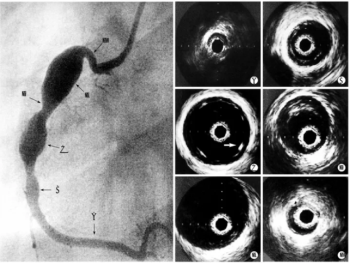
By ultrasound the sites of persistent aneurysms demon-strated a dilated lumen with a marked symmetrical (site B, C) or asymmetrical (site D, E) thickening of the intima-media complex: the images showed two layers of the vessel wall with no sonographic evidence of a distinct medial layer (Fig. 1). Within the aneurysms the thickness of the intima-media complex ranged from 0.4 to 1.4 mm. No thrombus was detected. This pathologic finding was also present in angiographically normal vessels near an aneurysm (site F) but with a mild thickening of 0.4 mm (normal, <0.3 mm). Coronary artery calcification was observed at one site in the aneurysms by IVUS. It was detected at the innermost lumen as a focal bright echo signal with acoustic shadowing. Far-ther distally, the right coronary artery showed no layered
ap-Fig. 1.A coronary angiogram and intravascular ultrasound (IVUS) images from the right coronary artery. The ultrasound image (far from an aneurysm) shows normal IVUS pattern without evidence of different layers (A). The ultrasound images (aneurysm) demon-strates a dilated lumen and a marked symmetrical (B, C) or asymmetrical (D, E) thickening of the intima-media complex with two-layer appearance. Note coronary artery calcification (arrow) with acoustic shadowing (C). The ultrasound image (angiographically normal) adjacent to a large aneurysm shows a mild thickening of the intima-media complex (F).
A B C D E F F E D C B A
Intracoronary Ultrasound in Kawasaki Disease 663 pearance with no measurable intima-media complex (site A).
DISCUSSION
Only 22 months after the onset of KD, IVUS revealed a pathologic thickening of the intima-media complex within the coronary aneurysms and in the adjacent angiographically normal coronary arteries. It also demonstrated calcification within the aneurysm, not detected by angiography. How-ever, apparently normal coronary arteries far from the aneurysm at the acute stage of the disease showed normal wall struc-ture, similar to those in normal children (4). We used the term of intima-media complex cited from Suzuki et al. (5) because medial layer could not be sonographically differen-tiated from the thickened intima. The thickness of the intima-media complex up to 1.4 mm was pathologic. The thick-ness of the intima >0.3 mm is considered abnormal in chil-dren and young adults (4, 5). In addition, as the normal coronary artery in childhood shows a single layered appear-ance in IVUS, layered appearappear-ance of the vessel wall itself indicates the presence of abnormal thickening of the intima, unless intimal thickness is <0.3 mm (4, 5). Intimal thick-ening of the coronary vessel wall is caused mainly by the proliferation of smooth muscle cells of the media (7), which is a possible mechanism of angiographic regression of aneu-rysms.
Sugimura et al. (4) demonstrated intimal thickening in regressed aneurysms but in only 1 of 5 persistent aneurysms at 7 to 15 yr from the onset of KD. Similarly, Iemura et al. (6) revealed intimal thickening in regressed aneurysms after 10 to 16 yr from disease onset, with more thickening in ini-tially large sized (≥4 mm) aneurysms than in small ones. However, Suzuki et al. (5) observed a remarkably thickened intima-media complex in persisting aneurysms as well as in regressed ones at 11.5±2.3 yr from the onset but with more thickening in the latter. In contrast, we observed intimal thickening within persistent aneurysms and in the adjacent angiographically normal vessels at 22 months after KD. This difference may be due to a shorter interval after the onset of KD and continuous healing process of the vessel wall. Our finding is compatible with that by von Xylander et al. (8) in terms of the presence of intimal thickening within a per-sistent aneurysm even a relatively short period of time after KD. Our case indicates that the healing process may contin-ue via cell proliferation, with extension to the proximity of the coronary aneurysms.
In addition to the evaluation of the coronary wall morphol-ogy in patients with KD, Iemura et al. (6) found that at the site of regressed aneurysms, there was significantly more vascular constriction with acetylcholine, and poorer dilata-tion with isosorbide dinitrate than in normal vessels. This result indicates that there is persistent functional impairment as well as abnormal vascular wall morphology in patients with KD.
It is not clear when the intimal thickening within aneu-rysms occurs during the course of KD, because it is difficult to perform IVUS study during the acute stage of the disease in children with small coronary arteries. With miniaturiza-tion of the ultrasound transducer, it may be possible to eval-uate the intimal changes in the early stage of development of the aneurysm.
REFERENCES
1. Kato H, Ichinose E, Yoshioka F, Takechi T, Matsunaga S, Suzuki K, Rikitake N. Fate of coronary aneurysms in Kawasaki disease: serial coronary angiography and long-term follow-up study. Am J Cardiol 1982; 49: 1758-66.
2. Suzuki A, Kamiya T, Kuwahara N, Ono Y, Kohata T, Takahashi O, Kimura K, Takamiya M. Coronary arterial lesions of Kawasaki dis-ease: cardiac catheterization findings of 1,100 cases. Pediatr Cardi-ol 1986; 7: 3-9.
3. Kato H, Ichinose E, Kawasaki T. Myocardial infarction in Kawasaki disease: clinical analyses in 195 cases. J Pediatr 1986; 108: 923-7. 4. Sugimura T, Kato H, Inoue O, Fukuda T, Sato N, Ishii M, Takagi J,
Akagi T, Maeno Y, Kawano T, Takagishi T, Sasaguri Y. Intravascu-lar ultrasound of coronary arteries in children. Assessment of the wall morphology and the lumen after Kawasaki disease. Circulation 1994; 89: 258-65.
5. Suzuki A, Yamagishi M, Kimura K, Sugiyama H, Arakaki Y, Kamiya T, Miyatake K. Functional behavior and morphology of the coronary artery wall in patients with Kawasaki disease assessed by intravascu-lar ultrasound. J Am Coll Cardiol 1996; 27: 291-6.
6. Iemura M, Ishii M, Sugimura T, Akagi T, Kato H. Long term conse-quences of regressed coronary aneurysms after Kawasaki disease: vascular wall morphology and function. Heart 2000; 83: 307-11. 7. Sasaguri Y, Kato H. Regression of aneurysms in Kawasaki disease: a
pathologic study. J Pediatr 1982; 100: 225-31.
8. von Xylander S, Mudra H, Rieber J, Klaus V, Dohlemann C. Intravas-cular ultrasonography of an adolescent boy with a coronary artery aneurysm due to Kawasaki disease. Pediatr Cardiol 1997; 18: 437-9.