Neurologic deficits resulting from stroke remain largely intractable, which has prompted thou-sands of studies aimed at developing methods for treating these neurologic sequelae. Endoge-nous neurogenesis is also known to occur after brain damage, including that due to cerebral in-farction. Focusing on this process may provide a solution for treating neurologic deficits caused by cerebral infarction. The phosphatidylinositol-3-kinase (PI3K) pathway is known to play im-portant roles in cell survival, and many studies have focused on use of the PI3K pathway to treat brain injury after stroke. Furthermore, since the PI3K pathway may also play key roles in the physiology of neural stem cells (NSCs), eliciting the appropriate activation of the PI3K pathway in NSCs may help to improve the sequelae of cerebral infarction. This review describes the PI3K pathway, its roles in the brain and NSCs after cerebral infarction, and the therapeutic pos-sibility of activating the pathway to improve neurologic deficits after cerebral infarction. Key Wordszz neural stem cells, stroke, phosphatidylinositol-3-kinases, regeneration.
The Role of the PI3K Pathway in the Regeneration
of the Damaged Brain by Neural Stem Cells
after Cerebral Infarction
INTRODUCTION
Stroke is one of the most common diseases and can induce several debilitating sequelae such
as hemiplegia, aphasia, and dementia. The most important treatment for stroke thus far is
thrombolysis, ideally within the so-called golden hour during which treatment is most likely
to be effective, or at least within around 4.5 hours of symptom onset of a cerebral infarction.
1However, not all patients with a cerebral infarction are candidates for thrombolysis. Due to
the risk of hemorrhage, there are strict inclusion and exclusion criteria for thrombolytic
therapy, and because of the nature of the disease and its treatment, many patients must
un-fortunately live with long-term neurologic deficits. Numerous clinical trials have tested
di-verse putative neuroprotective agents and stem cells as treatments for these deficits, but
al-most all of them have been found to be ineffective. Therefore, continued effort is necessary to
develop new therapeutic strategies for treating cerebral infarctions.
Ways of reversing the sequelae of cerebral infarction have been investigated, and various
molecular pathways have been found to play critical roles in the pathogenesis. Based on
these findings, several methods for blocking the pathogenic mechanisms have been
pro-posed and developed. One pathogenic mechanism is the pathway involving
phosphati-dylinositide 3-kinase (PI3K), which has been investigated intensively.
2-4Since this pathway is
important in cell survival and is significantly affected by ischemia, many attempts have been
made to modulate it for the treatment of cerebral infarction and to prevent the programmed
cell death caused by ischemic strokes.
Endogenous neural stem cells (NSCs) are well known to exist in the subventricular zone
Seong-Ho Koha,b Eng H. Loa
a Neuroprotection Research Laboratory, Massachusetts General Hospital, Harvard Medical School, Boston, MA, USA
b Department of Neurology, Hanyang University College of Medicine, Seoul, Korea
pISSN 1738-6586 / eISSN 2005-5013 / J Clin Neurol 2015;11(4):297-304 / http://dx.doi.org/10.3988/jcn.2015.11.4.297
Received February 16, 2015 Revised May 25, 2015 Accepted May 28, 2015 Correspondence Seong-Ho Koh, MD, PhD Department of Neurology,
Hanyang University College of Medicine, 153 Gyeongchun-ro, Guri 11923, Korea Tel +82-31-560-2267
Fax +82-31-560-2267 E-mail [email protected]
cc This is an Open Access article distributed under the terms of the Creative Commons Attribution Non-Com-mercial License (http://creativecommons.org/licenses/by-nc/3.0) which permits unrestricted non-comNon-Com-mercial use, distribution, and reproduction in any medium, provided the original work is properly cited.
Role of the PI3K Pathway in Neural Stem Cell Activity
JCN
of the ventricles and subgranular zone of the hippocampus.
5,6They can proliferate under appropriate stress and then
differ-entiate into various neuronal cells. Several studies have found
that mild-to-moderate ischemic strokes can induce
endoge-nous neurogenesis, and this neurogenesis may function to
reduce the severity of neurologic sequelae.
7-9Therefore,
methods for increasing endogenous neurogenesis have been
sought in numerous studies. It has been established that the
role of the PI3K pathway is crucial in the function of NSCs;
therefore, if the PI3K pathway can be activated in NSCs after
cerebral infarction, it could contribute to the recovery of
sub-sequent neurologic deficits.
This review describes the PI3K pathway itself, its roles in
the brain and in the activity of NSCs after cerebral infarction,
and the possibility of using methods for activating the PI3K
pathway in NSCs for the treatment of cerebral infarction.
WHAT IS THE PI3K PATHWAY?
It is well known that the PI3Ks are involved in various cellular
functions, such as cell proliferation, growth, differentiation,
motility, survival, and intracellular trafficking. The PI3K
path-way is necessary for the survival of both neurons and NSCs.
10,11PI3K is not a single enzyme, but rather a family of many
dif-ferent subtypes. The PI3K family is divided into three difdif-ferent
classes (Class I, Class II, and Class III) based on the primary
structure, regulation, and in vitro lipid substrate specificity.
12Among these, the Class I PI3Ks are the best understood and
can be divided into two groups: Class IA (p110α, p110β, and
p110δ) and Class IB (p110γ).
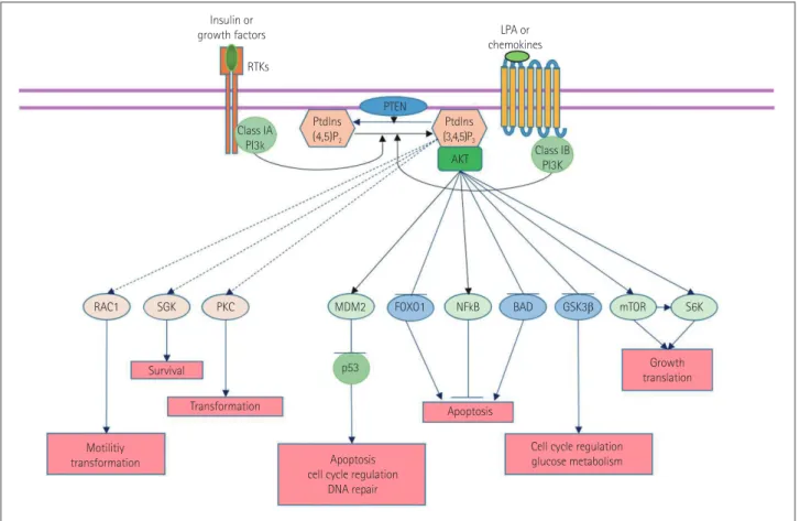
13The PI3K pathway is known
to interact with the insulin receptor substrate (IRS) and is
closely linked with the tumor suppressor phosphatase and
tensin homolog (PTEN), which inhibits PI3Ks. When the IRS
is activated by insulin, it activates PI3Ks and then regulates
glucose uptake through diverse phosphorylation events. In
detail, activated PI3Ks phosphorylate the 3-position hydroxyl
group of the inositol ring of phosphatidylinositol (Ptdlns) to
produce phosphoinositide Ptdlns(3,4,5)P
3from Ptdlns(4,5)
P
2.
14,15Ptdlns(3,4,5)P
3activates many different downstream
effectors, the most well known of which is Akt (protein kinase
B).
16Akt phosphorylated by PI3Ks (pAkt) affects many
im-portant downstream signals, including mouse double minute
Fig. 1. Role of the phosphatidylinositol-3-kinase (PI3K) pathway in cells. Akt: protein kinase B, BAD: Bcl-2-associated death promoter, FOXO1: forkhead box protein O1, GSK3β: glycogen synthase kinase 3β, MDM2: mouse double minute 2 homolog, mTOR: mammalian target of rapamycin, NF-kB: nuclear factor kappa-light-chain enhancer of activated B cells, PKC: protein kinase C, Ptdlns: phosphatidylinositol, PTEN: phosphatase and tensin homolog, RAC1: Ras-related C3 botulinum toxin substrate 1, SGK: serine/threonine-protein kinase, S6K: ribosomal protein S6 kinase.
RAC1 Class IA PI3k Class IB PI3K Ptdlns (4,5)P2 Insulin or
growth factors LPA or chemokines Ptdlns (3,4,5)P3 SGK Survival Transformation Apoptosis Motilitiy transformation Growth translation
Cell cycle regulation glucose metabolism Apoptosis
cell cycle regulation DNA repair PKC MDM2 p53 AKT S6K mTOR GSK3β BAD NFkB FOXO1 PTEN RTKs
Koh SH et al.
JCN
2 homolog (MDM2), nuclear factor
kappa–light-chain-en-hancer of activated B cells (NF-kB), endothelial nitric oxide
synthase (eNOS), mammalian target of rapamycin (mTOR),
and S6 kinase, and inhibits Forkhead box O (FOXO)s, BAD,
and glycogen synthase kinase (GSK)-3β.
16,17These effects
contribute to growth, translation, and cell-cycle regulation,
glucose metabolism, DNA repair, and inhibition of apoptosis
(Fig. 1).
16,17Many chemicals affecting the PI3K pathway have been
de-veloped and investigated for the possibility of clinical
applica-tions. In particular, PI3K inhibitors are being investigated for
the treatment of various cancers such as breast cancer and
colorectal cancer,
18and PI3K activators including an IRS-1
substrate and platelet-derived growth factor receptor
740Y-P
(PDGFR
740Y-P) have been produced and used to enhance
neuronal cell survival and differentiation.
19-21ROLE OF THE PI3K PATHWAY IN THE
BRAIN AFTER CEREBRAL INFARCTION
The protective role of the PI3K pathway in the brain after
ce-rebral infarction has been emphasized. It has been reported
that ischemia and reperfusion both inhibit the PI3K pathway
and induce apoptosis.
2,3On the other hand, activation of the
PI3K pathway after ischemic injury has also been reported.
4,22Considering our previous findings
19and those of other
inves-tigators,
2-4,22this discrepancy may be attributable to (among
other possibilities) differences in evaluation time after
isch-emia or reperfusion injury, ischemic duration, or the use of
different cell types such as neurons or astrocytes. The level of
pAkt has been found to decrease in the hyperacute phase (less
than 2 or 3 hours) after ischemic events, is slightly increased
in the acute phase (from 2 or 3 hours to several hours), and
then decreases 6–12 hours after an ischemic event.
19,23In
summary, ischemia or reperfusion inhibits the PI3K pathway
and then induces cell death after ischemic injury. Ischemia or
reperfusion inhibits PI3K and then reduces the
phosphoryla-tion of Akt. Decreased pAkt cannot inhibit BAD, caspase-3,
or GSK-3β, for example, and these changes are associated
with cell death after ischemia (Fig. 2).
These findings have prompted many studies demonstrating
that activation of the PI3K pathway can contribute to the
pro-tection of neurons and the brain from ischemic injury. For
ex-ample, vascular endothelial growth factor protects the brain
after focal cerebral ischemia through activation of the PI3K
pathway;
24the Ang1-Tie2-PI3K (angiopoietin-1-tunica
inti-ma endothelial kinase-2-PI3K) axis initiates survival
re-sponses in neural progenitor cells after oxygen and glucose
deprivation;
25and humanin enhances the PI3K pathway and
then contributes to protecting the brain against cerebral
isch-emia and reperfusion injury.
26Together these findings
indi-cate that appropriate activation of the PI3K pathway may be
useful for promoting brain cell survival and thus reducing cell
death after stroke.
ROLE OF THE PI3K PATHWAY IN NSCS
NSCs are very important in the regeneration of brain tissue
that has been damaged by cerebral infarction. It has been
es-tablished that endogenous NSCs are located in the
subven-tricular zone of the lateral ventricles and the subgranular zone
of the hippocampus, and that they contribute to
neurogene-sis.
5,6Endogenous NSCs can differentiate into neurons,
astro-cytes, and oligodendrocytes in the presence of exogenous
stimuli from their environment.
27It has also been reported
that the role of the PI3K pathway is very important for the
ac-tivity of NSCs. The PI3K/Akt pathway controls the
prolifera-tion, differentiaprolifera-tion, and migration of endogenous NSCs.
28For example, several kinds of neurotrophic factors such as
brain-derived neurotrophic factor (BDNF),
29fibroblast growth
factor (FGF),
28transforming growth factor β (TGF-β),
30in-sulin-like growth factor-1 (IGF1),
28and C-X-C motif
chemo-kine 12; also known as stromal-cell-derived factor 1α
(SDF-1α) (CXCL12)
31activate the PI3K pathway, and activated
PI3K/Akt affects mTORC1,
32Ras-related C3 botulinum
tox-Fig. 2. Alteration of the PI3K pathway after ischemia and reperfu-sion. Cas-3: caspase-3, Cyto C: cytochrome C, JNK: c-Jun N-terminal kinases.
BAD
GSK3β
β-catenin PI3K Reactive oxygen species
P-AKT (ser473) JNK Cyto C Cas-3 Mitochondria Apoptosis
Role of the PI3K Pathway in Neural Stem Cell Activity
JCN
in substrate 1 (Rac1),
33and Rho-kinase (ROCK)
34in NSCs.
In more detail, SDF-1α binds to C-X-C chemokine
recep-tor type 4 (CXCR4) and activates PI3K, which in turn
increas-es the phosphorylation of Akt-1.
31pAkt-1 induces the
phos-phorylation of the transcription factor FOXO3a,
31which
contributes to the proliferation of NSCs.
31It has also been
re-ported that activation of the PI3K and
extracellular-signal-reg-ulated kinase (ERK) pathways markedly enhances the
brain-ischemia-induced proliferation of NSCs.
35A link between Akt
and cAMP response-element-binding protein (CREB) also
plays important roles in the proliferation of NSCs.
28The role of Akt in NSC differentiation remains a matter of
controversy. Wang et al.
36and Zhang et al.
37reported that Akt
mediates proneuronal basic-loop-helix transcription factor
neurogenin 1 and another transcription factor, Brn-4, and
that these factors regulate neuronal differentiation and
neu-rite outgrowth. However, Jin et al.
38and Chan et al.
39suggest-ed that the PI3K pathway is not requirsuggest-ed for the
differentia-tion of NSCs. Therefore, further study is required to define
the exact role of the PI3K pathway in the differentiation of
NSCs.
Moreover, migration is one of the most important
charac-teristics of NSCs. Activation of the receptor tyrosine kinase
ErbB4 leads to activation of PI3K, and in turn increases the
migration of NSCs.
40Li et al.
41showed that up-regulation of
CXCR4 activates the PI3K pathways and then enhances the
migration of NSCs toward SDF-1α.
Based on all of the above findings, it can be concluded that
the PI3K pathway plays important roles in the proliferation,
differentiation, and migration of NSCs (Fig. 3). In addition,
there is indirect evidence supporting the importance of the
role of the PI3K pathway in NSC activity. Groszer et al.
42re-ported that PTEN deletion directly induces activation of the
PI3K pathway, thus increasing the proliferation of NSCs and
decreasing their death in the subventricular zone. Thus,
ulti-mately, the PI3K pathway plays key roles in the proliferation,
Fig. 3. Molecular events in the PI3K pathway. CXCR4: C-X-C chemokine receptor type 4, EGFR: epidermal growth factor receptor, ERK: extracellu-lar-signal-regulated kinase, FGFR: fibroblast growth factor receptor, Fkhr: forkhead transcription factor Foxo1, IR: insulin receptor, NGFR: nerve growth factor receptor, PDGFR: platelet-derived growth factor receptor, VEGFR: vascular endothelial growth factor receptor, XIAP: X-linked inhibi-tor of apoptosis protein.
P-AKT (ser473)
Apoptosis apoptosis blockCell survival Cell
cycle Translation, transcription,cell cycle progression, migration and survival
Cell cycle arrest, DNA repair and
apoptosis Glucose metabolism BAD XIAP FKHR NFkB Cyclin D1 GSK3β mTOR MDM2 p53 p70S6K VEGFR FGFR PDGFR IR RAS PI3K RAF MEK ERK NGFR EGFR CXCR4
Koh SH et al.
JCN
differentiation, migration, and survival of NSCs.
ROLE OF THE PI3K PATHWAY IN
NSC ACTIVITY AFTER CEREBRAL
INFARCTION
It is clear that ischemia induces neuronal cell death in the
brain via diverse mechanisms.
43-46However, it is interesting to
note that a small amount of ischemia, such as that resulting
from a transient cerebral ischemic attack, is also capable of
in-ducing neurogenesis.
47,48Ischemia transiently increases BDNF
and nerve growth factor (NGF) in the brain.
49Based on the
findings that epidermal growth factor (EGF), FGF-2, and
BDNF augment the proliferation of NSCs and their
differen-tiation to mature neurons in the brain,
50-52it was thought that
ischemia could transiently increase the expression of
neuro-trophic factors in the brain and then induce neurogenesis via
the activation of endogenous NSCs. This hypothesis was
indi-rectly supported by the finding that intraventricular injection
of FGF-2 or EGF after brain ischemia increased the number
of NSCs in the hippocampus.
53,54However, it has not yet been
established how these factors enhance neurogenesis in brain
tissue that has been damaged by ischemia.
As described in the sections above, those neurotrophic
fac-tors secreted in response to ischemia strongly stimulate
recep-tor tyrosine kinases and then activate PI3K/Akt. Activated
Akt plays several crucial roles in the stimulation of adult
neu-rogenesis from NSCs. This is supported by the findings that
PI3K inhibitors such as LY294002, wortmannin, and PD98059
block DNA synthesis in NSCs and inhibit their
prolifera-tion.
11,55In addition, the migration of NSCs to the lesion after
isch-emia is important for the regeneration of the damaged brain
tissue. The PI3K pathway plays a critical role during the
pro-cess of migration. PI3K/Akt activated by erythropoietin
en-hances the secretion of matrix metalloproteinase (MMP)-2
and MMP-9, which are very important in the migration of
NSCs.
56This was reconfirmed by the finding that MMP
in-hibitors significantly reduce the migration of neuroblasts in
the brain damaged by transient focal cerebral ischemia.
57A small degree of ischemia, such as that resulting from a
transient cerebral ischemia, may potentiate NSC proliferation,
differentiation, and migration. However, severe ischemia
in-duces endogenous NSC damage, and a recent study showed
that the clinical improvement after treatment with
mesenchy-mal stem cells (MSCs) was associated with the degree of
in-volvement of the subventricular zone of the lateral ventricle,
which is well known to have a relatively high concentration of
NSCs. This suggests that the treatment effect of MSCs is
ac-centuated due to a smaller contribution to the repair by
dam-aged endogenous NSCs.
58Given that prolonged and severe
hypoxia inhibits the PI3K pathway, it is possible that large and
severe infarcts inhibit the pathway in NSCs and then induce
the NSC death.
PI3K PATHWAY ACTIVATION TO
ENHANCE RECOVERY OR
REGENERATION OF DAMAGED
BRAIN TISSUE AFTER A CEREBRAL
INFARCTION
The findings described so far suggest that the PI3K pathway
plays several crucial roles in neurogenesis by activating the
proliferation, migration, and differentiation of NSCs. Recent
studies have shown that the PI3K pathway mediates
neuro-genesis of NSCs via various downstream activators.
59-61In
de-tail, PI3K activates mTORC2 in NSCs and then enhances their
survival, proliferation, and neurogenesis.
59Inactivation of
GSK-3 directly by PI3K induces mothers against
decapenta-plegic homolog 1 of Sma (SMAD-1) and then contributes to
axonal regeneration after axotomy.
60,61In addition, several
chemicals, drugs, and neurotrophic factors have been
report-ed to activate PI3K (Table 1).
62-68It is possible that appropriate activation of the PI3K
path-way and enhanced endogenous neurogenesis with these
acti-vators may help the regeneration of brain tissue damaged by
an ischemic stroke and the recovery of neurobehavioral
func-tions impaired by cerebral infarction. There is a significant
body of evidence indirectly supporting this hypothesis. For
example, repetitive ischemic preconditioning inhibits brain
damage after focal cerebral ischemia via activation of the PI3K
pathway,
69heme oxygenase-1 (HO-1) protects hippocampal
Table 1. Chemicals, neurotrophic factors, and other activators of the PI3K pathway
Activator of PI3K References
Insulin receptor substrate-1 (direct PI3K activator) 19
Coenzyme Q10 62
Propofol 67
Melatonin 63
Humanin 26
Endocannabinoid 64
Insulin-like growth factor-1 20
Vascular endothelial growth factor 24, 68
Brain-derived neurotrophic factor 29, 65
Epithelial growth factor 28, 66
Transforming growth factor β 30
Fibroblast growth factor 69
Postischemic intermittent hypoxia 70
Role of the PI3K Pathway in Neural Stem Cell Activity
JCN
neurons from ischemic stroke via activation of the PI3K
path-way,
70and formononetin activates the PI3K pathway and
pre-vents ischemia or reperfusion injury of the brain.
71These findings suggest that direct activation of PI3K can
contribute to protecting the brain after cerebral infarction and
that NSCs play an important role in this protection, although
there is no direct supporting evidence for this yet. From the
therapeutic perspective, early activation of the PI3K pathway
might be useful for protecting the brain in the hyperacute and
acute stages of ischemic stroke, and its delayed activation
would be helpful for restoring the ischemia-damaged brain in
the subacute and early chronic stages. Further studies
demon-strating the relationship between direct activation of the PI3K
pathway, the protection of the brain after cerebral infarction,
and regeneration by NSCs are necessary to confirm these
hy-potheses.
CONCLUSIONS
The PI3K signaling pathway plays several crucial roles in the
survival, proliferation, differentiation, and migration of NSCs.
This pathway also contributes to the protecting the brain after
cerebral infarction following stroke. Further investigation into
methods for enhancing endogenous neurogenesis via
activa-tion of the PI3K pathway is warranted.
Conflicts of Interest
The authors have no financial conflicts of interest. Acknowledgements
This work was supported by a grant from the Korea Research Foundation (no. 2012R1A1B3000473), and a grant from the NanoBio R&D Program of the Korea Science and Engineering Foundation, funded by the Ministry of Education, Science and Technology (no. 2007-04717).
REFERENCES
1. Del Zoppo GJ, Saver JL, Jauch EC, Adams HP Jr; American Heart As-sociation Stroke Council. Expansion of the time window for treat-ment of acute ischemic stroke with intravenous tissue plasminogen activator: a science advisory from the American Heart Association/ American Stroke Association. Stroke 2009;40:2945-2948.
2. Zhao H. Ischemic postconditioning as a novel avenue to protect against brain injury after stroke. J Cereb Blood Flow Metab 2009;29: 873-885.
3. Chalecka-Franaszek E, Chuang DM. Lithium activates the serine/ threonine kinase Akt-1 and suppresses glutamate-induced inhibition of Akt-1 activity in neurons. Proc Natl Acad Sci U S A 1999;96:8745-8750.
4. Miyawaki T, Ofengeim D, Noh KM, Latuszek-Barrantes A, Hem-mings BA, Follenzi A, et al. The endogenous inhibitor of Akt, CTMP, is critical to ischemia-induced neuronal death. Nat Neurosci 2009;12: 618-626.
5. Temple S. Division and differentiation of isolated CNS blast cells in microculture. Nature 1989;340:471-473.
6. Doetsch F. A niche for adult neural stem cells. Curr Opin Genet Dev 2003;13:543-550.
7. Yagita Y, Kitagawa K, Ohtsuki T, Takasawa Ki, Miyata T, Okano H, et al. Neurogenesis by progenitor cells in the ischemic adult rat hippo-campus. Stroke 2001;32:1890-1896.
8. Parent JM, Vexler ZS, Gong C, Derugin N, Ferriero DM. Rat fore-brain neurogenesis and striatal neuron replacement after focal stroke. Ann Neurol 2002;52:802-813.
9. Thored P, Arvidsson A, Cacci E, Ahlenius H, Kallur T, Darsalia V, et al. Persistent production of neurons from adult brain stem cells dur-ing recovery after stroke. Stem Cells 2006;24:739-747.
10. Crowder RJ, Freeman RS. Phosphatidylinositol 3-kinase and Akt pro-tein kinase are necessary and sufficient for the survival of nerve growth factor-dependent sympathetic neurons. J Neurosci 1998;18: 2933-2943.
11. Li BS, Ma W, Zhang L, Barker JL, Stenger DA, Pant HC. Activation of phosphatidylinositol-3 kinase (PI-3K) and extracellular regulated ki-nases (Erk1/2) is involved in muscarinic receptor-mediated DNA synthesis in neural progenitor cells. J Neurosci 2001;21:1569-1579. 12. Leevers SJ, Vanhaesebroeck B, Waterfield MD. Signalling through
phosphoinositide 3-kinases: the lipids take centre stage. Curr Opin Cell Biol 1999;11:219-225.
13. Vanhaesebroeck B, Guillermet-Guibert J, Graupera M, Bilanges B. The emerging mechanisms of isoform-specific PI3K signalling. Nat Rev Mol Cell Biol 2010;11:329-341.
14. Knight ZA, Gonzalez B, Feldman ME, Zunder ER, Goldenberg DD, Williams O, et al. A pharmacological map of the PI3-K family defines a role for p110alpha in insulin signaling. Cell 2006;125:733-747. 15. Auger KR, Serunian LA, Soltoff SP, Libby P, Cantley LC.
PDGF-de-pendent tyrosine phosphorylation stimulates production of novel polyphosphoinositides in intact cells. Cell 1989;57:167-175. 16. Liu P, Cheng H, Roberts TM, Zhao JJ. Targeting the phosphoinositide
3-kinase pathway in cancer. Nat Rev Drug Discov 2009;8:627-644. 17. Vivanco I, Sawyers CL. The phosphatidylinositol 3-Kinase AKT
path-way in human cancer. Nat Rev Cancer 2002;2:489-501.
18. Mahadevan D, Chiorean EG, Harris WB, Von Hoff DD, Stejskal-Bar-nett A, Qi W, et al. Phase I pharmacokinetic and pharmacodynamic study of the pan-PI3K/mTORC vascular targeted pro-drug SF1126 in patients with advanced solid tumours and B-cell malignancies. Eur J Cancer 2012;48:3319-3327.
19. Noh MY, Kim YS, Lee KY, Lee YJ, Kim SH, Yu HJ, et al. The early ac-tivation of PI3K strongly enhances the resistance of cortical neurons to hypoxic injury via the activation of downstream targets of the PI3K pathway and the normalization of the levels of PARP activity, ATP, and NAD+. Mol Neurobiol 2013;47:757-769.
20. Laurino L, Wang XX, de la Houssaye BA, Sosa L, Dupraz S, Cáceres A, et al. PI3K activation by IGF-1 is essential for the regulation of membrane expansion at the nerve growth cone. J Cell Sci 2005;118(Pt 16):3653-3662.
21. Cuesto G, Enriquez-Barreto L, Caramés C, Cantarero M, Gasull X, Sandi C, et al. Phosphoinositide-3-kinase activation controls synapto-genesis and spinosynapto-genesis in hippocampal neurons. J Neurosci 2011; 31:2721-2733.
22. Hasegawa Y, Suzuki H, Altay O, Zhang JH. Preservation of tropomy-osin-related kinase B (TrkB) signaling by sodium orthovanadate at-tenuates early brain injury after subarachnoid hemorrhage in rats. Stroke 2011;42:477-483.
23. Noshita N, Sugawara T, Lewén A, Hayashi T, Chan PH. Copper-zinc superoxide dismutase affects Akt activation after transient focal cere-bral ischemia in mice. Stroke 2003;34:1513-1518.
24. Kilic E, Kilic U, Wang Y, Bassetti CL, Marti HH, Hermann DM. The phosphatidylinositol-3 kinase/Akt pathway mediates VEGF’s neuro-protective activity and induces blood brain barrier permeability after focal cerebral ischemia. FASEB J 2006;20:1185-1187.
25. Bai Y, Meng Z, Cui M, Zhang X, Chen F, Xiao J, et al. An Ang1-Tie2-PI3K axis in neural progenitor cells initiates survival responses against oxygen and glucose deprivation. Neuroscience
2009;160:371-Koh SH et al.
JCN
381.26. Xu X, Chua CC, Gao J, Chua KW, Wang H, Hamdy RC, et al. Neuro-protective effect of humanin on cerebral ischemia/reperfusion injury is mediated by a PI3K/Akt pathway. Brain Res 2008;1227:12-18. 27. Clarke DL, Johansson CB, Wilbertz J, Veress B, Nilsson E, Karlström
H, et al. Generalized potential of adult neural stem cells. Science 2000;288:1660-1663.
28. Peltier J, O’Neill A, Schaffer DV. PI3K/Akt and CREB regulate adult neural hippocampal progenitor proliferation and differentiation. Dev Neurobiol 2007;67:1348-1361.
29. Nguyen N, Lee SB, Lee YS, Lee KH, Ahn JY. Neuroprotection by NGF and BDNF against neurotoxin-exerted apoptotic death in neural stem cells are mediated through Trk receptors, activating PI3-kinase and MAPK pathways. Neurochem Res 2009;34:942-951.
30. Park SM, Jung JS, Jang MS, Kang KS, Kang SK. Transforming growth factor-beta1 regulates the fate of cultured spinal cord-derived neural progenitor cells. Cell Prolif 2008;41:248-264.
31. Wu Y, Peng H, Cui M, Whitney NP, Huang Y, Zheng JC. CXCL12 in-creases human neural progenitor cell proliferation through Akt-1/ FOXO3a signaling pathway. J Neurochem 2009;109:1157-1167. 32. Sato A, Sunayama J, Matsuda K, Tachibana K, Sakurada K, Tomiyama
A, et al. Regulation of neural stem/progenitor cell maintenance by PI3K and mTOR. Neurosci Lett 2010;470:115-120.
33. Kendall SE, Najbauer J, Johnston HF, Metz MZ, Li S, Bowers M. Neu-ral stem cell targeting of glioma is dependent on phosphoinositide 3-kinase signaling. Stem Cells 2008;26:1575-1586.
34. Leong SY, Faux CH, Turbic A, Dixon KJ, Turnley AM. The Rho ki-nase pathway regulates mouse adult neural precursor cell migration. Stem Cells 2011;29:332-343.
35. Shioda N, Han F, Fukunaga K. Role of Akt and ERK signaling in the neurogenesis following brain ischemia. Int Rev Neurobiol 2009;85: 375-387.
36. Wang L, Zhang ZG, Zhang RL, Jiao ZX, Wang Y, Pourabdollah-Nejad DS, et al. Neurogenin 1 mediates erythropoietin enhanced differenti-ation of adult neural progenitor cells. J Cereb Blood Flow Metab 2006; 26:556-564.
37. Zhang X, Zhang L, Cheng X, Guo Y, Sun X, Chen G, et al. IGF-1 pro-motes Brn-4 expression and neuronal differentiation of neural stem cells via the PI3K/Akt pathway. PLoS One 2014;9:e113801.
38. Jin L, Hu X, Feng L. NT3 inhibits FGF2-induced neural progenitor cell proliferation via the PI3K/GSK3 pathway. J Neurochem 2005;93: 1251-1261.
39. Chan WS, Sideris A, Sutachan JJ, Montoya G JV, Blanck TJ, Recio-Pinto E. Differential regulation of proliferation and neuronal differen-tiation in adult rat spinal cord neural stem/progenitors by ERK1/2, Akt, and PLCγ. Front Mol Neurosci 2013;6:23.
40. Gambarotta G, Garzotto D, Destro E, Mautino B, Giampietro C, Cu-trupi S, et al. ErbB4 expression in neural progenitor cells (ST14A) is necessary to mediate neuregulin-1beta1-induced migration. J Biol Chem 2004;279:48808-48816.
41. Li S, Deng L, Gong L, Bian H, Dai Y, Wang Y. Upregulation of CXCR4 favoring neural-like cells migration via AKT activation. Neurosci Res 2010;67:293-299.
42. Groszer M, Erickson R, Scripture-Adams DD, Lesche R, Trumpp A, Zack JA, et al. Negative regulation of neural stem/progenitor cell pro-liferation by the Pten tumor suppressor gene in vivo. Science 2001;294: 2186-2189.
43. Garcia JH, Lossinsky AS, Kauffman FC, Conger KA. Neuronal isch-emic injury: light microscopy, ultrastructure and biochemistry. Acta Neuropathol 1978;43:85-95.
44. Rothman S. Synaptic release of excitatory amino acid neurotransmit-ter mediates anoxic neuronal death. J Neurosci 1984;4:1884-1891. 45. Deshpande JK, Siesjö BK, Wieloch T. Calcium accumulation and
neuronal damage in the rat hippocampus following cerebral ischemia. J Cereb Blood Flow Metab 1987;7:89-95.
46. Chen YH, Chiang YH, Ma HI. Analysis of spatial and temporal pro-tein expression in the cerebral cortex after ischemia-reperfusion inju-ry. J Clin Neurol 2014;10:84-93.
47. Endoh M, Pulsinelli WA, Wagner JA. Transient global ischemia in-duces dynamic changes in the expression of bFGF and the FGF re-ceptor. Brain Res Mol Brain Res 1994;22:76-88.
48. Zhang RL, Chopp M, Roberts C, Liu X, Wei M, Nejad-Davarani SP, et al. Stroke increases neural stem cells and angiogenesis in the neuro-genic niche of the adult mouse. PLoS One 2014;9:e113972.
49. Lindvall O, Ernfors P, Bengzon J, Kokaia Z, Smith ML, Siesjö BK, et al. Differential regulation of mRNAs for nerve growth factor, brain-derived neurotrophic factor, and neurotrophin 3 in the adult rat brain following cerebral ischemia and hypoglycemic coma. Proc Natl Acad Sci U S A 1992;89:648-652.
50. Craig CG, Tropepe V, Morshead CM, Reynolds BA, Weiss S, van der Kooy D. In vivo growth factor expansion of endogenous subependy-mal neural precursor cell populations in the adult mouse brain. J Neurosci 1996;16:2649-2658.
51. Kuhn HG, Winkler J, Kempermann G, Thal LJ, Gage FH. Epidermal growth factor and fibroblast growth factor-2 have different effects on neural progenitors in the adult rat brain. J Neurosci 1997;17:5820-5829.
52. Pencea V, Bingaman KD, Wiegand SJ, Luskin MB. Infusion of brain-derived neurotrophic factor into the lateral ventricle of the adult rat leads to new neurons in the parenchyma of the striatum, septum, thalamus, and hypothalamus. J Neurosci 2001;21:6706-6717. 53. Nakatomi H, Kuriu T, Okabe S, Yamamoto S, Hatano O, Kawahara N,
et al. Regeneration of hippocampal pyramidal neurons after ischemic brain injury by recruitment of endogenous neural progenitors. Cell 2002;110:429-441.
54. Teramoto T, Qiu J, Plumier JC, Moskowitz MA. EGF amplifies the re-placement of parvalbumin-expressing striatal interneurons after isch-emia. J Clin Invest 2003;111:1125-1132.
55. Jin K, Mao XO, Del Rio Guerra G, Jin L, Greenberg DA. Heparin-binding epidermal growth factor-like growth factor stimulates cell proliferation in cerebral cortical cultures through phosphatidylinosi-tol 3’-kinase and mitogen-activated protein kinase. J Neurosci Res 2005; 81:497-505.
56. Wang L, Zhang ZG, Zhang RL, Gregg SR, Hozeska-Solgot A, LeTour-neau Y, et al. Matrix metalloproteinase 2 (MMP2) and MMP9 secret-ed by erythropoietin-activatsecret-ed endothelial cells promote neural pro-genitor cell migration. J Neurosci 2006;26:5996-6003.
57. Lee SR, Kim HY, Rogowska J, Zhao BQ, Bhide P, Parent JM, et al. In-volvement of matrix metalloproteinase in neuroblast cell migration from the subventricular zone after stroke. J Neurosci 2006;26:3491-3495.
58. Lee JS, Hong JM, Moon GJ, Lee PH, Ahn YH, Bang OY; STARTING collaborators. A long-term follow-up study of intravenous autologous mesenchymal stem cell transplantation in patients with ischemic stroke. Stem Cells 2010;28:1099-1106.
59. Wahane SD, Hellbach N, Prentzell MT, Weise SC, Vezzali R, Kreutz C, et al. PI3K-p110-alpha-subtype signalling mediates survival, prolifer-ation and neurogenesis of cortical progenitor cells via activprolifer-ation of mTORC2. J Neurochem 2014;130:255-267.
60. Zhang BY, Saijilafu, Liu CM, Wang RY, Zhu Q, Jiao Z, et al. Akt-inde-pendent GSK3 inactivation downstream of PI3K signaling regulates mammalian axon regeneration. Biochem Biophys Res Commun 2014; 443:743-748.
61. Saijilafu, Hur EM, Liu CM, Jiao Z, Xu WL, Zhou FQ. PI3K-GSK3 sig-nalling regulates mammalian axon regeneration by inducing the ex-pression of Smad1. Nat Commun 2013;4:2690.
62. Choi H, Park HH, Lee KY, Choi NY, Yu HJ, Lee YJ, et al. Coenzyme Q10 restores amyloid beta-inhibited proliferation of neural stem cells by activating the PI3K pathway. Stem Cells Dev 2013;22:2112-2120. 63. Song J, Kang SM, Lee KM, Lee JE. The protective effect of melatonin
Role of the PI3K Pathway in Neural Stem Cell Activity
JCN
on neural stem cell against LPS-induced inflammation. Biomed Res Int 2015;2015:854359.
64. Sütterlin P, Williams EJ, Chambers D, Saraf K, von Schack D, Reisen-berg M, et al. The molecular basis of the cooperation between EGF, FGF and eCB receptors in the regulation of neural stem cell function. Mol Cell Neurosci 2013;52:20-30.
65. Dolcet X, Egea J, Soler RM, Martin-Zanca D, Comella JX. Activation of phosphatidylinositol 3-kinase, but not extracellular-regulated ki-nases, is necessary to mediate brain-derived neurotrophic factor-in-duced motoneuron survival. J Neurochem 1999;73:521-531. 66. Tsai YW, Yang YR, Sun SH, Liang KC, Wang RY. Post ischemia
inter-mittent hypoxia induces hippocampal neurogenesis and synaptic al-terations and alleviates long-term memory impairment. J Cereb Blood Flow Metab 2013;33:764-773.
67. Wang H, Wang G, Wang C, Wei Y, Wen Z, Wang C, et al. The early stage formation of PI3K-AMPAR GluR2 subunit complex facilitates
the long term neuroprotection induced by propofol post-condition-ing in rats. PLoS One 2013;8:e65187.
68. Zhu Y, Jin K, Mao XO, Greenberg DA. Vascular endothelial growth factor promotes proliferation of cortical neuron precursors by regu-lating E2F expression. FASEB J 2003;17:186-193.
69. Tu XK, Yang WZ, Chen JP, Chen Y, Chen Q, Chen PP, et al. Repetitive ischemic preconditioning attenuates inflammatory reaction and brain damage after focal cerebral ischemia in rats: involvement of PI3K/Akt and ERK1/2 signaling pathway. J Mol Neurosci 2015;55:912-922. 70. Qi D, Ouyang C, Wang Y, Zhang S, Ma X, Song Y, et al. HO-1
attenu-ates hippocampal neurons injury via the activation of BDNF-TrkB-PI3K/Akt signaling pathway in stroke. Brain Res 2014;1577:69-76. 71. Liang K, Ye Y, Wang Y, Zhang J, Li C. Formononetin mediates
neuro-protection against cerebral ischemia/reperfusion in rats via downreg-ulation of the Bax/Bcl-2 ratio and upregdownreg-ulation PI3K/Akt signaling pathway. J Neurol Sci 2014;344:100-104.