방사선과 촬영실 장비의 세균오염도 측정
최은진1· 송현제2· 동경래3· 김창복3· 류재광4· 곽종길1,5,*1동신대학교 보건의료학과, 2광주보건대학교 임상병리과, 3광주보건대학교 방사선과
4서울아산병원 핵의학과, 5KS 병원 종합건진센터
A Study Regarding Measurements of Bacterial Contamination
Levels in Radiology Room Equipment
Eun-Jin Choi
1, Hyeon-Je Song
2, Kyung-Rae Dong
3, Chang-Bok Kim
3,
Jae-Kwang Ryu
4and Jong-Gil Kwak
1,5,*
1Department of Public Health and Medicine, Dongshin University Graduate School,
185, Geonjae-ro, Naju-si, Jeollanam-do 58245, Republic of Korea
2Department of Clinical Pathology, Gwangju Health University, 683-3 Shinchang-Dong,
Gwangsan-Gu, Gwangju, 62287, Republic of Korea
3Department of Radiological Technology, Gwangju Health University, 683-3 Shinchang-Dong,
Gwangsan-Gu, Gwangju 62287, Republic of Korea
4Department of Nuclear Medicine, Asan Medical Center, 88, Olympic-ro 43-gil,
Songpa-gu, Seoul 05505, Republic of Korea
5Comprehensive Medical Examination Center, KS Hospital, 220, Wangbeodeul-ro,
Gwangsan-gu, Gwangju 62248, Republic of Korea
Abstract - Reported some level of bacteria in areas that are well made contact in Radiology imaging room evaluate the importance of cleanliness in the hospital management of equipment to check for the presence of pathogenic bacteria. Gwang-ju and Jeol-la city and medium-sized hospitals in the material with a cotton swab and rub evenly Radiology selection cassette, a handle, Apron of the imaging apparatus having the most contact with patients from July 2016 to August 2016 as a target in place and special studios 6, and saline solution will placed in a test tube containing. The swab sample was diluted 1,000 times, you can see the bacteria and the intestinal bacterial selective medium Trypticase Soy Agar(TSA), Muller-Hinton Agar(MHA), Eosin-Methylene Blue(EMB), ENDO(BD, NJ, USA) then incubated smear to. In the incubator(incubator, SANYO, Japan) was observed after incubation of bacteria and counting the total number of bacteria also Colonies(colony) suspected intestinal bacteria were isolated and cultured on KIA medium(BD, NJ, USA). As a result, it was found that this came Gram positive Coccus A hospital handle the F hospital, from the C Gram positive Coccus cassette and handle the F hospital. The striking yellow coloring Staphylococcus aureus 110 agar(STA 110) in the medium sample, but it is suspected staphylococcal Coccus to the final identification in the laboratory is not a single specimen of the two samples from Gram positive Coccus biochemical identification Identification Kit is an API could not, it was thought to be non-Staphylococcus aureus was cultured on blood
─ 1 ─ Technical Paper
* Corresponding author: Jong-Gil Kwak, Tel. +82-10-3617-8534, Fax. +82-62-958-7669, E-mail. [email protected]
서 론
의료 기술의 발달과 소득 수준의 향상으로 국민 대다수가
건강에 대한 관심이 높아졌고, 그로 인하여 질병의 조기 진
단과 치료가 현저히 많아졌다(Rutala and Weber 1999; Wu
et al. 2008; Lee et al. 2011). 병원은 다양한 질병을 대상으
로 진단과 치료를 하는 공간이기에 여러 종류의 병원균이
사실상 존재한다. 따라서 병원은 환자를 이러한 환경에서
세균을 포함한 병원균으로부터 보호해야 할 의무가 있다 (Bergogne-Bérézin and Towner 1996; Kim 2008; Han et al. 2010). 그러나 이러한 병원의 대형화에 비해, 면역력이 약한 많은 사람들의 치료 과정에서 의료기기 사용 및 감염에 대 한 신체적 저항력을 감소시키는 약제의 사용 등에서 발생하
는 병원 감염률은 지속적으로 증가하는 추세이며, 또한 의
료의 질을 저하시키며 의료비의 상승을 가져와 보건 및 사
회적인 문제점으로 부각되고 있다(Shin 1999; Kweon et al.
2000, 2001; Dong et al. 2009). 병원감염(Hospital acquired infection or nosocomial infection)은 병원 입원과 관련하여 입원기간 중 발생하거나 퇴원 후 발생하는 감염증을 말한
다. 병원감염에 대한 정의로 가장 널리 사용되는 국제적이
고 표준화된 개념은 미국 질병관리센터(Center for Disease
Control and Prevention; CDC)에서 발표한 정의로 ‘병원감 염이란 국소적인 또한 또는 전신적인 증상으로서 그것이
감염성 물질(infectious agents)이나 그로 인한 독소(toxin)
에 의해서 야기되는 반응 결과이어야 하며, 입원 당시에 감
염이 있거나 잠복기에 있지 않아야 한다’는 조건을 만족해
야 한다(Hughes et al. 1988; Merrer et al. 2000; Kweon et
al. 2001; Bae et al. 2008). 영상의학과 내 촬영실의 경우 다
양한 질환을 가진 환자가 방문하게 되는데 안전하고 정확한
검사를 위해 환자와의 접촉이 불가피한 경우와 촬영장치 등
에 몸을 의지하는 경우가 발생한다. 이 경우 방사선사와 촬
영장비, 도구는 병원균으로부터 자유롭지 못하다(Horan and
Emori 1997; Bae et al. 2008). 물론 밝혀진 환자의 감염에 대해서는 병원의 철저한 관리가 이루어져 문제가 되지 않으 나, 공개되지 않은 검사 결과 전의 방문에 대해서는 오염도 를 현실적으로 내포하고 있다(Kim 2008). 이에 본 연구에서 는 영상의학과 촬영실 내 접촉이 잘 이루어지는 부위에 어 느 정도의 세균이 있는지를 조사하고, 병원성 세균의 유무 를 확인하여 병원 장비에 대한 청결관리의 중요성을 알아보 고자 한다.
실험대상 및 방법
1. 실험대상 광주광역시 및 전라도 소재의 중소형병원 영상의학과 내 촬영실 6곳을 대상으로 2016년 7월부터 2016년 8월까지 일 반촬영실 내의 테이블, 촬영장치 손잡이, Apron 등 각 부위 의 병원성 세균의 유무를 확인하였다(Table 1). 2. 실험방법 환자 접촉이 가장 많이 이루어지는 촬영장치의 손잡이, 카세트, Apron을 선택하여 각각의 중앙 20×20cm2에 해당 하는 부위를 면봉으로 골고루 문지른 후, 10ml 생리식염수 가 들어 있는 테스트튜브에 넣어 세균배양 실험실로 운반하 여 실험에 이용하였다. 면봉 검체를 1,000배까지 희석시키 고, 일반세균과 장내세균을 볼 수 있는 선택 배지 Trypticaseagar suggesting that(BAP) blood of dance. Dynamic tests were conducted biochemical API kit of the two samples were identified from Gram positive Coccus bacteria Escherichia coli(E. coli) is F hospital cassette was confirmed Eenterobacter cloaca in A hospital possession. Did not aggregate O-26, O-111, O-157 and the serum test was conducted in the laboratory from the E. coli F cassette hospital.
Key words : Handle, Bacillary, Clean administration, Cassette
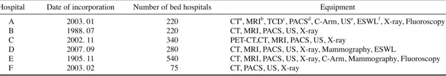
Table 1. Hospital status
Hospital Date of incorporation Number of bed hospitals Equipment
A B C D E F 2003. 01 1988. 07 2002. 11 2007. 09 1905. 11 2003. 02 220 220 340 280 540 75
CTa, MRIb, TCDc, PACSd, C-Arm, USe, ESWLf, X-ray, Fluoroscopy
CT, MRI, PACS, US, X-ray PET-CT,CT, MRI, PACS, US, X-ray
CT, MRI, PACS, US, X-ray, Mammography, ESWL
CT, MRI, PACS, US, X-ray, C-Arm, Mammography, Fluoroscopy CT, PACS, US, X-ray
aComputed tomography, bmagnetic resonance imaging, ctranscranial doppler, dpicture archiving and communication system, eultra sonography, fextracorporeal shock wave lithotripsy.
Soy Agar(TSA), Muller-Hinton Agar(MHA),
Eosin-Meth-ylene Blue(EMB), ENDO(BD, NJ, USA)에 도말 후 배양시
켰다. 배양은 배양기(incubator, SANYO, Japan)에서 36℃에
서 18시간 배양 후 세균을 관찰하고 총 세균 수를 counting
하였다. 장내세균이 의심되는 집락(colony)을 분리하고 KIA
배지(Kligler Iron Agar, BD, NJ, USA)에 배양시켰다. KIA
배지에서 자라난 세균을 Gram 염색과 세균의 생화학적 동
정 kit인 API 20E(bioMerieux, France)를 이용하여 동정을
하였고, 디스크 확산법에 의한 약제감수성검사를 통하여 세
균의 특성을 파악하였다.
결 과
검체를 1,000배까지 희석시키고 일반세균을 알 수 있는
배지(TSA, MHA)와 장내세균을 알 수 있는 배지(EMB,
ENDO)에 36℃에서 18시간 배양 후 세균의 수를 측정하 였다.
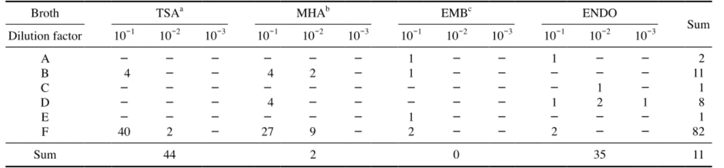
1. 카세트의 세균 수
카세트에서 채취한 세균을 배양기(incubator)에서 36℃
에서 18시간 배양 후 총 세균 수를 counting한 결과 F병원
과 C병원에서 일반세균이 각각 46CFU(Colony Forming
Unit)ml-1, 193CFUml-1로 가장 많이 검출되었고, A병원과
E병원에서 4CFUml-1, 1CFUml-1가 검출되었다. 장내세균
이 의심되는 것으로는 C병원에서 4CFUml-1, B병원에서 2
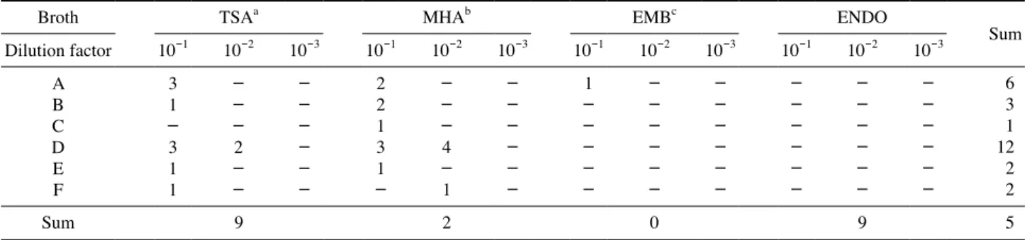
CFUml-1, F병원에서 1CFUml-1가 검출되었다(Table 2). 2. 촬영장치 손잡이의 세균 수 손잡이에서 채취한 세균을 배양기(incubator)에서 배양 후 세균을 관찰하여 총 세균 수를 counting한 결과 F병원에 서 일반세균이 78CFUml-1로 가장 많이 검출되었고, A병 원과 D병원, E병원에서는 검출되지 않았다. 장내세균이 의 심되는 것으로는 D병원과 A병원에서 각각 4CFUml-1와 2
CFUml-1가 검출되었고, B병원, C병원, E병원에서 1CFU
ml-1가 검출되었다(Table 3). 3. Apron의 세균 수 Apron에서 채취한 세균을 배양기(incubator)에서는 총 세 균 수를 counting한 결과 D병원과 A병원에서 일반세균이 각각 12CFUml-1, 5CFUml-1로 가장 많이 검출되었고, C 병원에서는 1CFUml-1가 검출되었다. 장내세균이 의심되는
것으로는 A병원에서 1CFUml-1가 관찰되었다(Table 4).
4. 종합적인 일반세균과 장내세균
카세트와 촬영장치 손잡이, Apron의 세균 수를 counting
Table 2. Number of bacteria in the cassette
Broth TSAa MHAb EMBc ENDO
Sum Dilution factor 10-1 10-2 10-3 10-1 10-2 10-3 10-1 10-2 10-3 10-1 10-2 10-3 A B C D E F 4 59 1 1 19 -11 1 3 -1 1 -1 4 5 79 6 -19 -43 2 4 -2 -2 -1 -1 -1 4 11 197 11 1 47 Sum 102 162 2 5 271
aTrypticase soy agar, bmuller-hinton Agar, ceosin-methylene blue.
Table 3. Patient table handle bacteria count of X-ray equipment
Broth TSAa MHAb EMBc ENDO
Sum Dilution factor 10-1 10-2 10-3 10-1 10-2 10-3 10-1 10-2 10-3 10-1 10-2 10-3 A B C D E F 4 -40 -2 4 4 -27 -2 -9 -1 1 -1 2 -1 -1 -2 -1 2 -1 2 11 1 8 1 82 Sum 44 2 0 35 11
한 결과 환자와의 접촉이 가장 쉬운 카세트에 많이 분포되
어 있음을 알 수 있었다. 세균의 수를 counting하여 종합한
결과, 일반세균은 C병원 194CFUml-1, F병원 126CFUml-1, D병원 27CFUml-1, B병원 22CFUml-1, A병원 9CFUml-1,
E병원 3CFUml-1가 검출되었다. 장내세균으로 의심되는 것
으로는 C병원 5CFUml-1, F병원 5CFUml-1, D병원 4CFU ml-1, A병원 3CFUml-1, B병원 2CFUml-1, E병원 1CFU ml-1가 검출되었다(Table 5).
5. 병원성 세균 분리
장내세균이 가장 의심되는 A병원 손잡이, C병원 카세트
와 F병원 카세트와 손잡이에 분리된 균이었다(Figs. 1~3).
의심되는 병원성 세균을 스트레킹을 하고 KIA에 배양시
켜 Gram 염색을 실시하였다. Gram 염색 결과 A병원 손잡이
와 F병원에는 그람음성막대균이 나왔고, C카세트와 F병원
손잡이에는 그람양성알균이 나온 것을 알 수 있었다. 그람양
성알균이 나온 2개의 검체 중 한 개의 검체가 Staphylococcus
aureus 110 agar(STA 110)배지에서 노란 색소를 띄는 검체
가 있어 포도상구균이 의심되나 알균을 동정하는 생화학동
정 kit인 API가 실험실에 없어 최종적인 동정을 할 수 없었
고, 혈액배지(BAP)에 배양하였는데 무용혈인 것으로 보아
포도상구균이 아닌 것으로 나타났다. 그람음성막대균이 나
온 2개의 검체에 생화학동적검사 kit인 API를 실시하였는데
F병원 카세트에는 대장균(E. coli)이 확인되었고, A병원 손
잡이에서는 Enterobacter cloaca가 확인되었다(Figs. 4, 5). F
병원 카세트에서 나온 E. coli는 혈청검사를 실시했는데 실
험실에 있는 O26, O111, O157과 응집하지 않았다.
Table 4. Number of bacteria in the Apron
Broth TSAa MHAb EMBc ENDO
Sum Dilution factor 10-1 10-2 10-3 10-1 10-2 10-3 10-1 10-2 10-3 10-1 10-2 10-3 A B C D E F 3 1 -3 1 1 -2 -2 2 1 3 1 -4 -1 -1 6 3 1 12 2 2 Sum 9 2 0 9 5
aTrypticase soy agar, bmuller-hinton Agar, ceosin-methylene blue.
Table 5. Numbers of general bacteria and Enterobacteriaceae
Hospital General bacteria Enterobacteriaceae Sum
A B C D E F 9 22 194 27 3 126 3 2 5 4 1 5 12 24 199 31 4 131
Fig. 1. A Hospital X-ray equipment patient table handle
Entero-bacteriaceae.
Fig. 2. Enterobacteriaceae number of C hospital cassette.
고 찰
국민 대다수가 건강에 대한 관심이 높아졌고, 질병의 조
기 진단과 치료가 현저히 많아졌다(Rutala and Weber 1999;
Wu et al. 2008; Lee et al. 2011). 그로 인해 병원 의료기기에 대한 청결관리가 중요시됨에 따라 영상의학과 촬영실 내 접 촉이 잘 이루어지는 부위 테이블, 촬영장치 손잡이, Apron에
어느 정도의 세균이 있는지 관찰하였다. 그 결과 C병원 199
CFUml-1, F병원 131CFUml-1, D병원 31CFUml-1, B병원 24CFUml-1, A병원 12CFUml-1, E병원 4CFUml-1의 세균
이 분리되었다. 김선칠의 연구에 따르면 총 세균 수는 265 CFUml-1이었는데 본 실험에서 C병원은 199CFUml-1, F병 원은 131CFUml-1로 세균 수가 적게 나와 위생관리가 양 호한 것으로 생각되었다(Kim 2008). 그리고 영상의학과 촬 영실 내 접촉이 잘 이루어지는 카세트와 촬영장치 손잡이, Apron의 각각의 세균 수를 비교해 보았을 때 C병원, F병원,
D병원, B병원, A병원, E병원의 카세트 세균 수는 271CFU
ml-1이었고, 촬영장치 손잡이는 105CFUml-1, Apron은 28 CFUml-1이었는데 김선칠의 연구에 따르면 카세트 세균 수 는 41CFUml-1이었고, 촬영장치 손잡이는 167CFUml-1, Apron은 57CFUml-1인 것으로 보고하여 다른 장소에 비해 보다 더 많은 세균이 분리되어 촬영실 내 카세트와 촬영장 치 손잡이의 위생관리 필요성이 있음을 알 수 있었다. 세균을 counting한 후, 장내세균이 의심되는 검체들을 따 로 모아 병원성 세균의 유무를 확인하여 병원 장비에 대한 위생관리의 중요성을 알아보고자 하였다. Gram 염색을 실 시한 결과 A병원 손잡이와 F병원에는 그람음성간균이 나왔 고, C카세트와 F병원 손잡이에는 그람양성알균이 나온 것 을 알 수 있었다. 그람음성균이 나온 두 개의 검체에 생화
학동적검사 kit인 API를 실시하였다. 그 결과 A병원 촬영장
치 손잡이에는 Enterobacter cloaca가 검출되었고, F병원 카 세트에는 E. coli가 검출되어 인체 감염의 가능성이 있을 것 으로 판단되어 촬영실 내 카세트와 촬영장치 손잡이에 대해 더 집중적인 살균처리 및 예방이 요구됨을 알 수 있었다. 대 표적인 살균처리로는 70% 알코올을 예를 들 수 있다. 70% 알코올로 소독한 후 세균을 동정한 결과, 70% 알코올 소독 군에서는 카세트와 테이블 그룹 모두에서 100% 세균이 사
멸하였다(Bae et al. 2008; Han and Moon 2009). 예방법으로
는 첫 번째가 올바른 손 씻기이다. 손 씻기의 방법은 첫째 두 손바닥을 마찰하여 닦고, 둘째 손바닥으로 다른 손등을 문지르며, 셋째 손바닥을 마주대고 손가락 사이를 문지른다. 이는 환자와 접촉하기 전과 후에 실시하며 장갑을 끼기 전 후, 오염물(혈액, 체액, 점막, 손상된 피부, 오염된 기구 등) 과 접촉하기 전후에 꼭 실시하여야 하며 마찰 시간은 10~ 15초간 실시하여야 한다. 손을 씻은 후 꼭 종이 타월이나 hand dryer로 건조를 하여 손에 수분이 없도록 하여야 한다. 두 번째는 환자 중 점염성이 강한 질병을 가진 환자의 방문 후에는 꼭 소독제를 사용하여 전염성 병원균을 제거해야 한 다(Shin et al. 1999; Kweon et al. 2000; Kweon et al. 2001). 이러한 방법을 통하여 우리 방사선사는 환자들을 촬영하는 것뿐만 아니라 촬영실 내 및 방사선사 개개인의 위생관리를 철저히 하여 환자가 검사받음으로 촬영실 내의 위생 상태로 인해 병원성 감염에 위협받지 않도록 하여야 한다.
결 론
광주광역시 및 전라남도 소재의 중소형병원 영상의학과 내 촬영실 6곳의 촬영실 내 테이블, 촬영장치 손잡이, Apron 을 대상으로 세균 분포와 병원성 세균을 조사하였다. 조사 병원의 테이블에서 271CFUml-1, 촬영장치 손잡이에서 105CFUml-1의 균이 검출되어 Apron의 28CFUml-1보다 많아
이 장소의 위생관리 필요성을 알 수 있었다. A병원의 손잡
이와 F병원 카세트에서 장내세균인 E. coli와 Enterobacter
cloaca가 관찰되어 환자의 접촉이 가장 많은 카세트와 방사
선사의 접촉이 많은 촬영장치 손잡이에 대해서 70% 알코올
Fig. 4. A Hospital X-ray equipment patient table handle API test.
소독과 방사선사의 손 씻기를 통해 보다 청결관리를 철저히 하여 방사선과 촬영실에 대한 세균오염도를 낮게 유지해야
될 것으로 사료된다.
사 사
The Research has been conducted by the Research Grant of Gwangju Health University in 2015(3015013).
참 고 문 헌
Bergogne-Bérézin E and Towner KJ. 1996. Acinetobacter spp. as nosocomial pathogens: microbiological, clinical, and ep-idemiological features. Microbiol. Rev. 9(2):148-165. Bae SH, Lee MS, Lim CS and Kim GJ. 2008. A Study on the
Measurement of the Pollution Level of Bacteria and Dis-infection of Table and IP Cassette. J. Radiol. Sci. Technol.
31(3):229-237.
Dong KR, Ro SH, Kweon DC, Ryu YH, Dong CB, Yu EY and Cho YK. 2009. A Study Regarding Measurements of Bac-terial Contamination Levels in Radiology Room within the Department of Radiological Technology. J. Korean Soc.
Indoor Environ. 6(4):275-284.
Han EO and Moon IO. 2009. A model for protective behavior against the harmful effects of radiation for radiological technologists in medical centers. J. Korea Asso. Radiat.
Prot. 34(3):95-101.
Han EO, Kwon DM, Dong KR and Han SM. 2010. A Model for Protective Behavior against the Harmful Effects of Ra-diation based on Medical Institution Classifications. J.
Ko-rea Asso. Radiat. Prot. 35(4):157-162.
Horan TC and Emori TG. 1997. Definitions of key terms used in the NNIS System. Am. J. Infect. Control. 25(2):112-116. Hughes JM. 1988. Study on the efficacy of nosocomial infec-tion control(SENIC Project): results and implications for the future. Chemotherapy 34(6):553-561.
Kim SC. 2008. Bacteriological Monitoring of Radiology Room Apparatus in the Department of Radiological Technology and Contamination on Hands of Radiological Technologists.
J. Radiol. Sci. Technol. 31(4):329-335.
Kweon DC, Chung KM and Choi JW. 2000. A Study on Con-tamination and Disinfection of Film Cassette. J. Radiol.
Sci. Technol. 23(2):55-61.
Kweon DC, Jeon YW and Cho A. 2001. Disinfection Efficacy of an Ultraviolet Light on Film Cassettes for Preventive of the Nosocomial Infection. J. Radiol. Sci. Technol. 24(1):27-32.
Kweon DC, Kim MS, Kim DS and Park B. 2001. A Survey of the Radiographic Cassettes Disinfection of University Hos-pitals in Seoul. J. Radiol. Sci. Technol. 24(2):65-70. Kweon DC and Park B. 2001. Disinfection Effect of Film
Cas-settes by Ultraviolet Irradiation. J. Korea Asso. Radiat.
Prot. 26(4):425-432.
Lee JS, Jeong KH, Kim KH, Im IC, Kweon DC, Goo EH, Dong KR and Chung WK. 2011. Radiology Department Infection Control According to Radiography Frequency and Disinfection Period. J. Korean Soc. Radiol. 5(2):73-80. Merrer J, Santoli F, Appéré de Vecchi C, Tran B, De Jonghe B
and Outin H. 2000. Colonization pressure and risk of ac-quisition of methicillin-resistant Staphylococcus aureus in a medical intensive care unit. Infect. Control. Hosp.
Epide-miol. 21(11):718-723.
Rutala WA and Weber DJ. 1999. Infection control: the role of disinfection and sterilization. J. Hosp. Infect. 43(1):S43-55. Shin MG, Park YK, Kim KK, Shin JH, Suh SP and Ryang DW.
1999. Molecular Epidemiology of Staphylococcus aureus Isolated in the Hospital Environments. Korean J. Infect.
Dis. 31(4):332-340.
Wu BU, Johannes RS, Kurtz S and Banks PA. 2008. The im-pact of hospital-acquired infection on outcome in acute pancreatitis. Gastroenterology 135(3):816-820.
Received: 22 September 2016 Revised: 21 November 2016 Revision accepted: 25 January 2017