Clinicopathological Characteristics of Patients with Gastric Cancer
according to the Expression of LIN28A
Chan Hyuk Park1, Jung Hwa Lee2, Na Keum Lee2, Yong Chan Lee2, and Sang Kil Lee2
1Department of Internal Medicine, Hanyang University Guri Hospital, Hanyang University College of Medicine, Guri, and 2Division of Gastroenterology, Department of Internal Medicine, Severance Hospital, Institute of Gastroenterology, Yonsei University College of Medicine, Seoul, Korea
Correspondence to: Sang Kil Lee
Division of Gastroenterology, Department of Internal Medicine, Yonsei University College of Medicine, 50-1 Yonsei-ro, Seodaemun-gu, Seoul 03722, Korea
Tel: +82-2-2228-1996, Fax: +82-2-393-6884, E-mail: [email protected]
Received on June 26, 2015. Revised on September 5, 2015. Accepted on September 8, 2015. Published online February 22, 2016 pISSN 1976-2283 eISSN 2005-1212 http://dx.doi.org/10.5009/gnl15283
Background/Aims: Although LIN28A is known to potentially play a role in the oncogenesis of various cancers, whether LIN28A expression is a predictor of poor prognosis in pa-tients with gastric cancer has not been fully explored. We sought to evaluate clinicopathological characteristics accord-ing to the expression of LIN28A in numerous gastric cancer tissue samples. Methods: LIN28A expression was evaluated by immunohistochemical (IHC) analysis of a tissue microar-ray comprising 288 gastric cancer tissues and 288 adjacent normal tissues. Clinicopathological characteristics, including overall survival, were compared according to LIN28A expres-sion. Results: The IHC staining score was lower for the can-cer tissues than the normal tissues (p<0.001). However, no significant differences were observed in the clinicopathologi-cal characteristics between the low and high LIN28A expres-sion groups. In addition, the 5-year overall survival rate did not differ between the two groups: 75.3% (95% confidence interval [CI], 69.3% to 81.7%) versus 71.6% (95% CI, 63.3% to 80.9%) for low versus high expression, respectively. Con-clusions: The expression of LIN28A did not appear to play a distinct role in predicting the clinicopathological charac-teristics of patients with gastric cancer. In addition, LIN28A expression was not an independently associated factor for overall survival in patients with gastric cancer. (Gut Liver 2016;10:714-718)
Key Words: LIN28A; Stomach neoplasms; Survival
INTRODUCTION
While the survival rates of patients with early gastric cancer have improved with the increase of earlier diagnoses in Korea
and Japan due to the use of endoscopy screening,1-4 the
long-term oncologic outcomes of treatment in patients with advanced gastric cancer are still relatively poor.5,6 Although ongoing
col-laborative sequencing efforts have highlighted recurrent somatic genomic aberrations in gastric cancer, the outcomes of patients have not sufficiently improved.7
Better understanding of the molecular pathogenesis of gastric carcinogenesis may be needed for the advancement of gastric cancer treatments.
LIN28A is a highly conserved RNA-binding protein that was originally recognized as a key regulator of developmental tim-ing in Caenorhabditis elegans.8 Recently, a study on LIN28A
showed that it is overexpressed in various human cancers and functions as an oncogenes.9 p53 activates the expression of
miR-34a and miR-145, which inhibit the expression of several stem-cell factors including OCT4, KLF4, LIN28A, and SOX2.10
Additionally, it has been shown that tristetraprolin induced by p53 in cancer cells increases the level of let-7, a known tumor suppressor, via downregulation of LIN28A.11
Blockade of the processing of various microRNAs, including let-7, has been sug-gested as one of the pathogenetic mechanisms of LIN28A in hu-man carcinogenesis.12-15 In addition, LIN28A has been reported
to suppress many other factors including OCT4, SOX2, and NANOG, which are involved in reprogramming human somatic cells, by post-transcriptional regulation.16,17
Expression of LIN28A has been identified in various human cancers, including breast cancer,18
ovarian cancer,19
hepatocel-lular carcinoma,9 and colorectal cancer.20 As for gastric
can-cer, one study from China showed that positive expression of LIN28A was correlated with poor overall survival.21 However,
expression of LIN28A was found to be more common in cor-responding normal tissue than in gastric cancer tissue in that study. In addition, the study did not fully adjust for TNM stages
This is an Open Access article distributed under the terms of the Creative Commons Attribution Non-Commercial License (http://creativecommons.org/licenses/by-nc/4.0) which permits unrestricted non-commercial use, distribution, and reproduction in any medium, provided the original work is properly cited.
in the survival analysis. Therefore, we cannot be certain wheth-er LIN28A expression is a poor prognostic factor in patients with gastric cancer. In the current study, we aimed to evaluate clinicopathological characteristics according to the expression of LIN28A by using a large number of gastric cancer tissue samples.
MATERIALS AND METHODS
1. Study design and patient population
In order to evaluate the relationship between the clinicopath-ological characteristics of gastric cancers and the expression of LIN28A, we performed immunohistochemical (IHC) staining for LIN28A using tissue microarrays (TMAs). The TMAs were constructed from 288 pairs of gastric cancer tissue samples and corresponding normal tissues that were obtained from patients who had undergone curative surgery for gastric cancer at the same center between April 2001 and December 2003. We col-lected the following data from the patients’ medical records: de-mographic information, tumor location, histology, TNM staging, and overall survival duration. This study was approved by the Institutional Review Board of Severance Hospital.
2. Tissue microarray construction
The TMAs were constructed as previously described.22 On
hematoxylin and eosin-stained slides of tumors, a representa-tive area was selected, and the corresponding spot was marked on the surface of the paraffin block. Using a biopsy needle, the selected area was punched out, and a 2-mm tissue core was placed into an 8×6 recipient block. Gastric cancer tissues and corresponding normal tissues were then extracted. Each tissue core was assigned a unique TMA location number that was linked to a database containing other clinicopathological data.
3. Immunohistochemistry
IHC analyses were performed on formalin-fixed, paraffin-embedded tissues, using the TMA. The slides were deparaf-finized in xylene and rehydrated through an ethanol gradient. Endogenous peroxidase was blocked with 3% H2O2 for 10
minutes. The slides were immersed in 10 mM citrate buffer (pH 6.0) and heated for 10 minutes for antigen retrieval. Nonspecific binding was blocked by preincubation with a protein blocking agent (Immunotech Laboratories Inc., Monrovia, CA, USA), and the slides were incubated at room temperature for 1 hour with antibody against LIN28A (rabbit polyclonal, 1:500; Abcam, Cambridge, MA, USA). The samples were washed three times with phosphate-buffered saline (PBS) and then incubated with secondary antibodies for 30 minutes. After an additional three washes in PBS, the slides were incubated with the streptavidin-horseradish peroxidase complex (Dako, Glostrup, Denmark) for 30 minutes. The slides were visualized with diaminobenzidine (Dako) and then counterstained with hematoxylin. The
expres-sion of the antibodies was assessed semiquantitatively by esti-mating the percentage of tumor cells with positive cytoplasm staining among the entire population of tumor cells. All slides were examined and scored independently by two experienced investigators to avoid subjective biases. Each slide was exam-ined in its entirety under a light microscope, and a proportion score was initially assigned, which represented the estimated proportion of positive tumor cells (0, none; 1, 0% to 10%; 2, 10% to 50%; and 3, 50% to 100%). Next, an intensity score was assigned, which represented the average intensity of the positive tumor cells (0, none; 1, weak; 2, intermediate; and 3, strong). The proportion and intensity scores were then multiplied to obtain a total score, which ranged from 0 to 9, and high expres-sion of LIN28A was defined as a total score ≥2.21
4. Statistical analysis
Statistical tests used to compare the measured results included the t-test, Mann-Whitney U test, chi-square test, and Fisher ex-act test. The Kaplan-Meier method and log-rank test were used for survival analysis. In addition, the Cox proportional hazards model was used to adjust for possible confounding variables including TNM stage. A value of p less than 0.05 was regarded as a statistically significant difference for comparisons between groups. All statistical procedures were conducted using the statistical software SPSS for Windows version 18.0 (SPSS Inc., Chicago, IL, USA) with the exception of the survival analysis, which was performed using R version 2.15.3 (R Foundation for Statistical Computing, Vienna, Austria).
RESULTS
1. Comparison of LIN28A expression between cancer tissues and corresponding normal tissues
IHC staining was performed on 288 pairs of normal and can-cer tissue samples to analyze the difference in LIN28A expres-sion. The mean IHC staining score was higher in the normal tis-sues than in the cancer tistis-sues (normal vs cancer [mean±standard deviation], 2.0±1.6 vs 1.1±1.5; p<0.001). The proportion of tissues with LIN28A expression was higher in the normal tissues than in the cancer tissues (normal vs cancer, 63.5% vs 35.4%; p<0.001).
2. LIN28A expression and clinicopathological characteristics of gastric cancer
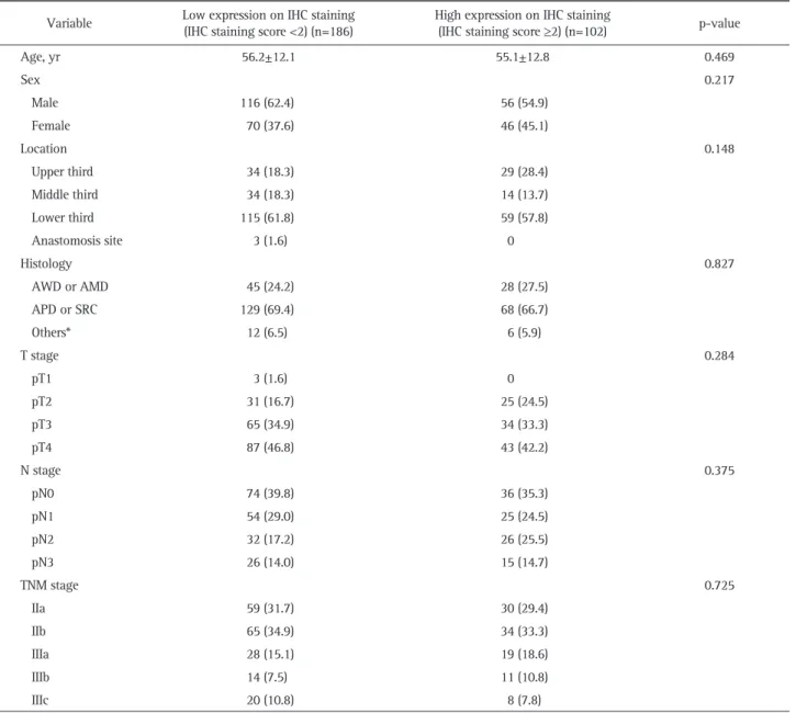
The clinicopathological characteristics of the enrolled patients according to the LIN28A expression of cancer tissues on IHC staining are shown in Table 1. High expression of LIN28A was shown in 102 of 288 patients (35.4%). The mean patient age and the proportion of men did not differ between the groups (p=0.469 and p=0.217, respectively). Neither tumor location nor histology differed between the groups (p=0.148 and p=0.827, respectively). In addition, T, N, and TNM stages did not differ
between the groups (p=0.284, p=0.375, and p=0.725, respec-tively).
3. Survival analysis according to LIN28A expression Kaplan-Meier plots were used to illustrate overall survival ac-cording to the expression of LIN28A (Fig. 1). The median follow-up durations were 69.4 months (interquartile range [IQR], 60.6 to 77.4 months) and 66.9 months (IQR, 40.8 to 76.7 months) in the low and high expression groups, respectively (p=0.158). The 5-year overall survival was 75.3% (95% confidence interval [CI], 69.3% to 81.7%) in the low expression group and 71.6% (95% CI, 63.3% to 80.9%) in the high expression group. The duration of overall survival did not differ between the groups (p=0.405).
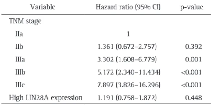
A Cox proportional hazards model showed that high LIN28A expression was not an independently associated factor for death after adjusting for TNM stage (hazard ratio, 1.191; 95% CI, 0.758 to 1.872) (Table 2).
DISCUSSION
LIN28A has been considered an oncogene in the development of various human cancers.9,18-20
One of the possible mechanisms of LIN28A as an oncogene is downregulation of the let-7 fam-ily, well-known microRNAs that function as tumor suppres-sors.12-14 Under this hypothesis, Xu et al.21 performed a study
on the expression of LIN28A in gastric cancer patients via
real-Table 1. Clinicopathological Characteristics according to LIN28A Expression via Immunohistochemical Staining
Variable Low expression on IHC staining (IHC staining score <2) (n=186) High expression on IHC staining (IHC staining score ≥2) (n=102) p-value
Age, yr 56.2±12.1 55.1±12.8 0.469 Sex 0.217 Male 116 (62.4) 56 (54.9) Female 70 (37.6) 46 (45.1) Location 0.148 Upper third 34 (18.3) 29 (28.4) Middle third 34 (18.3) 14 (13.7) Lower third 115 (61.8) 59 (57.8) Anastomosis site 3 (1.6) 0 Histology 0.827 AWD or AMD 45 (24.2) 28 (27.5) APD or SRC 129 (69.4) 68 (66.7) Others* 12 (6.5) 6 (5.9) T stage 0.284 pT1 3 (1.6) 0 pT2 31 (16.7) 25 (24.5) pT3 65 (34.9) 34 (33.3) pT4 87 (46.8) 43 (42.2) N stage 0.375 pN0 74 (39.8) 36 (35.3) pN1 54 (29.0) 25 (24.5) pN2 32 (17.2) 26 (25.5) pN3 26 (14.0) 15 (14.7) TNM stage 0.725 IIa 59 (31.7) 30 (29.4) IIb 65 (34.9) 34 (33.3) IIIa 28 (15.1) 19 (18.6) IIIb 14 (7.5) 11 (10.8) IIIc 20 (10.8) 8 (7.8)
Data are presented as mean±SD or number (%).
IHC, immunohistochemical; AWD, adenocarcinoma well differentiated; AMD, adenocarcinoma moderated differentiated; APD, adenocarcinoma poorly differentiated; SRC, signet ring cell carcinoma.
time polymerase chain reaction analysis and IHC staining. In their study, however, the expression level of LIN28A in normal tissues was higher than that in cancer tissues (normal vs cancer, 62.8% vs 46.0%). Although it was shown that LIN28A expres-sion was independently associated with poor overall survival, the authors could not clearly explain how LIN28A expression affected the overall survival of patients with gastric cancer. We questioned whether the expression of LIN28A really has a nega-tive influence on overall survival in patients with gastric cancer. In our study, similar proportions of cancer tissues and cor-responding normal tissues showed expression of LIN28A on IHC staining (normal vs cancer, 63.5% vs 35.4%) compared to the results of Xu et al.21
However, no associated clinicopathological factor for the high LIN28A expression in gastric cancers was identified. Unlike Xu et al.,21
we classified gastric cancers into four or five groups according to T stage (pT1, pT2, pT3, and pT4), N stage (pN0, pN1, pN2, and pN3), and TNM stage (IIa, IIb, IIIa, IIIb, and IIIc). These detailed classifications were selected to stratify the gastric cancers more accurately for survival analy-sis. As a result, we showed that LIN28A expression was not an independently associated factor for overall survival. The expres-sion of LIN28A seemed to have no distinct role in predicting clinicopathological characteristics including survival outcomes in patients with gastric cancer.
Our results, which are in conflict with the usual expectation that LIN28A may have a role as an oncogene, are likely due to the various roles of LIN28A aside from repressing let-7. First, overexpression of LIN28A induces the expression of several transcription factors that are involved in early embryonic-cell fate decisions. Moreover, LIN28A functions independently of let-7.23
Second, LIN28A directly regulates various metabolisms
including oxidative phosphorylation or glucose metabolism.24,25
Third, LIN28A may facilitate the global translational suppres-sion of endoplasmic reticulum-associated mRNAs in undifferen-tiated stem cells.26
We believe that LIN28A may have a role not only in gastric cancer cells but also in normal gastric cells. In addition, the heterogeneity of gastric cancers could contribute to the discrepancy between the results of Xu et al.’s study and ours.27,28
A better understanding of the molecular biology of gastric cancers and appropriate selection of the patient popula-tion might be necessary to clarify the role of LIN28A in gastric carcinogenesis.
Although this was the largest study to date that evaluated clinicopathological characteristics according to the expres-sion of LIN28A in patients with gastric cancer, it has several limitations. First, we did not assess the lesion characteristics according to well-known associated factors for gastric cancer, including Helicobacter pylori infection status and family history of gastric cancer. Unfortunately, H. pylori infection status and family history were unavailable in our gastric cancer cohort for TMAs. Second, various associated factors for LIN28A including p53, Ki-67, miR-34a, miR-145, and let-7, were not analyzed in the study. Evaluating these factors might support the negative results of our study in patients with gastric cancer. The lack of an analysis of LIN28B was another limitation of the study. Although the study was performed to validate the results of the previous study,21
expression of LIN28B should be investigated to better understand LIN28 in patients with gastric cancer.29,30
Despite these limitations, our data may form the basis of a system that can be used to understand the clinicopathological characteristics based on LIN28A expression.
The expression of LIN28A seemed to have no distinct role in predicting clinicopathological characteristics in gastric cancer. Additionally, LIN28A expression was not an independently asso-ciated factor for overall survival in patients with gastric cancer.
CONFLICTS OF INTEREST
No potential conflict of interest relevant to this article was reported.
Fig. 1. Kaplan-Meier plots for the overall survival rate according to LIN28A expression. The median follow-up durations of the low- and high-expression groups were 69.4 months (interquartile range [IQR], 60.6 to 77.4 months) and 66.9 months (IQR, 40.8 to 76.7 months), respectively. 0 12 24 36 48 60 72 84 100 80 60 40 20 96 5-Y ear overall survival (%) Time (mo) 0
Low LIN28A expression 5-year overall survival 75.3% (95% CI, 69.3%-81.7%)
High LIN28A expression 5-year overall survival 71.6% (95% CI, 63.3%-80.9%) p=0.405 No. at risk No expression Expression 186 102 178 96 23 11 78 37 140 73 146 73 154 81 163 85
Table 2. Cox Proportional Hazards Model for Predicting Death Variable Hazard ratio (95% CI) p-value TNM stage IIa 1 IIb 1.361 (0.672–2.757) 0.392 IIIa 3.302 (1.608–6.779) 0.001 IIIb 5.172 (2.340–11.434) <0.001 IIIc 7.897 (3.826–16.296) <0.001
High LIN28A expression 1.191 (0.758–1.872) 0.448 CI, confidence interval.
ACKNOWLEDGEMENTS
This research was supported by the Basic Science Research Program through the National Research Foundation of Korea funded by the Ministry of Education, Science and Technology (2012R1A1A2001479).
REFERENCES
1. Lee HJ, Yang HK, Ahn YO. Gastric cancer in Korea. Gastric Cancer 2002;5:177-182.
2. Isobe Y, Nashimoto A, Akazawa K, et al. Gastric cancer treatment in Japan: 2008 annual report of the JGCA nationwide registry. Gastric Cancer 2011;14:301-316.
3. Carter KJ, Schaffer HA, Ritchie WP Jr. Early gastric cancer. Ann Surg 1984;199:604-609.
4. Shimizu S, Tada M, Kawai K. Early gastric cancer: its surveillance and natural course. Endoscopy 1995;27:27-31.
5. Cunningham D, Starling N, Rao S, et al. Capecitabine and ox-aliplatin for advanced esophagogastric cancer. N Engl J Med 2008;358:36-46.
6. Waddell T, Chau I, Cunningham D, et al. Epirubicin, oxaliplatin, and capecitabine with or without panitumumab for patients with previously untreated advanced oesophagogastric cancer (REAL3): a randomised, open-label phase 3 trial. Lancet Oncol 2013;14:481-489.
7. Yang W, Raufi A, Klempner SJ. Targeted therapy for gastric can-cer: molecular pathways and ongoing investigations. Biochim Biophys Acta 2014;1846:232-237.
8. Moss EG, Lee RC, Ambros V. The cold shock domain protein LIN-28 controls developmental timing in C. elegans and is regulated by the lin-4 RNA. Cell 1997;88:637-646.
9. Viswanathan SR, Powers JT, Einhorn W, et al. Lin28 promotes transformation and is associated with advanced human malignan-cies. Nat Genet 2009;41:843-848.
10. Jain AK, Allton K, Iacovino M, et al. p53 regulates cell cycle and microRNAs to promote differentiation of human embryonic stem cells. PLoS Biol 2012;10:e1001268.
11. Lee JY, Kim HJ, Yoon NA, et al. Tumor suppressor p53 plays a key role in induction of both tristetraprolin and let-7 in human cancer cells. Nucleic Acids Res 2013;41:5614-5625.
12. Nam Y, Chen C, Gregory RI, Chou JJ, Sliz P. Molecular basis for interaction of let-7 microRNAs with Lin28. Cell 2011;147:1080-1091.
13. Leung AK, Sharp PA. MicroRNA functions in stress responses. 2010;40:205-215.
14. Newman MA, Thomson JM, Hammond SM. Lin-28 interaction with the Let-7 precursor loop mediates regulated microRNA pro-cessing. RNA 2008;14:1539-1549.
15. Mao XG, Hütt-Cabezas M, Orr BA, et al. LIN28A facilitates the
transformation of human neural stem cells and promotes glio-blastoma tumorigenesis through a pro-invasive genetic program. Oncotarget 2013;4:1050-1064.
16. Qiu C, Ma Y, Wang J, Peng S, Huang Y. Lin28-mediated post-transcriptional regulation of Oct4 expression in human embryonic stem cells. Nucleic Acids Res 2010;38:1240-1248.
17. Yu J, Vodyanik MA, Smuga-Otto K, et al. Induced pluripotent stem cell lines derived from human somatic cells. Science 2007; 318:1917-1920.
18. Lv K, Liu L, Wang L, et al. Lin28 mediates paclitaxel resistance by modulating p21, Rb and Let-7a miRNA in breast cancer cells. PLoS One 2012;7:e40008.
19. Peng S, Maihle NJ, Huang Y. Pluripotency factors Lin28 and Oct4 identify a sub-population of stem cell-like cells in ovarian cancer. Oncogene 2010;29:2153-2159.
20. Saiki Y, Ishimaru S, Mimori K, et al. Comprehensive analysis of the clinical significance of inducing pluripotent stemness-related gene expression in colorectal cancer cells. Ann Surg Oncol 2009; 16:2638-2644.
21. Xu C, Shen J, Xie S, Jiang Z, Huang L, Wang L. Positive expres-sion of Lin28 is correlated with poor survival in gastric carcinoma. Med Oncol 2013;30:382.
22. Rimm DL, Camp RL, Charette LA, Costa J, Olsen DA, Reiss M. Tissue microarray: a new technology for amplification of tissue resources. Cancer J 2001;7:24-31.
23. Balzer E, Heine C, Jiang Q, Lee VM, Moss EG. LIN28 alters cell fate succession and acts independently of the let-7 microRNA during neurogliogenesis in vitro. Development 2010;137:891-900. 24. Zhu H, Shyh-Chang N, Segrè AV, et al. The Lin28/let-7 axis
regu-lates glucose metabolism. Cell 2011;147:81-94.
25. Shyh-Chang N, Zhu H, Yvanka de Soysa T, et al. Lin28 enhances tissue repair by reprogramming cellular metabolism. Cell 2013; 155:778-792.
26. Cho J, Chang H, Kwon SC, et al. LIN28A is a suppressor of ER-associated translation in embryonic stem cells. Cell 2012;151:765-777.
27. Das K, Gunasegaran B, Tan IB, Deng N, Lim KH, Tan P. Mutually exclusive FGFR2, HER2, and KRAS gene amplifications in gastric cancer revealed by multicolour FISH. Cancer Lett 2014;353:167-175.
28. Nishida Y, Kuwata T, Nitta H, et al. A novel gene-protein assay for evaluating HER2 status in gastric cancer: simultaneous analyses of HER2 protein overexpression and gene amplification reveal in-tratumoral heterogeneity. Gastric Cancer 2015;18:458-466. 29. Wang T, Wang G, Hao D, et al. Aberrant regulation of the
LIN28A/LIN28B and let-7 loop in human malignant tumors and its effects on the hallmarks of cancer. Mol Cancer 2015;14:125. 30. Piskounova E, Polytarchou C, Thornton JE, et al. Lin28A and
Lin28B inhibit let-7 microRNA biogenesis by distinct mechanisms. Cell 2011;147:1066-1079.