DOI 10.17480/psk.2016.60.3.128
L1 Cell Adhesion Molecule에 의한 대식세포 매개 염증반응의
억제 기전 분석
이 영 수#
청주대학교 이공대학 제약공학과
(Received April 14, 2016; Revised May 16, 2016; Accepted May 23, 2016)
L1 Cell Adhesion Molecule Suppresses Macrophage-mediated Inflammatory Responses
Young-Su Yi#
Department of Pharmaceutical Engineering, Cheongju University, Cheongju 28503, Korea
Abstract — L1 cell adhesion molecule (L1CAM) is a cell surface molecule to initiate a variety of cellular responses through interacting with other cell adhesion molecules in a homophilic or heterophilic manner. Although its expression was found to be upregulated in some tumor cells, including cholangiocarcinomas, and ovarian cancers, and many studies have inves-tigated the role of L1CAM in these cancers, its role in inflammatory responses has been poorly understood. In this study, we explored the role of L1CAM in macrophage-mediated inflammatory responses. L1CAM significantly suppressed the pro-duction of nitric oxide (NO), but induced cell proliferation in RAW264.7 cells. L1CAM expression was detectable, but its expression was markedly decreased by lipopolysaccharide (LPS) in RAW264.7 cells. In addition, the expression of pro-inflammatory genes, such as tumor necrosis factor (TNF)-α, cyclooxygenase (COX)-2, and inducible nitric oxide synthase (iNOS) induced by LPS was dramatically suppressed by L1CAM in RAW264.7 cells. L1CAM inhibited the transcriptional activities of NF-κB and AP-1 while its cytoplasmic domain deletion form, L1ΔCD did not suppressed their activities in RAW264.7 cells. Moreover, L1CAM suppressed nuclear translocation of p65 and p50 as well as c-Jun, c-Fos and p-ATF2 which are transcription factors of NF-κB and AP-1, respectively. In conclusion, L1CAM suppressed inflammatory responses in macrophages through inhibiting NF-κB and AP-1 pathways.
Keywords □ L1CAM, inflammatory response, macrophage, signal transduction, NF-κB, AP-1
염증은 외부 병원균의 감염으로부터 우리 몸을 방어하는 면역 계 최전선의 신체방어 기전으로, 통증, 발열, 부종, 발적, 신체의 기능저하 등의 증상을 나타낸다.1) 염증이 신체의 방어 기전임에 도 불구하고, 만성 염증은 염증성 자가면역질환 및 암 등 다양한 질병의 주요 요인으로 생각 되어져왔다.2)이러한 염증반응은 주 로 선천성 면역세포인 대식세포, 단구 및 중성구 등에 의해 발생 되는데, 이는 톨-유사 수용체(toll-like receptors) 등과 같은 세포 표면 수용체가 감염균을 인식하면서 시작된다. 톨-유사 수용체의 감염균 인식을 통해 대식세포는 활성화되고, 그 결과 산화질소 (NO), 활성산소/활성질소(ROS/RNS), 프로스타글란딘(PG)-E2, 종 양괴사인자(TNF)-α, 인터루킨(IL)-1β 및 인터루킨-6 등과 같은 다양한 염증성 물질을 발생한다.3-5)위에서 언급한 선천성 면역 세포 중, 염증반응에 가장 중요한 역할을 하는 세포는 대식세포 로서, 대식세포에서의 염증반응에 관한 연구는 분자세포학적으 로 오랜 기간 동안 진행되어왔다. 대식세포가 표면 수용체를 통 해 병원균을 인식하게 되면 염증반응이 개시되며, 수용체로부터 전해진 염증반응 신호를 시작으로 단백질 티로신 키나아제(protein tyrosine kinase; e.g. Src 및 Syk) 및 미토젠 활성화 단백질 키 나아제(mitogen-activated protein kinase; e.g. p38, ERK1/2, 및 JNK) 등과 같은 세포 내 신호전달 분자들의 순차적인 활성화를 통해 핵인자 카파 B(NF-κB) 및 활성화 단백질-1(AP-1) 등과 같 은 전사인자들이 활성화되어 결과적으로 염증성 물질이 생성된
#Corresponding Author Young-Su Yi
Department of Pharmaceutical Engineering, Cheongju University, Cheongju 28503, Korea
Tel.: 043-229-7852 Fax.: 043-229-8577 E-mail: [email protected]
종설
다.6-9)따라서, 염증반응 중 활성화되는 대부분의 세포 내 신호 전달 분자들은 염증반응 억제 및 염증성 자가면역 질환의 증상 완화를 위한 잠재적 약물 타겟이 될 수 있으며, 실제로 다양한 종류의 물질을 이용한 염증억제 연구가 활발히 진행되어 오고 있 다. 그러나 이러한 연구에도 불구하고, 아직 다양한 종류의 항염 후보물질에 대한 연구가 필요한 상황이며, 특히 만성염증 및 궤 양이 암의 직접적인 주요 원인 중 하나라는 점을 감안해 볼 때, 항암 관련 후보물질 및 분자를 이용한 염증반응 연구는 아직 많 은 연구가 진행되지는 않았으나 매우 흥미롭고 심도 깊은 다양 한 연구가 필요한 분야라 할 수 있다.
L1 세포부착분자(L1 cell adhesion molecule; L1CAM)는 면 역글로불린 슈퍼패밀리에 속하는 약 200~220 kDa 크기의 막관 통 당단백질로, 신경계 발생에 중요한 역할을 하는 분자로 처음 발견이 되었다.10,11)세포부착분자는 다양한 생물학적 활성에 중 요한 역할을 하는 것으로 알려져 있으며, 특히 암세포의 성장 및 전이 등과 연관된 연구가 활발히 진행되어왔다.12-15) L1CAM 역 시 다양한 암세포에서 그 발현이 비정상적으로 변화되어 있음이 보고되었으며, 암과 관련된 기능에 대한 많은 연구가 현재까지 활발히 진행되어 오고 있다.16-22)그러나, 세포부착분자는 암세포 의 성장 및 전이 등 암세포와 연관된 역할만 하는 것이 아니다. 세포부착분자는 다양한 면역반응, 특히 대식세포 매개 선천성 면 역반응에서도 면역세포의 염증부위로의 이동, 면역세포의 혈관 벽 투과, 면역세포 간 신호전달 및 활성화 등과 같은 중요한 역 할을 한다. 그러나, 현재까지 대부분의 L1CAM 관련 연구는 암 과 관련된 연구에만 집중되어 왔으며, 면역반응, 특히 대식세포 매개 염증반응에서의 연구는 그 중요성이 매우 높음에도 불구하 고 거의 연구되어오지 않았다. 따라서, 이 연구에서는 다양한 생물학적 기능을 지닌 세포부 착분자 중 하나인 L1CAM이 대식세포 매개 염증반응에서 어떠 한 역할을 하는지에 대하여 연구를 수행하였다.
실험 방법
시약L1CAM(pCMV-L1; phL1A-pcDNA3; Fig. 1A) 및 L1CAM 세 포내 도메인이 제거된 돌연변이인 L1CAMΔCD(pCMV-L1ΔCD; L1-1147)를 발현하는 플라스미드를 Addgene(Cambridge, MA, USA)으로부터 구입하였다. RAW264.7 세포는 American Type Culture Collection(Manassas, VA, USA)에서 구입하였다. Roswell Park Memorial Institute 1640(RPMI 1640), Dulbecco’s Modified Eagle’s medium(DMEM), fetal bovine serum(FBS), phosphate-buffered saline(PBS), streptomycin, penicillin, and L-glutamine는 Gibco(Grand Island, NY, USA) 구매하였다. Lipopolysaccharide(LPS), Tumor necrosis factor-α, phorbol 12-myristate 13-acetate(PMA), 3-(4,5-dimethylthiazol-2-yl)-2,5-diphenyltetrazolium bromide(MTT), 그리고 polyethylenimine (PEI)은 Sigma(St Louis, NO, USA)에서 구매하였다. AP-1 및 NF-κB luciferase 플라스미드는 조재열교수(성균관대학교, 수원, 대한민국)로부터 제공받았다. TNF-α, cyclooxygenase(COX)-2, inducible nitric oxide synthase(iNOS) 및 GAPDH PCR primers 및 PCR pre-mix는 Bioneer(대전, 대한민국)로부터 합성 및 구매 하였다. 인산화 형태(phosphorylated) 및 전체 형태(total form)의 p65, p50, c-Jun, c-Fos, ATF2 및 LaminA/C 항체는 Cell signaling Technology(Beverly, MA, USA)에서 구매하였다. TRI reagent®는 Molecular Research Center Inc.(Cincinnati, OH, USA)에서 구입 하였다. MuLV 역전사효소는 Thermo Fisher Scientific(Waltham, MA, USA)에서 구입하였다. Enhanced chemiluminescence system 은 AbFrontier(Seoul, Korea)에서 구입하였다.
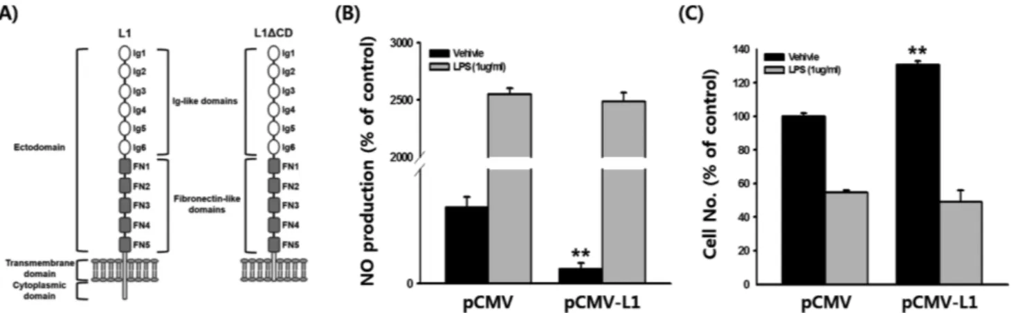
Fig. 1 − Effect of L1CAM on NO production and cell proliferation. (A) Structure of L1CAM (L1) and cytoplasmic domain deleted L1CAM (L1ΔCD) (B) RAW264.7 cells transfected with either pCMV or pCMV-L1 were treated with LPS for 24 h, and NO production was determined by Griess assay. (C) RAW264.7 cells transfected with either pCMV or pCMV-L1 were treated with LPS for 24 h, and cell viability was determined by MTT assay. **P<0.01.
세포배양
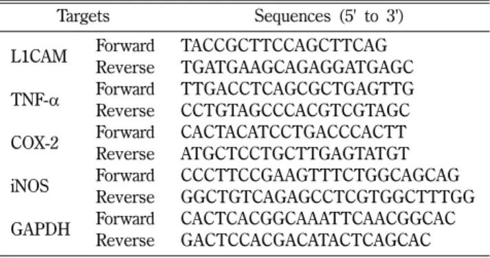
RAW264.7 세포는 10% FBS와 항생제(100 U/ml penicillin, 100μg/ml strwptomycin)가 포함된 RPMI1640 배지에서 5% CO2, 37oC의 환경에서 배양하였다. 최상의 세포 상태의 유지를 위해 1주일에 3회 배지 교환 및 1회 세포 계대를 실시하였다. NO 생성 측정 NO 측정은 Griess 시약을 이용하여 측정하였다.23) RAW264.7 세포(1×10⁶ cells/ml)에 pCMV 또는 pCMV-L1를 transfection 시킨 후, 24시간 동안 배양하였다. 세포 배양액(100 μl)에 생성된 NO에 100 μl Griess 시약(1% sulphanilamide, 0.1% N-[1-naphthyl]-ethylenediamine in 5% phosphoric acid)을 첨가한 후, 상온에서 10분간 반응시켰다. OD 값을 550 nm에서 측정하여 세 포 생존율을 결정하였고, 100%로 표시한 대조군에 대하여 백분 율로 표시하였다. 세포 분열능 측정 세포생존률 측정을 위해 MTT assay를 이용하였다.24) RAW264.7 세포(1×10⁶ cells/ml)에 pCMV 또는 pCMV-L1를 transfection 시킨 후, 24시간 동안 배양하였다. 각 well의 세포 배양액(100 μl)에 10 μl MTT 용액(10 mg/ml in PBS)을 첨가하 고 4시간 동안 37oC에서 배양한 후, 15% sodium dodecyl sulfate(SDS)를 첨가하여 24시간 동안 37oC에서 배양하였다. OD 값을 490 nm에서 측정하여 세포 생존율을 결정하였고, 100%로 표시한 대조군에 대하여 백분율로 표시하였다. 반정량적 중합효소 연쇄반응(Semi-quantitative polymerase chain reaction; PCR) RAW264.7 세포(1×10⁶ cells/ml)에 LPS를 6시간 처리 후, TRI reagent®를 이용하여 총 RNA를 추출하였다. MuLV 역전사 효소를 이용해 1 μg의 총 RNA로부터 cDNA를 합성한 후, 이를 이용하여 PCR을 수행하였다. 각 타겟의 PCR에 이용한 primer 서열은 Table I에 정리하였다.
정량적 실시간 중합효소 연쇄반응(Quantitative real time PCR)
pCMV 또는 pCMV-L1를 transfection 시킨 RAW264.7 세포(1 ×10⁶ cells/ml)에 LPS를 6시간 처리 후, TRI reagent®를 이용 하여 총 RNA를 추출하였다. MuLV 역전사효소를 이용해 1 μg 의 총 RNA로부터 cDNA를 합성한 후, 이 전에 이용한 방법25)
으로 실시간 PCR을 수행하였다. 각 타겟의 PCR에 이용한 primer 서열은 Table II에 정리하였다.
루시퍼라제 리포터 유전자 분석(Luciferase reporter gene assay)
pCMV 또는 pCMV-L1를 transfection 시킨 RAW264.7 세포(1 ×10⁶ cells/ml), 그리고 pCMV, pCMV-L1 또는 pCMV-L1ΔCD 를 transfection 시킨 RAW264.7 세포(1×10⁶ cells/ml)에 TNF-α
(15 ng/ml) 또는 PMA(100 nM)를 처리한 후, 이 세포로부터 이 전에 이용한 방법23)으로 루시퍼라제 리포터 유전자 분석을 수행
하였다. 모든 RAW264.7 세포는 NF-κB-Luc 또는 AP-1-Luc 플 라스미드를 transfection하였으며, transfection 보정을 위해 β-galactosidase 플라스미드를 동시에 transfection하였다.
세포의 핵 분획(nuclear fraction) 분리 및 Western blot 분석
pCMV, pCMV-L1 또는 pCMV-L1ΔCD를 transfection 시킨 RAW264.7 세포(1×10⁶ cells/ml)에 LPS(1 μg/ml)를 1시간 처리 한 후, 핵 분획을 이 전에 이용한 방법26)으로 분리하였다. Western blot 분석을 위해 분리한 핵분획을 SDS polyacrylamide 전기영 동으로 분리 후, polyvinylidene fluoride membrane로 이동시켰 다. 인산화 및 전체 형태의 p65, p50, c-Jun, c-Fos, ATF2 및 Lamin A/C 항체를 이용해 타겟 단백질을 탐지하였고, enhanced chemiluminescence system을 이용해 이 단백질들을 확인하였다.
통계 분석
모든 실험에서 얻은 결과는 평균±표준편차로 표시하였다. 통 계적 유의성 분석은 Student’s t-test 또는 one-was ANOVA 방법 을 사용하였으며, P<0.05인 값에 대하여 유의적으로 처리하였다.
Table II − Sequences of primers used for quantitative real time PCR Targets Sequences (5' to 3')
TNF-α Forward TGCCTATGTCTCAGCCTCTT Reverse GAGGCCATTTGGGAACTTCT COX-2 Forward CACTACATCCTGACCCACTT
Reverse ATGCTCCTGCTTGAGTATGT iNOS Forward GGAGCCTTTAGACCTCAACAGA
Reverse TGAACGAGGAGGGTGGTG GAPDH Forward CAATGAATACGGCTACAGCAAC
Reverse AGGGAGATGCTCAGTGTTGG
Table I − Sequences of primers used for semi-quantitative PCR Targets Sequences (5' to 3')
L1CAM Forward TACCGCTTCCAGCTTCAG Reverse TGATGAAGCAGAGGATGAGC TNF-α Forward TTGACCTCAGCGCTGAGTTG Reverse CCTGTAGCCCACGTCGTAGC COX-2 Forward CACTACATCCTGACCCACTT Reverse ATGCTCCTGCTTGAGTATGT iNOS Forward CCCTTCCGAAGTTTCTGGCAGCAG
Reverse GGCTGTCAGAGCCTCGTGGCTTTGG GAPDH Forward CACTCACGGCAAATTCAACGGCAC
실험 결과 및 고찰
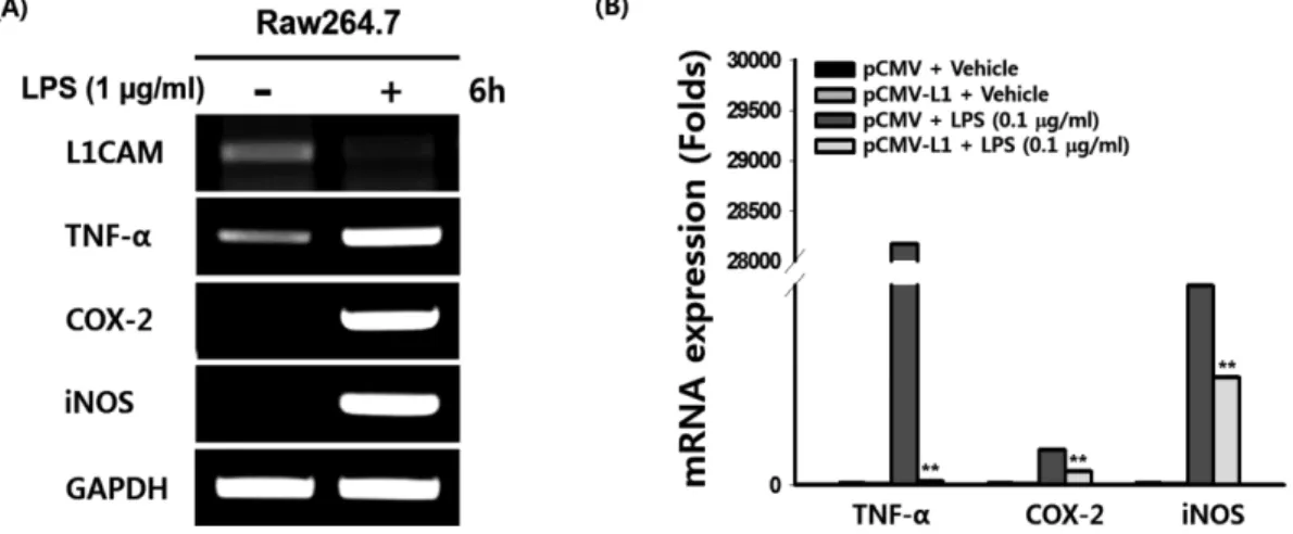
세포표면분자인 L1CAM은 여러 종류의 암세포에서 그 발현이 증가되는 것으로 관찰되어 이러한 암종에서 그 기능 및 면역치 료 연구가 활발히 수행되어왔다.12,13,15-21,27-30)그러나 암발병이 만성염증 및 궤양으로부터 진행됨이 잘 알려져 있음에도 불구하 고, 염증반응에서의 L1CAM 기능 연구는 거의 이루어지지 않은 상황이다. 따라서 이 연구에서는 염증성 자가면역질환의 직접적 발병 요인인 대식세포 매개 염증반응에서 L1CAM 분자의 기능 및 그 기전에 대한 연구를 마우스 대식세포주인 RAW264.7 세 포를 이용하여 분자 및 세포 수준에서 수행하였다. L1CAM 분 자는 다양한 세포부착분자와의 상호작용을 통해 세포 외부 신호 를 세포 내로 전달하는 역할을 하며,26,31-33)크게 세포 외 도메인 과 세포내 도메인 그리고 세포막 투과 도메인의 구조로 이루어 졌 있다(Fig. 1A). 이 중 세포 외 도메인은 다른 세포부착분자와 의 상호작용에 그리고 세포내 도메인은 세포 외부 신호를 세포 내부로 전달 시키는데 중요한 역할을 하는 부분으로, 세포내 도 메인의 제거는 신호전달을 차단하여 다양한 세포 반응을 저해하 는 것으로 알려져 있다.34)따라서 염증반응에서의 L1CAM 기능 을 연구하기 위해, 온전한 형태의 L1CAM(pCMV-L1)과 세포내 도메인이 제거된 L1CAM(pCMV-L1ΔCD; Fig. 1A)을 발현하는 플라스미드를 이용하였다. 먼저 염증반응에서 L1CAM의 역할을 알아보기 위해 먼저 NO의 생성을 살펴보았다. RAW264.7 세포 에 pCMV-L1을 transfection 시킨 후, NO 생성을 확인한 결과, 빈 플라스미드(pCMV)만을 transfection 시킨 대조군에 비해 그 수준이 현저히 감소함(81%)을 확인하였다(Fig. 1B). 그러나 이 두 그룹의 세포들을 LPS로 자극했을 경우에는 NO 생성량이 통 계적으로 유의한 차이를 보이지 않았다(Fig. 1B). 대식세포는 염 증성 자극을 주었을 때 세포증식(cell proliferation)이 감소하는 것으로 잘 알려져 있다. 따라서 대식세포 증식에 있어 L1CAM 의 역할을 알아보기 위해 RAW264.7 세포에 pCMV-L1을 transfection 시킨 후, 세포 증식을 살펴본 결과, pCMV만을 transfection 시킨 대조군에 비해 그 수준이 현저히 증가함(31%) 을 확인하였다(Fig. 1C). 그러나 이 두 그룹의 세포들을 LPS로 자극했을 경우, NO 생성과 마찬가지로 세포 증식이 통계적으로 유의한 차이를 보이지 않았다(Fig. 1C). 그 결과를 통해, L1CAM 은 NO의 생성을 감소시킴과 동시에 세포 증식을 증가시키는 등 항염증반응의 기능을 확인하였다. 그러나, NO 생성 및 세포 증 식이 LPS 자극에도 두 그룹 간 큰 차이가 없는 것으로 관찰 된 이유는 LPS는 매우 강력한 염증반응 유도물질이기 때문에 L1CAM에 의한 효과를 넘어선 것으로 생각 되어지며 이에 대한 추가적인 연구가 필요할 것으로 판단된다. 다음으로는 대식세포에서의 염증반응과 L1CAM 발현의 연관 성을 PCR 분석을 통해 살펴보았다. RAW264.7 세포에 LPS를 처리한 후, 먼저 염증반응이 잘 유도 되었는지를 확인하기 위하 여 다양한 염증성 마커 유전자(e.g. TNF-α, COX-2, 및 iNOS)를 확인한 결과, 그 발현이 현저히 증가되는 것이 확인되어 대식세 포에서 LPS에 의한 염증반응이 성공적으로 유도되었음을 확인 하였다(Fig. 2A). 이어서 LPS를 처리한 RAW264.7 세포에서 L1CAM 발현을 확인한 결과 그 발현이 LPS를 처리하지 않은 대 조군에 비해 현저히 감소함을 확인하였다(Fig. 2A). 염증반응 시, L1CAM의 발현이 감소하는 앞선 결과에 근거하여, L1CAM의 발 현을 증가시킬 경우 이러한 염증성 유전자의 발현이 감소될 것 이라 가정하였고, 이를 확인하기 위해 L1CAM을 발현시킨 RAW264.7 세포에 LPS을 이용해 염증반응을 유도한 후, 염증성 유전자의 발현을 실시간 PCR을 통해 정량적으로 분석하였다. 예 상대로, LSP를 처리한 RAW264.7 세포에서는 염증성 유전자의 발현이 크게 증가했음에도 불구하고, L1CAM을 transfection한Fig. 2 − Effect of L1CAM on mRNA expression of inflammatory genes. (A) RAW264.7 cells were treated with LPS for 6 h and mRNA levels of the indicated genes were determined by semi-quantitative PCR. (B) RAW264.7 cells transfected with either pCMV or pCMV-L1 were treated with LPS for 6 h and mRNA levels of the indicated genes were determined by quantitative real time PCR. **P<0.01.
RAW264.7 세포에서는 LPS를 처리하였음에도 불구하고 이러한 염증성 유전자의 발현이 현저하게 감소됨을 확인하였다(Fig. 2B). 이러한 결과는 L1CAM이 염증성 유전자이 발현을 감소시킴으로 염증반응을 억제함을 의미한다. 다음으로, 앞선 L1CAM의 항염증반응 기능에 대한 분자 기전 (molecular mechanism) 연구를 수행하였다. 대식세포 매개 염증 반응 시, 활성화되는 가장 대표적인 세포 내 기전은 NF-κB와 AP-1 신호전달과정(signaling pathways)의 활성화이며, 이러한 활 성화에 의해 염증성 유전자의 발현 및 염증성 물질의 생성이 크 게 증가된다.6-9)따라서, 염증반응에 의한 NF-κB와 AP-1 신호전 달과정의 활성화에서 L1CAM의 기능을 확인하기 위하여 NF-κB 및 AP-1 루시퍼라제 리포터 유전자 분석을 수행하였다. RAW264.7 세포에 pCMV-L1을 transfection 시킨 후, NF-κB 및 AP-1 루시퍼라제 리포터 유전자 활성을 확인한 결과, pCMV만 을 transfection 시킨 대조군에 비해 NF-κB 및 AP-1 두 가지 모 두의 루시퍼라제 활성이 현저히 감소됨을 확인하였으며(Fig. 3A~ B), pCMV-L1에 의해 감소된 NF-κB 및 AP-1 루시퍼라제 활성 은 pCMV-L1ΔCD를 transfection 시킨 경우 다시 회복되었다(Fig. 3C~D). 이러한 결과는 L1CAM에 의한 염증성 유전자의 발현 감소(Fig. 2B) 및 염증성 물질의 생성 억제(Fig. 1B)가 NF-κB 및 AP-1의 활성을 억제함으로 유도됨을 강하게 시사한다. NF-κB 및 AP-1 신호전달 활성화에 중요한 전사인자는 각각 p65 및 p50과 c-Jun, c-Fos 및 ATF2이다. 염증반응에 의한 NF-κB와 AP-1 전사인자의 활성화에서 L1CAM의 기능을 확인하기 위하여 염증반응 시, L1CAM에 의한 이들 전사인자의 핵내 이 동의 변화를 살펴보았다. 먼저 NF-κB 전사인자의 핵내 이동을 확인한 결과, LPS에 의해 유도된 p65 및 p50의 핵내 이동이 pCMV-L1을 transfection 시킨 후 현저히 감소된 반면, pCMV-L1ΔCD를 transfection 시킨 경우 다시 증가됨을 확인하였다(Fig. 4A). AP-1 전사인자의 핵내 이동 또한 NF-κB 전사인자의 핵내
Fig. 4 − Effect of L1CAM on nuclear translocation of NF-κB and AP-1 transcription factors. RAW264.7 cells transfected with either pCMV, pCMV-L1 or pCMV-L1ΔCD for 24 were treated with LPS for 1 h and nuclear translocation of (A) phosphorylated and total forms of p65 and p50 and (B) c-Jun, c-Fos and ATF2 was determined by Western blot analysis. Lamin A/C was used as an internal control. Fig. 3 − Effect of L1CAM on luciferase activities of NF-κB and AP-1. (A~B) RAW264.7 cells were transfected with either pCMV or pCMV-L1 for 24 h, and luciferase activities of NF-κB and AP-1 were determined using luminometer. (C~D) RAW264.7 cells transfected with either pCMV, pCMV-L1 or pCMV-L1ΔCD for 24 were treated with TNF-α or PMA for 18 h, and luciferase activities of NF-κB and AP-1 were determined using luminometer. **P<0.0AP-1.
이동 결과와 동일한 양상으로 관찰되었다. LPS에 의해 유도된 c-Jun, c-Fos 및 p-ATF2의 핵내 이동이 pCMV-L1을 transfection 시킨 후 현저히 감소되었으며, pCMV-L1ΔCD를 transfection 시 킨 경우에는 다시 증가됨을 확인하였다(Fig. 4B). 이 두 가지 결 과는 L1CAM이 NF-κB 및 AP-1 전사인자의 핵내 이동을 감소 시켜 염증반응에 의해 유도되는 NF-κB 및 AP-1 신호전달 활성 화를 억제함을 의미한다.
결
론
이 연구를 통해, 이 전에는 주로 암에서 연구되었던 L1CAM 의 기능을 대식세포 매개 염증반응에서 규명하였다. L1CAM은 NO의 생성 억제, 세포 증식 유도 및 염증성 마커 유전자의 발현 을 현저히 감소시킴으로 대식세포 매개 염증반응을 억제하였다. 이러한 L1CAM의 항염 효과는 염증반응 시, 활성화되는 것으로 잘 알려진 NF-κB와 AP-1 신호전달과정을 억제함으로써 이루어 졌고, NF-κB와 AP-1 신호전달 억제효과는 각각의 대표적 전사 인자인 p65와 p50 그리고 c-Jun, c-Fos와 p-ATF2의 핵내 이동 이 억제된 결과로 나타났다. 이 연구는 L1CAM 관련 새로운 분 야인 대식세포 매개 염증반응에서 그 기능을 규명하였으며, 염 증반응의 분자세포학적 기전 및 L1CAM을 활용한 대식세포 매 개 염증성 자가면역질환에 대한 치료제 개발에 활용될 수 있을 것으로 기대한다.감사의 말씀
이 논문은 2016학년도 청주대학교 산업과학연구소가 지원한 학술연구조성비(특별연구과제)에 의해 연구되었음.References
1) Ferrero-Miliani, L., Nielsen, O. H., Andersen, P. S. and Girardin, S. E. : Chronic inflammation: importance of NOD2 and NALP3 in interleukin-1beta generation. Clin. Exp. Immunol. 147, 227 (2007).
2) Kaur, M., Singh, M. and Silakari, O. : Inhibitors of switch kinase ‘spleen tyrosine kinase’ in inflammation and immune-mediated disorders: a review. Eur. J. Med. Chem. 67, 434 (2013).
3) Massarotti, E. M. : Clinical and patient-reported outcomes in clinical trials of abatacept in the treatment of rheumatoid arthritis. Clin. Ther. 30, 429 (2008).
4) Taylor, P. R., Martinez-Pomares, L., Stacey, M., Lin, H. H., Brown, G. D. and Gordon, S. : Macrophage receptors and immune recognition. Annu. Rev. Immunol. 23, 901 (2005).
5) Vo, V. A., Lee, J. W., Chang, J. E., Kim, J. Y., Kim, N. H., Lee, H. J., Kim, S. S., Chun, W. and Kwon, Y. S. : Avicularin inhibits lipopolysaccharide-induced inflammatory response by suppressing ERK phosphorylation in RAW 264.7 macrophages. Biomol. Ther. (Seoul) 20, 532 (2012).
6) Yi, Y. S., Son, Y. J., Ryou, C., Sung, G. H., Kim, J. H. and Cho, J. Y. : Functional roles of Syk in macrophage-mediated inflammatory responses. Mediators Inflamm. 2014, 270302 (2014).
7) Yang, Y., Kim, S. C., Yu, T., Yi, Y. S., Rhee, M. H., Sung, G. H., Yoo, B. C. and Cho, J. Y. : Functional roles of p38 mitogen-activated protein kinase in macrophage-mediated inflammatory responses. Mediators Inflamm. 2014, 352371 (2014). 8) Byeon, S. E., Yi, Y. S., Oh, J., Yoo, B. C., Hong, S. and Cho,
J. Y. : The role of Src kinase in macrophage-mediated inflammatory responses. Mediators Inflamm. 2012, 512926 (2012).
9) Yu, T., Yi, Y. S., Yang, Y., Oh, J., Jeong, D. and Cho, J. Y. : The pivotal role of TBK1 in inflammatory responses mediated by macrophages. Mediators Inflamm. 2012, 979105 (2012). 10) Rathjen, F. G. and Schachner, M. : Immunocytological and
biochemical characterization of a new neuronal cell surface component (L1 antigen) which is involved in cell adhesion. EMBO J. 3, 1 (1984).
11) Brummendorf, T. and Rathjen, F. G. : Cell adhesion molecules 1: immunoglobulin superfamily. Protein Profile 2, 963 (1995). 12) Siesser, P. F. and Maness, P. F. : L1 cell adhesion molecules as regulators of tumor cell invasiveness. Cell. Adh. Migr. 3, 275 (2009).
13) Zecchini, S. and Cavallaro, U. : Neural cell adhesion molecule in cancer: expression and mechanisms. Adv. Exp. Med. Biol. 663, 319 (2010).
14) Bergom, C., Gao, C. and Newman, P. J. : Mechanisms of PECAM-1-mediated cytoprotection and implications for cancer cell survival. Leuk. Lymphoma 46, 1409 (2005).
15) Weichert, W., Knosel, T., Bellach, J., Dietel, M. and Kristiansen, G. : ALCAM/CD166 is overexpressed in colorectal carcinoma and correlates with shortened patient survival. J. Clin. Pathol. 57, 1160 (2004).
16) Choi, S. Y., Jo, Y. S., Huang, S. M., Liang, Z. L., Min, J. K., Hong, H. J. and Kim, J. M. : L1 cell adhesion molecule as a novel independent poor prognostic factor in gallbladder carcinoma. Hum. Pathol. 42, 1476 (2011).
17) Li, S., Jo, Y. S., Lee, J. H., Min, J. K., Lee, E. S., Park, T., Kim, J. M. and Hong, H. J. : L1 cell adhesion molecule is a novel independent poor prognostic factor of extrahepatic cholangiocarcinoma. Clin. Cancer Res. 15, 7345 (2009). 18) Raveh, S., Gavert, N. and Ben-Ze'ev, A. : L1 cell adhesion
(2009).
19) Gast, D., Riedle, S., Schabath, H., Schlich, S., Schneider, A., Issa, Y., Stoeck, A., Fogel, M., Joumaa, S., Wenger, T., Herr, I., Gutwein, P. and Altevogt, P. : L1 augments cell migration and tumor growth but not beta3 integrin expression in ovarian carcinomas. Int. J. Cancer 115, 658 (2005).
20) Gavert, N., Conacci-Sorrell, M., Gast, D., Schneider, A., Altevogt, P., Brabletz, T. and Ben-Ze'ev, A. : L1, a novel target of beta-catenin signaling, transforms cells and is expressed at the invasive front of colon cancers. J. Cell. Biol. 168, 633 (2005).
21) Min, J. K., Kim, J. M., Li, S., Lee, J. W., Yoon, H., Ryu, C. J., Jeon, S. H., Lee, J. H., Kim, J. Y., Yoon, H. K., Lee, Y. K., Kim, B. H., Son, Y. S., Choi, H. S., Lim, N. K., Kim, D. G. and Hong, H. J. : L1 cell adhesion molecule is a novel therapeutic target in intrahepatic cholangiocarcinoma. Clin. Cancer Res. 16, 3571 (2010).
22) Silletti, S., Mei, F., Sheppard, D. and Montgomery, A. M. : Plasmin-sensitive dibasic sequences in the third fibronectin-like domain of L1-cell adhesion molecule (CAM) facilitate homomultimerization and concomitant integrin recruitment. J. Cell. Biol. 149, 1485 (2000).
23) Jeong, D., Yi, Y. S., Sung, G. H., Yang, W. S., Park, J. G., Yoon, K., Yoon, D. H., Song, C., Lee, Y., Rhee, M. H., Kim, T. W., Kim, J. H. and Cho, J. Y. : Anti-inflammatory activities and mechanisms of Artemisia asiatica ethanol extract. J. Ethnopharmacol. 152, 487 (2014).
24) Dung, T. T., Yi, Y. S., Heo, J., Yang, W. S., Kim, J. H., Kim, H. G., Park, J. G., Yoo, B. C., Cho, J. Y. and Hong, S. : Critical role of protein L-isoaspartyl methyltransferase in basic fibroblast growth factor-mediated neuronal cell differentiation. BMB Rep. (2016).
25) Yang, W. S., Jeong, D., Yi, Y. S., Park, J. G., Seo, H., Moh, S. H., Hong, S. and Cho, J. Y. : IRAK1/4-targeted anti-inflammatory action of caffeic acid. Mediators Inflamm. 2013, 518183 (2013). 26) Yi, Y. S., Baek, K. S. and Cho, J. Y. : L1 cell adhesion molecule induces melanoma cell motility by activation of mitogen-activated protein kinase pathways. Pharmazie 69, 461 (2014).
27) Jung, J., Son, Y. S., Park, H., Jeon, S. K., Lee, J. W., Choi, S. Y., Kim, J. M., Kwon, Y. G., Hong, H. J. and Min, J. K. : The cell adhesion molecule L1 promotes gallbladder carcinoma progression in vitro and in vivo. Oncol. Rep. 25, 945 (2011). 28) Kim, H. S., Yi, S. Y., Jun, H. J., Ahn, J. S., Ahn, M. J., Lee, J., Kim, Y., Cui, Z. Y., Hong, H. J., Kim, J. M., Li, S., Hwang, I. G. and Park, K. : L1 cell adhesion molecule as a predictor for recurrence in pulmonary carcinoids and large-cell neuroendocrine tumors. APMIS 117, 140 (2009).
29) Doberstein, K., Milde-Langosch, K., Bretz, N. P., Schirmer, U., Harari, A., Witzel, I., Ben-Arie, A., Hubalek, M., Muller-Holzner, E., Reinold, S., Zeimet, A. G., Altevogt, P. and Fogel, M. : L1CAM is expressed in triple-negative breast cancers and is inversely correlated with androgen receptor. BMC Cancer 14, 958 (2014).
30) Ben, Q., An, W., Fei, J., Xu, M., Li, G., Li, Z. and Yuan, Y. : Downregulation of L1CAM inhibits proliferation, invasion and arrests cell cycle progression in pancreatic cancer cells. Exp. Ther. Med. 7, 785 (2014).
31) Yoon, H., Min, J. K., Lee, D. G., Kim, D. G., Koh, S. S. and Hong, H. J. : L1 cell adhesion molecule and epidermal growth factor receptor activation confer cisplatin resistance in intrahepatic cholangiocarcinoma cells. Cancer Lett. 316, 70 (2012).
32) Son, Y. S., Seong, R. H., Ryu, C. J., Cho, Y. S., Bae, K. H., Chung, S. J., Lee, B., Min, J. K. and Hong, H. J. : Brief report: L1 cell adhesion molecule, a novel surface molecule of human embryonic stem cells, is essential for self-renewal and pluripotency. Stem Cells 29, 2094 (2011).
33) Anderson, H. J. and Galileo, D. S. : Small-molecule inhibitors of FGFR, integrins and FAK selectively decrease L1CAM-stimulated glioblastoma cell motility and proliferation. Cell Oncol. (Dordr) (2016).
34) Shtutman, M., Levina, E., Ohouo, P., Baig, M. and Roninson, I. B. : Cell adhesion molecule L1 disrupts E-cadherin-containing adherens junctions and increases scattering and motility of MCF7 breast carcinoma cells. Cancer Res. 66, 11370 (2006).