Korean Circulation Journal
Introduction
The long-term success of coronary artery bypass surgery depends on the patency of the employed conduits. Numerous previous stud-Print ISSN 1738-5520 • On-line ISSN 1738-5555
Comparison of the Radial Artery and Saphenous Vein
as Composite Grafts in Off-Pump Coronary Artery Bypass
Grafting in Elderly Patients: A Randomized Controlled Trial
Suk-Won Song, MD
1, Soon-Young Sul, RN
2, Hee-Jung Lee, RN
2, and Kyung-Jong Yoo, MD
2 1Department of Thoracic and Cardiovascular Surgery, Gangnam Severance Hospital, Yonsei University College of Medicine, Seoul, 2Department of Thoracic and Cardiovascular Surgery, Severance Hospital, Yonsei University College of Medicine, Seoul, KoreaBackground and Objectives: Arterial grafts have a better long-term patency rate than saphenous vein (SV) when used in off-pump coro-nary artery bypass surgery (OPCAB). However, arterial grafts in elderly patients are often diseased. We sought to compare the early outcomes achieved by using the two different types of composite grafts.
Subjects and Methods: We conducted a randomized trial to compare radial artery (RA) and SV composite grafts based on the in situ left
internal mammary artery in 60 elderly (>70 years old) patients, who were scheduled to undergo OPCAB. Clinical outcomes and 1-year post-operative CT angiography results were compared. The quality of the conduit was evaluated by employing vascular ultrasonography, optical co-herence tomography (OCT), and histologic examination.
Results: No differences in immediate postoperative morbidity and mortality were observed between the two groups. Early postoperative CT angiography revealed a SV patency rate of 100%, which was not different from that of RA composite grafts (99.1%). CT angiography after a year showed an overall patency rate of 96.3%. The overall patency rate of the SV group at 1 year was 94.7%, which was similar to that of the RA group (97.4%). Also, there was no difference in overall survival rate between the two groups. Vascular ultrasonographic images showed strong correlations between OCT and histopathology.
Conclusion: Our analysis of early outcomes revealed that the SV could be used as an alternative composite graft to the RA in elderly pa-tients. Vascular ultrasonography is an accurate, real-time, and reproducible method for assessing the quality of the RA conduit. (Korean Circ J 2012;42:107-112)
KEY WORDS: Coronary artery bypass grafting; Saphenous vein; Radial artery.
Received: August 1, 2011
Revision Received: September 22, 2011 Accepted: September 22, 2011
Correspondence: Kyung-Jong Yoo, MD, Department of Thoracic and
Cardio-vascular Surgery, Severance Hospital, Yonsei University College of Medicine, 50 Yonsei-ro, Seodaemun-gu, Seoul 120-752, Korea
Tel: 82-2-2228-2485, Fax: 82-2-313-2992 E-mail: [email protected]
• The authors have no financial conflicts of interest.
This is an Open Access article distributed under the terms of the Creative Commons Attribution Non-Commercial License (http://creativecommons. org/licenses/by-nc/3.0) which permits unrestricted non-commercial use, distribution, and reproduction in any medium, provided the original work is properly cited.
ies have reported that the long-term patency rates of saphenous vein (SV) grafts are lower than those of internal mammary artery (IMA) grafts.1)2) Furthermore, there is a constant increase in the use of bilateral IMA, the radial artery (RA), or other arterial conduits to achi-eve total arterial myocardial revascularization.3-9) However, because of preexisting atherosclerosis in elderly patients, arterial conduits such as the RA are often diseased. Increasing age is strongly relat-ed to intimal thickening and mrelat-edial thinning of the arterial con-duits.10)11) In elderly patients, the SV is still widely used in grafts be-cause of its accessibility, length, and ease of manipulation.
The lack of a convenient method in objectively assessing the in-timal quality in real-time has created difficulties in establishing the effects of conduit selection practices. Vascular ultrasonography or catheter-based optical coherence tomography (OCT) have been shown to provide vascular images that yield morphologic informa-tion about the tissue that approaches histologic resoluinforma-tion.11)12)
Our aims in this study were to compare the outcomes of RA ver-sus SV grafts in elderly patients (older than 70 years) scheduled to undergo isolated off-pump coronary artery bypass surgery (OPCAB), and to determine the feasibility of applying vascular ultrasonogra-phy or OCT preoperatively to screen conduits.
Subjects and Methods
Study designThe present randomized single center trial was conducted from a period of March 2008 to December 2009 in the Department of Car-diovascular Surgery, Severance Hospital, Yonsei University Health System, Seoul, Korea. Sixty patients were allocated in a random ra-tio of 1 : 1 to two groups, according to the nature of composite gr-aft used (35 patients in RA group, and 25 patients in SV group). This study was approved by the institutional review board (Yonsei IRB number: 4-2007-0249), and patients provided informed consent. Inclusion criteria were age ≥70 years and primary isolated OPCAB. Exclusion criteria were single-vessel disease, emergent surgery, a positive Allen test, or acute or chronic renal failure. Primary endpo-ints were early and 1-year graft patencies of the RA and SV in elder-ly patients who underwent OPCAB. Secondary endpoints were the incidence of diseased RA and SV in elderly patients, and the feasibi-lity of preoperative vascular ultrasonography for RA when compar-ed with OCT and histopathology.
Surgical procedures
All the patients underwent general endotracheal anesthesia with continuous Swan-Ganz catheter monitoring, transesophageal echo-cardiography, and arterial pressure monitoring.
After median sternotomy, the left IMA was harvested in all pa-tients by using a skeletonization technique. At the same time, the RA or SV was harvested by using an open technique. The RA or SV was exposed by a longitudinal incision and all visible side branches were ligated. The SV was isolated together with a pedicle of sur-rounding fatty tissues (no-touch technique). After removal, the SV or RA was stored in heparinized blood. To check for leakage from the side branch, the grafts were flushed or distended manually. He-parin was administered just before Y anastomosis at a calculated dose to obtain an activated clotting time of greater than 350 sec-onds. Coronary artery bypass graft (CABG) was performed without cardiopulmonary bypass. The target arteries were stabilized by us-ing a tissue stabilizer. In most instances, the left IMA was first an-astomosed to the left anterior descending artery by using intra-coronary shunts. A proximal silastic snare was used to anastomose other coronary arteries. Blood was removed from the sites of arterio-tomy by using a misted CO2 blower and irrigation with warm saline.
Construction of composite grafts
Left IMA-RA or left IMA-SV Y anastomosis was initially perform-ed for each RA or SV graft. Basperform-ed on the requirement, sequential an-astomosis or double Y anan-astomosis was performed on a case-by-case basis (Fig. 1).
Patency follow-up
Follow-up coronary CT and echocardiograms were performed on the seventh postoperative day and after a year of surgery. If newly developed angina or myocardial infarction occurred, additional cor-onary CT scans or angiograms were performed.
Vascular ultrasonography
Bilateral evaluation of the radial arteries was performed by du-plex ultrasound scanning (Acuson Sequoia C512; Siemens Medical Systems, Issaquah, WA, USA), using a 15 MHz probe after induction of general anesthesia. Each artery was imaged from the antecubital fossa to the wrist. The maximal intima-medial thickness was de-termined after sonography.
Ex vivo optical coherence tomography analysis
Discarded segments of the SVs or RA from the distal end of the conduit were stored in Hank’s balanced salt solution at 4°C. Ex vivo
evaluations were performed by using OCT (LightLab Imaging, Inc., Westford, MA, USA) within 2 hours of removal from the operative field. For the examination, a cannula was inserted into one end of the segment, and the other end was occluded with a heavy silk knot.
The OCT probe was introduced into the vessel though a Y connec-tor attached to the cannula, allowing for gentle infusion of Hank’s balanced salt solution during imaging, and automated pullback ages were obtained. Plaques visualized in OCT cross-sectional im-ages were categorized as fibrous, lipid-laden, or calcified based on prior reported criteria for OCT.12) Intimal disease within the analyz-ed conduit was quantifianalyz-ed by determining the maximum intima-to-medial thickness ratio, and the severity of calcification (none, mild, and severe) was quantified by using intravascular ultrasonography as described previously.11)
Histologic examination
Biopsy specimens for histologic processing were procured at the completion of the ex vivo scan. For exact matching of the OCT
im-ages with the corresponding histopathologic sections, the vessel sites at which the biopsy specimens were obtained were externally marked at the location of the catheter, as visualized by the rotat-ing infrared light at the catheter tip. These image-guided biopsy specimens were then stored in solution before being embedded and frozen in a cutting compound. Additional sections were embedded
in paraffin, sectioned at 5 μm, and stained with hematoxylin and eosin, Verhoeff-Van Gieson elastic fiber stain, and Masson’s trich-rome stain. The quality of the graft was graded in terms of the inti-ma-media thickness ratio as determined by the image analyzer as
follows: grade 0, intima-to-media ratio less than or equal to 0.25; gr-ade 1, intima-to-media ratio greater than 0.25, but less than or eq-ual to 0.5; grade 2, intima-to-media ratio greater than 0.5, but less than or equal to 0.75; grade 3, intima-to-media ratio greater than
Fig. 1. Construction of a composite graft with either the radial artery or saphenous vein based on the left internal mammary artery. LIMA: left internal mammary artery, LAD: left anterior descending artery, OM: obtuse marginal branch, Dx: diagonal branch, VD: vessel disease, RCA: right coronary artery.
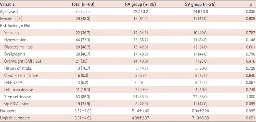
Table 1. Preoperative patient characteristics
Variable Total (n=60) RA group (n=35) SV group (n=25) p
Age (years) 73.5±3.5 72.7±3.2 74.6±3.8 0.035 Female, n (%) 29 (48.3) 18 (51.4) 11 (44.0) 0.609 Risk factors, n (%) Smoking 22 (36.7) 12 (34.3) 10 (40.0) 0.787 Hypertension 44 (73.3) 23 (65.7) 21 (84.0) 0.146 Diabetes mellitus 28 (46.7) 15 (42.9) 13 (52.0) 0.601 Dyslipidemia 28 (46.7) 17 (48.6) 11 (44.0) 0.796 Overweight (BMI >25) 21 (35) 14 (40.0) 7 (28.0) 0.416 History of stroke 10 (16.7) 5 (14.3) 5 (20.0) 0.728
Chronic renal failure 5 (8.3) 2 (5.7) 3 (12.0) 0.640
LVEF <35% 3 (5.2) 0 (0) 3 (13.0) 0.057
Left main disease 11 (18.3) 7 (20.0) 4 (16.0) 0.748
3-vessel disease 53 (88.3) 31 (88.6) 22 (88.0) 1.000
s/p PTCA c stent 19 (31.9) 8 (22.9) 11 (44.0) 0.099
Euroscore 5.52±1.88 5.14±1.40 6.04±2.24 0.095
Logistic euroscore 5.51±4.62 4.38±2.37 7.10±6.38 0.051
0.75; grade 4, completely obliterated lumen due to thickening or thrombosis or both.13)
Statistical analysis
All values are expressed as means±standard deviations. Between-group differences in clinical variables were analyzed by the χ2 test, Fisher’s exact test, unpaired t-test, or Mann-Whitney U test. The time-related events that we studied included major adverse cardio-vascular events and death of the patient after being discharged from the hospital. Freedom from these time-related events was estimated by the nonparametric actuarial Kaplan-Meier method. All statistical analyses were performed by using Statistical Package for the Social Sciences (SPSS) 12.0.1 for Windows (SPSS, Inc., Chicago, IL, USA).
Results
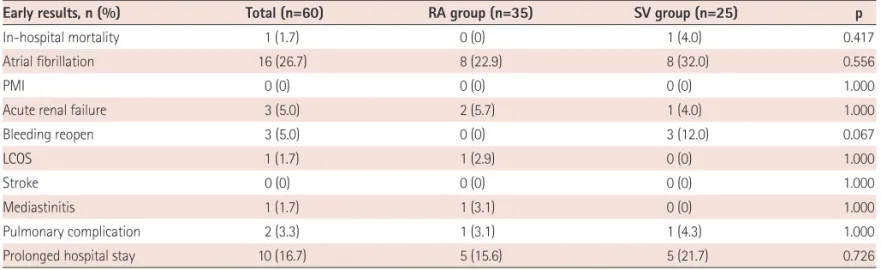
Early clinical outcomes
Preoperative characteristics of the patients (Table 1) and number of distal anastmosis according to the territories are described in detail (Table 2). The in-hospital mortality in the SVG and RA groups was 4.0 (1/25) and 0% (p=0.417), respectively. The incidence of post-operative morbidities including atrial fibrillation (n=16, 26.7%), acu-te renal failure (n=3, 5.0%), reoperation due to postoperative bleed-ing (n=3, 5.0%), low cardiac output syndrome (n=1, 1.7%),
media-stinitis (n=1, 1.7%), pulmonary complications (n=2, 3.3%), and pro-longed hospital stay (n=10, 16.7%) was not significantly different between the two groups (Table 3).
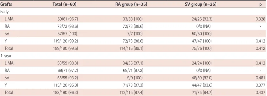
Early and one-year graft patency outcomes
Early postoperative CT angiography demonstrated patency rates of 100 (75/75) and 99.1% (114/115) in the SVG and RA groups, re-spectively (p=0.412). In situ left IMA grafts showed a 100% (58/58)
patency rate. The patency rate of SVG was 100% (50 of 50), which was not different from the overall patency rate of RA grafts {98.6% (72 of 73)} (Table 4).
One-year postoperative CT angiography demonstrated an overall graft patency rate of 96.3% (183/190). No significant difference was observed in the patency rate of the left IMA between the two groups (100%). The overall patency rate of the SVG group was 94.7% (71/75), which was not significantly different from that of the RA group {97.4% (112/115)}.
Preexisting pathologic conditions
Histopathologic evaluation was performed with 23 RAs (66%), and 22 SVs (88%). Histopathologic examination revealed that the overall incidence of intimal hyperplasia of the radial arteries was 74% (17/23). One specimen (4.3%) showed an evidence of medial calcification. Five specimens exhibited an intima-to-media ratio of less than or equal to 0.25 (Kobayashi grade 0). Overall, 34.8% (8/23) of the radial arteries exhibited an intima-to-media ratio greater than 50% (Kobayashi grade 2 and above). According to the histopa-thology findings, fifty percent (11/22) of the SVs were reported to have diffused intimal hyperplasia.
Correlations of vascular ultrasonography, optical coherence tomography findings, and histopathology
The mean RA intima-to-media thickness (IMT) was found to be
Table 2. Number of distal anastomoses
Territory (n=60)Total RA group(n=35) SV group(n=25) p
Total 3.2±0.8 3.3±0.8 3.1±0.8 0.447
LAD 1.4±0.5 1.4±0.6 1.4±0.5 0.773
LCX 1.0±0.5 1.0±0.5 0.9±0.5 0.279
RCA 0.9±0.6 0.9±0.6 0.8±0.5 0.330
LAD: left anterior descending artery, LCX: left circumflex artery, RCA: right coronary artery, RA: radial artery, SV: saphenous vein
Table 3. Comparison of early clinical outcomes between the RA and SV groups
Early results, n (%) Total (n=60) RA group (n=35) SV group (n=25) p
In-hospital mortality 1 (1.7) 0 (0) 1 (4.0) 0.417
Atrial fibrillation 16 (26.7) 8 (22.9) 8 (32.0) 0.556
PMI 0 (0) 0 (0) 0 (0) 1.000
Acute renal failure 3 (5.0) 2 (5.7) 1 (4.0) 1.000
Bleeding reopen 3 (5.0) 0 (0) 3 (12.0) 0.067
LCOS 1 (1.7) 1 (2.9) 0 (0) 1.000
Stroke 0 (0) 0 (0) 0 (0) 1.000
Mediastinitis 1 (1.7) 1 (3.1) 0 (0) 1.000
Pulmonary complication 2 (3.3) 1 (3.1) 1 (4.3) 1.000
Prolonged hospital stay 10 (16.7) 5 (15.6) 5 (21.7) 0.726
0.511±0.046 (range, 0.233-0.7671).
The IMT as assessed by vascular ultrasonography was in strong correlation with that determined by OCT determination for the ex vivo RA segments (r=0.80, p<0.001), and histopathology (r=0.75,
p<0.001).
Discussion
The IMA graft is known to possess the best long-term patency rates of all conduits currently used in coronary artery bypass sur-gery, because of its resistance to atherosclerosis.14)15) Histologic ch-aracteristics, arterial wall metabolism, and tone regulation capabili-ty give rise to high resistance to atherosclerosis of the IMA. Although RA is generally successful as a graft, significant differences between the RA and the IMA have been noted in vasoreactivity and anatomy, which may explain the higher degree of atherosclerosis of the RA.16-18)
Total arterial coronary revascularization strategies were adapted to overcome the problems of the vein graft atherosclerosis and oc-clusion. Utilization of RA has increased up to 75% in all myocardial revascularization procedures. Therefore, atherosclerotic change in the RA has become crucial. Previous reports have stressed on the importance of the RA for total arterial coronary revascularization, by using Y- and T-grafting methods; overcoming RA spasm and probable etiologic factors for string sign have also been discussed, but preoperative RA atherosclerosis and postoperative patency were not mentioned.19) We believe that this could be a probable rea-son for the lower early rate of RA patency. It is important that va-sospasm is resolved, but existing atherosclerosis cannot be evalu-ated after grafting.
Our results indicate that preoperative vascular ultrasonography can be used to assess the quality of the RA conduit before use, as
the vascular ultrasonography results were in well correlation with the OCT and histopathology findings. Furthermore, we found that the early and 1-year CT angiographic patency rates of OPCAB by us-ing the SV as a composite graft based on the in situ left IMA were
similar to those of OPCAB by using a RA composite graft.
Previous studies have reported that in patients who underwent CABG, the long-term patency rate of SV grafts was lower than that of IMA grafts.1)2) However, SV grafts remain the most widely used graft because of the accessibility, length, and ease of use of the SV. The recent use of no-touch techniques to harvest the SV has been reported to significantly improve long-term graft patency.20)
In the present prospective randomized controlled study, either a RA or SV graft was used as a composite graft based on the left IMA. The preoperative characteristics of the RA and SV groups were simi-lar. Early clinical outcomes, including hospital mortality and postop-erative morbidity, as well as 1-year follow-up CT angiographic pat-ency rates, were also similar between the two groups.
We used the SV as a composite graft on the left IMA based on the assumption that a vein graft anastomosed to the left IMA might be exposed to less pressure trauma or shearing stress than a graft an-astomosed to the ascending aorta. We also hypothesized that the quality of the SV would be identical to that of the RA in elderly pa-tients because the RA might have intimal hyperplasia or fibrous or calcified plaques in elderly patients.
We harvested the SV after systemic heparinization to prevent th-rombus formation in the venous endothelium during vein harvest. Manipulation and tension were minimized and dilatation using a pressure syringe was avoided during harvest. These measures may have decreased intimal injury during harvest, thereby explaining the good patency rates.
Table 4. Early and 1-year CT angiographic patency rates
Grafts Total (n=60) RA group (n=35) SV group (n=25) p
Early LIMA 59/61 (96.7) 33/33 (100) 24/26 (92.3) 0.328 RA 72/73 (98.6) 72/73 (98.6) 0/0 (NA) -SV 57/57 (100) 7/7 (100) 50/50 (100) -Y 119/120 (99.2) 72/73 (98.6) 47/47 (100) 0.412 Total 189/190 (99.5) 114/115 (99.1) 75/75 (100) 0.412 1-year LIMA 58/59 (98.3) 34/35 (97.1) 24/24 (100) 0.412 RA 69/71 (97.2) 69/71 (97.2) 0/0 (NA) -SV 55/59 (93.2) 9/9 (100) 46/50 (92.0) 0.481 Y 115/120 (95.8) 71/73 (97.3) 44/47 (93.6) 0.377 Total 183/190 (96.3) 112/115 (97.4) 71/75 (94.7) 0.437
Study limitations
Our study possesses the following limitations; the follow-up peri-od of our study was relatively short. The mean follow-up periperi-od was only 8 months; a longer follow-up period is mandatory for accu-rate evaluation of the patency accu-rates of the two types of graft con-duits. Further studies involving large numbers of patients with a longer follow-up period are required.
In conclusion, based on the early clinical outcomes, our results in-dicate that the SVG could be used as an alternative composite graft to the RA in elderly patients.
Furthermore, vascular ultrasonography is an accurate, real-time, and reproducible means for assessing the quality of RA conduits.
Acknowledgments
The authors are deeply grateful to Sun-Hee Lim, RN. for collecting the data used in this study. This study was supported by a grant from the Korean Society of Cardiology for 2007 (7-2007-0382).
References
1. Loop FD, Lytle BW, Cosgrove DM, et al. Influence of the internal-mam-mary-artery graft on 10-year survival and other cardiac events. N Engl J Med 1986;314:1-6.
2. Cameron A, Davis KB, Green G, Schaff HV. Coronary bypass surgery with internal-thoracic-artery grafts: effects on survival over a 15-year period. N Engl J Med 1996;334:216-9.
3. Tagusari O, Kobayashi J, Bando K, et al. Total arterial off-pump coro-nary artery bypass grafting for revascularization of the total corocoro-nary system: clinical outcome and angiographic evaluation. Ann Thorac Surg 2004;78:1304-11.
4. Fukui T, Takanashi S, Hosoda Y, Suehiro S. Total arterial myocardial revascularization using composite and sequential grafting with the off-pump technique. Ann Thorac Surg 2005;80:579-85.
5. Kim WS, Lee J, Lee YT, et al. Total arterial revascularization in triple-ves-sel disease with off-pump and aortic no-touch technique. Ann Thorac Surg 2008;86:1861-5.
6. Glineur D, Hanet C, Poncelet A, et al. Comparison of saphenous vein graft versus right gastroepiploic artery to revascularize the right coro-nary artery: a prospective randomized clinical, functional, and angio-graphic midterm evaluation. J Thorac Cardiovasc Surg 2008;136:482-8.
7. Zacharias A, Schwann TA, Riordan CJ, Durham SJ, Shah AS, Habib RH. Late results of conventional versus all-arterial revascularization based
on internal thoracic and radial artery grafting. Ann Thorac Surg 2009;
87:19-26. e2.
8. Tatoulis J, Buxton BF, Fuller JA, et al. Long-term patency of 1108 radial arterial-coronary angiograms over 10 years. Ann Thorac Surg 2009;88:
23-9.
9. Halbersma WB, Arrigoni SC, Mecozzi G, et al. Four-year outcome of OP-CAB no-touch with total arterial Y-graft: making the best treatment a daily practice. Ann Thorac Surg 2009;88:796-801.
10. Osika W, Dangardt F, Gronros J, et al. Increasing peripheral artery inti-ma thickness from childhood to seniority. Arterioscler Thromb Vasc Biol
2007;27:671-6.
11. Ozkan S, Akay TH, Gultekin B, et al. Atherosclerosis of radial and inter-nal thoracic arteries used in coronary bypass: atherosclerosis in arte-rial grafts. J Card Surg 2007;22:385-9.
12. Burris N, Schwartz K, Tang CM, et al. Catheter-based infrared light scanner as a tool to assess conduit quality in coronary artery bypass surgery. J Thorac Cardiovasc Surg 2007;133:419-27.
13. Kobayashi H, Kitamura S, Kawachi K, Morita R, Konishi Y, Tsutsumi M. A pathological and biochemical study of arteriosclerosis in the internal thoracic artery, a vessel commonly used as a graft in coronary artery bypass surgery. Surg Today 1993;23:697-703.
14. Singh RA, Sosa JA, Green GE. Long-term fate of the internal mammary artery and saphenous vein graft. J Thorac Cardiovasc Surg 1983;86:
359-63.
15. Sims FH. A comparison of coronary and internal mammary arteries and implications of the results in the etiology of arteriosclerosis. Am Heart J 1983;105:560-6.
16. Sperti G, Manasse E, Kol A, et al. Comparison of response to serotonin of radial artery grafts and internal mammary grafts to native coronary arteries and the effect of diltiazem. Am J Cardiol 1999;83:592-6.
17. Segarra G, Medina P, Vila JM, et al. Contractile effects of arginine ana-logues on human internal thoracic and radial arteries. J Thorac Cardio-vasc Surg 2000;120:729-36.
18. Van Son JA, Smedts F, Vincent JG, van Lier HJ, Kubat K. Comparative anatomic studies of various arterial conduits for myocardial revascu-larization. J Thorac Cardiovasc Surg 1990;99:703-7.
19. Tatoulis J, Buxton BF, Fuller JA, Royse AG. Total arterial coronary re-vascularization: techniques and results in 3,220 patients. Ann Thorac Surg 1999;68:2093-9.
20. Souza DS, Johansson B, Bojö L, et al. Harvesting the saphenous vein with surrounding tissue for CABG provides long-term graft patency comparable to the left internal thoracic artery: results of a randomiz-ed longitudinal trial. J Thorac Cardiovasc Surg 2006;132:373-8.