Kor J Oral Maxillofac Pathol 2013;37(2):83-88
하악 전방부에 발생한 소세포 골육종의 증례보고
양동현
1), 김형준
2), 박혁
3), 양우익
4), 김현실
1)*
연세대학교 치과대학 구강병리학교실, 구강종양연구소1), 구강악안면외과학교실2), 구강악안면방사선학교실3), 연세대학교 의과대학 병리학교실4) <Abstract>A Case Report of Small Cell Osteosarcoma of the Anterior Mandible
Dong Hyun Yang
1), Hyung Jun Kim
2), Hyok Park
3), Woo Ick Yang
4), Hyun Sil Kim
1)*
Department of Oral Pathology, Oral Cancer Research Institute1), Department of Oral and maxillofacial Surgery2), Department of Oral and Maxillofacial Radiology3), College of Dentistry, Department of Pathology,
College of Medicine4), Yonsei University
Small cell osteosarcoma of bone, which was first described in 1979, is an unusual variant of osteosarcoma. Osteoid production by tumor cells is frequently focal or minimal, making the differential diagnosis with other small round cell tumors of bone difficult. Here, we present a rare case of small cell osteosarcoma of the mandible appearing as bony bulging mass in 31-year-old male who has neither tenderness nor paresthesia. Histologically, the tumor contains hypercellular cartilage and abnormal osteoid associated with small round to ovoid malignant cells. Awareness of small cell osteosarcoma should be emphasized because it has worse prognosis than both other small round cell tumor and conventional osteosarcoma.
Key words:Small cell osteosarcoma, Osteoid, Mandible
* Correspondence: Hyun Sil Kim, Department of Oral Pathology, Oral Cancer Research Institute College of Dentistry, Yonsei University 50 Yonsei-ro, Seodaemun-gu, Seoul 120-752, Korea. Tel: +82-2-2228-3033, Fax: +82-2-392-2959 E-mail: khs@yuhs.ac
* Acknowledgements: 본 연구는 2012년도 정부(교육과학기술부) 의 재원으로 한국연구재단의 지원을 받아 수행된 연구임 (2012M3A9B2052523).
Received: Mar 15, 2013; Revised: Mar 18, 2013; Accepted: Mar 22, 2013
Ⅰ. 서론
소세포 골육종은 골육종의 드문 변이형태로, Sim등에 의해 서 1979년에 처음으로 보고되었다1). 발생 빈도는 전체 골육종 의 약 1-4%를 차지하며, 골육종과 동일한 위치와 연령, 성별의 분포를 보인다2). 조직학적으로 소세포 골육종은 세포질이 적으며 과염색성의 둥근 핵을 가지는 소세포로 이루어져 있으며, 이러한 특징은뼈에 발생하는 Ewing’s sarcoma/primitive neuroectodermal tumor (ES/PNET), mesenchymal chondrosarcoma, malignant lymphoma와 같은 small round cell tumor 그룹에서 관찰되
는 공통된 조직학적 소견이다3). 생검 조직에서 악성 유골
조직이 관찰되지 않는다면, 소세포 골육종을 앞서 언급한 다른 종류의 small round cell tumor와 감별하기란 매우 어렵다.
대부분의 소세포 골육종은 femur, tibia, humerus와 같 은 장골에서 보고되었으며, 악골에서 발생하는 경우는 매
우 드물어, 현재까지 4증례만이 보고되었다4-7). 본 연구에
서는 하악골에서 발생한 소세포 골육종의 증례를 보고하 고자 한다.
Fig. 1. Clinical photo show ill‐defined bulging mass with rubbery tendency on the anterior labial vestibular area of the mandible.
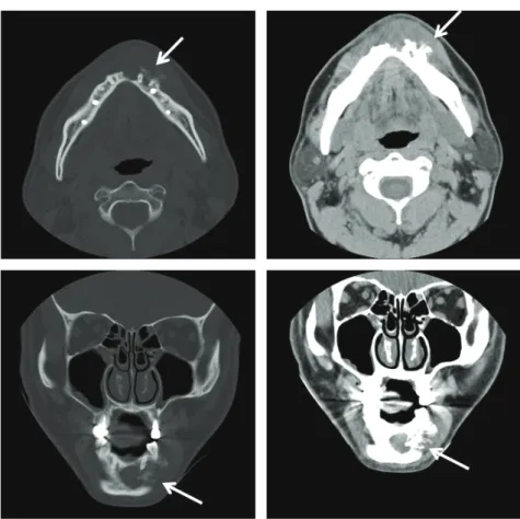
Fig. 3. Computed tomographic image shows intrabony osteolytic and osteoblastic admixed lesion which has irregular margin. Buccal cortical bone perforation with new bone formation is also identified (arrow).
Fig. 2. Panoramic radiographic view shows ill‐defined bony destructive lesion ranging from the left body to right body of mandible.
II. 증례보고
2011년 4월, 31세 남자 환자가 두 달 전부터 발생하여 점점 커지는 하악 전치부 종창에 대한 평가를 위해 연세대학교 치 과대학병원 구강악안면외과에 내원하였다. 환자는 특기할 만 한 전신질환을 가지고 있지 않았으며, 주소부위에 대한 외상 병력도 없었다. 초진 임상검사 상 하악 전치부 순측 전정부위에 주변과 경 계가 불명확한 5x3cm 가량의 rubbery tendency를 가지는 bulging mass가 관찰되었다 (Fig. 1). 촉진 시에 환자가 호소85
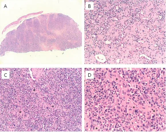
Fig. 4. Histologic findings of incisional biopsy specimen. A. High density cellular clusters are observed beneath the oral epithelium. B. Tumor cells are arranged around vascular channels. C. and D. Tumor consist of monotonous small round to ovoid cells showing diffuse infiltrating pattern. Eosinophilic osteoid‐like materials (*) exist between tumor cells.
Fig. 5. Immunohistochemical staining results. A. The tumor cells are positive membranous staining pattern for synaptophysin. B. The tumor cells are focal positive for S‐100. C. The tumor cells are focal positive for neuron specific enolase.
Fig. 6. Histologic findings of surgical specimen. A. Lace‐ like malignant osteoid (*) and definite bone formation is observed between the tumor cells. B. The tumors cells have considerable size variation and prominent nucleoli.
하는 통증이나 감각이상의 증상은 없었으며, 병소 주변 치아 의 동요도나 타진 시의 통증도 없었다. 혈액 및 혈청검사 소견 상 alkaline phosphatase level을 포함한 모든 검사결과가 정 상범위였다. 파노라마 방사선 사진상 주변조직과 경계가 불분명한 다방 성의 골파괴성 병소가 하악 좌측 체부에서 우측 체부에 걸쳐 서 광범위하게 관찰되었으며 (Fig. 2), 전산화 단층 영상에서 병소는 주변골과의 경계가 불분명하였고 골용해성과 골형성 병소가 융합된 특징을 보였으며, 협측 피질골의 파괴와 함께 햇살 모양의 신생골 형성 또한 관찰되었다 (Fig. 3). 조직검사 결과 세포밀도가 높은 간엽세포기원의 종양세포 가 결합조직 내부에 광범위하게 침윤하고 있는 것이 관찰되 었다. 종양세포는 둥글거나 타원형의 모양이었으며, 과염색 성의 핵이 종양세포의 대부분을 차지하고 세포질은 거의 없 는 것이 특징이었다 (Fig. 4). 면역조직화학염색결과 종양세 포들은 synaptophysin에 양성소견, S-100과 neuron specific enolase에 부분적으로 양성소견을 보였으며, 국소적인 유골 조직 형성부위로 판단되는 부분이 관찰되어 잠정적으로 소세 포 골육종으로 진단하였다 (Fig. 5).
생검 후 시행한 PET-CT 소견상 FGT uptake가 증가한 골용 해성 병소가 하악골에서 관찰되었으며, 우측 폐의 하엽에서 small tiny nodule이 관찰되었으나 PET-CT 상에서 평소를 평 가하기에는 매우 작아서 further evaluation을 위해서 Chest-CT를 촬영하였다. Chest-CT 상에서 right lower lobe (RLL)에 non-calcified polygonal shape의 결절이 관찰되었으 며, 종양세포의 전이보다는 육아종 또는 intrapulomonary lymph node로 판단하였다. Fibular osteocutaneous flap reconstruction을 동반한 하악골의 분절골 절제술이 시행되었 으며, 수술 검체에 대한 조직검사소견상 악성간엽세포와 연관 된 세포밀도가 높은 연골조직 및 비정상적인 레이스 패턴의 악성 유골조직이 관찰되었다. 결합조직 내에 높은 혈관 증식 및 주변 치아 및 뼈조직에 대한 종양세포의 공격적인 파괴양 상 또한 관찰되었으며, 위와 같은 조직병리소견을 바탕으로 최종적으로 소세포 골육종으로 진단하였다 (Fig. 6). 수술 후 adramycin, ifosfamide를 포함한 항암요법이 시행되었으며, 술 후 2년이 경과한 현재 재발소견은 보이지 않으며, 환자의 상태는 양호한 편이다.
III. 고찰
소세포 골육종은 1979년에 Sim 등에 의해서 보고된 이후 지금까지 약 100여 증례가 보고된 드문 골육종의 변이형으로 전체 골육종의 약 1-4%의 빈도를 차지한다. 발생하는 연령, 성별, 부위 등의 양상은 골육종과 유사한 패턴을 보이며, 이 종양은 distal femur, proximal tibia, proximal humerus 등의장골에 많이 발생하는 것으로 보고되고 있다1-9). 저자들이
조사해 본 바에 의하면 악골에서는 현재까지 총 4증례가 보
고되었다4-7). 조직학적으로 악성유골조직을 생성하므로, 골
육종으로 분류되지만 특징적으로 종양세포들이 small round cell로 이루어져 있어서 골에서 발생하는 Ewing’s sarcoma /
primitive neuroectodermal tumor (ES/PNET), mesenchmyal chondrosarcoma, malignant lymphoma과 같은 다양한 종류
의 small cell malignancy와의 감별진단이 필요하다3).
유잉육종은 특정조직으로의 분화를 보이지 않는 종양세포 가 세포 사이 간질이 거의 없이 소엽의 형태로 배열되어 있다. 각각의 세포는 둥근 형태의 핵을 가지고 있으며 크기는 약 10~15um 정도로 보고되고 있어 소세포 골육종에서 관찰되는 6.7~10um의 크기에 비해서 상대적으로 큰 편이며, 소세포 골 육종에 비해서 핵이 좀더 획일적인 모양을 가진다. 유잉육종 은 때때로 섬유혈관성 중격에 의해 분리되어 엽상형태를 이루 지만, 개개의 종양세포 사이에 소세포 골육종에서 관찰되는 유골조직이 없는 점으로 소세포 골육종과 감별할 수 있다. 악골에서 발생하는 악성 림프종은 악성 림프구 세포에 의 한 골소주나 피질골 층판의 광범위한 침윤을 특징으로 하며 때때로, 경화성이나 섬유소성의 간엽조직을 보일 수 있다. 면 역조직화학염색에서 T-세포 표지자인 CD3 및 B-세포 표지자 인 CD20이 모두의 음성을 보이는 것으로 악성 림프종을 감별 할 수 있으며, 또한 악성 유골조직형성을 보이지 않는 점이 소세포 골육종과의 감별점이다.
87 간엽성 연골육종 역시 소세포 골육종과 구별이 어려울 정 도로 유사한 핵을 가지는 종양세포로 구성되어 있으나, 연골 분화를 보이는 부위와 연골분화가 없는 부분의 급격한 전이
(abrupt transition)를 보이는 biphasic pattern이 특징이다1-9).
본 환자의 경우 임상적 및 방사선학적 소견상으로는 전형 적인 골육종과 유사하였다. 전형적인 골육종에 비해 소세포 골육종의 예후는 매우 나쁜 것으로 알려져 있는데, Sim등에 의하면 소세포 골육종으로 진단된 24명의 환자 중 9명의 환자 가 진단 후 1년 이내에 사망하였다고 하며1), Bertoni 등은 소 세포 골육종으로 진단된 보고 하였다9). Nakajima 등에 의하 면 소세포 골육종의 5년 생존율은 28.6%로 patric등에 의해서 보고된 전형적인 골육종의 5년 생존율 55% 및 유잉육종의 5년 생존율 50%에 비해서 매우 저조하다8,10). 소세포 골육종의 치료에 있어서도 수술만 시행한 경우의 환자의 평균 생존기간은 1.4년으로, 수술과 화학요법을 같이 시행한 경우의 평균 생존기간인 13.4년에 비하여 현저하게 불량하였다8).
병리의사는 생검 조직에서 small round cell 만이 관찰되고 악성 유골조직이 관찰되지 않는 경우에는 소세포 골육종을 진단하기가 용이하지 않으므로, 이러한 사실을 감별진단에 염 두에 두어야 할 것이다. 몇몇 전자현미경 연구에 따르면, 소세포 골육종에서 관찰 되는 조직소견이 미분화된 유잉육종이나 간엽성 연골육종과 중복된다는 결론을 내리고 있다11-13). 또한 면역조직화학염색 에서 neuroendocrine marker에 양성 소견을 보이는 점은 이 종양이 미분화된 간엽성 종양으로부터 분화하였으며, 유잉육 종과 같은, small round cell tumor와 특징을 공유한다는 사실 을 시사한다3,14,15).
결론적으로, 악골에서 발생하는 소세포 골육종은 현재까지 단지 4증례만이 보고된 매우 드문 증례이며, 임상의사는 본 종양의 예후가 다른 small round cell tumor 및 골육종에 비해 서 훨씬 좋지 않다는 사실을 인지하는 것이 매우 중요하다. 이러한 사실은 본 종양의 병리학적 미분화 정도와 연관이 있 을 것으로 생각되며, 본 종양의 본질 (nature)에 대한 보다 많은 연구가 수행되어야 할 것으로 판단된다.
IV. 참고문헌
1. Sim FH, Unni KK, Beabout JW, Dahlin DC: Osteosarcoma with small cells simulating Ewing's tumor. J Bone Joint Surg Am 1979;61:207-215. 2. Ayala AG, Ro JY, Raymond AK, et al: Small cell
osteosarcoma. A clinicopathologic study of 27 cases. Cancer 1989;64:2162-2173.
3. Hameed M: Small round cell tumors of bone. Arch Pathol Lab Med 2007;131:192-204. Review.
4. Giangaspero F, Stracca V, Visonà A, Eusebi V: Small-cell osteosarcoma of the mandible. Case report. Appl Pathol 1984;2:28-31.
5. Kim YM, Yang SW, Cho MY, Hong SW, Choi BH: Small cell osteosarcoma similar to Ewing's sarcoma in histo-logic findings and MIC2 expression. Korean J Pathol 1999;33:204-209.
6. Sethi A, Rehani S, Arya K: Small cell osteosarcoma of mandible. A rare case report and review of literature. J Clin Exp Dent 2010;2:e96-99.
7. Uma K, Cherian G, Nayak V, Patil S: Small cell osteo-sarcoma of the mandible: Case report and review of its diagnostic aspects. J Oral Maxillofac Pathol 2011 Sep;15:330-334.
8. Nakajima H, Bond JR, Unni KK: Small cell osteosarcoma of bone. Review of 72 cases Cancer. 1997;79:2095-2106.
9. Bertoni F, Present D, Bacchini P, et al: The Istituto Rizzoli experience with small cell osteosarcoma. Cancer 1989;64:2591-2599.
10. Ha PK, Eisele DW, Frassica FJ, et al: Osteosarcoma of
the head and neck: a review of the Johns Hopkins experience. Laryngoscope 1999;109:964-969.
11. Roessner A, Immenkamp M, Hiddemann W, et al: Case
report 331: Small cell osteosarcoma of the tibia with diffuse metastatic disease. Skeletal Radiol 1985;14: 216-225.
12. Dickersin GR, Rosenberg AE: The ultrastructure of small-cell osteosarcoma, with a review of the light mi-croscopy and differential diagnosis. Hum Pathol 1991;22:267-275.
13. Kyriakos M, Gilula LA, Becich MJ, Schoenecker PL: Intracortical small cell osteosarcoma. Clin Orthop Relat Res 1992;279:269-280.
14. Llombart-Bosch A, Contesso G, Peydro-Olaya A: Small round blue cell sarcoma of bone mimicking atypical
Ewing's sarcoma with neuroectodermal features. An analysis of five cases with immunohistochemical and electron microscopic support. Cancer 1987;60:1570-1582.
15. Pinto A, Grant LH, Schell MJ, Parham DM: Immunohistochemical expression of neuron-specific enolase and Leu 7 in Ewing's sarcoma of bone. Cancer 1989;64:1266-1273.