저작자표시-비영리-변경금지 2.0 대한민국 이용자는 아래의 조건을 따르는 경우에 한하여 자유롭게 l 이 저작물을 복제, 배포, 전송, 전시, 공연 및 방송할 수 있습니다. 다음과 같은 조건을 따라야 합니다: l 귀하는, 이 저작물의 재이용이나 배포의 경우, 이 저작물에 적용된 이용허락조건 을 명확하게 나타내어야 합니다. l 저작권자로부터 별도의 허가를 받으면 이러한 조건들은 적용되지 않습니다. 저작권법에 따른 이용자의 권리는 위의 내용에 의하여 영향을 받지 않습니다. 이것은 이용허락규약(Legal Code)을 이해하기 쉽게 요약한 것입니다. Disclaimer 저작자표시. 귀하는 원저작자를 표시하여야 합니다. 비영리. 귀하는 이 저작물을 영리 목적으로 이용할 수 없습니다. 변경금지. 귀하는 이 저작물을 개작, 변형 또는 가공할 수 없습니다.
Identifying the role of ornithine
decarboxylase1 (ODC1) in breast cancer
Yun Jin Shin
Department of Medicine
Identifying the role of ornithine
decarboxylase (ODC1) in breast cancer
Directed by Professor Joon Jeong
The Master's Thesis
submitted to the Department of Medicine
the Graduate School of Yonsei University
in partial fulfillment of the requirements for the degree of
Master of Medical Science
Yun Jin Shin
This certifies that the Master's Thesis of
Yun Jin Shin is approved.
---
Thesis Supervisor : Joon Jeong
---
Thesis Committee Member#1 : Joo Hyuk Sohn
---
Thesis Committee Member#2 : Yoon Jin Cha
The Graduate School
Yonsei University
ACKNOWLEDGEMENTS
Many people helped me to finish my dissertation. I would like to
take this opportunity to thank you.
First of all, I would like to express my sincere gratitude to Professor
Joon Jeong for giving me advice and guidance in the research
process.
I would also like to thank Doctor Hye kyung Shin and Jung Hee Lim
for their help with many things, such as trouble shooting as well as
experimental methods.
Many thanks to the breast cancer center teachers and the lab staff.
Thanks to them, I was able to enjoy my time as a master’s student.
Lastly, I would like to thank my family and friends for their support
and encouragement.
With the help of many people, I was able to graduate.
I will work hard to make their kind help shine.
TABLE OF CONTENTS
ABSTRACT··· 1
I. INTRODUCTION ··· 2
II. MATERIALS AND METHODS
1. Cell culture ··· 8
2. Western blot analysis ··· 8
3. Cell viability assay ··· 9
4. Colony formation assay ··· 9
5. Wound healing assay ··· 10
6.
siRNA transfection··· 10
7. qPCR ··· 11
8. Apoptosis analysis ··· 12
9. Cell cycle analysis ··· 12
III. RESULTS
1. ODC1 is highly expressed in breast cancer. ··· 14
2. DFMO treatment highly reduced breast cancer cell growth. ··· 15
3.DFMO treatment decreased colony formation ability and cell migration.
··· 18
4.Decreased ODC1 expression through siRNA transfection ··· 20
5. Reduced ODC1 expression in breast cancer cell induces apoptosis.
··· 21
6.
Downregulation of ODC1 in breast cancer cell induces cell cycle arrest at
s phase. ··· 23
IV. DISCUSSION ··· 25
V. CONCLUSION ··· 28
REFERENCES ··· 29
ABSTRACT (IN KOREAN) ··· 35
LIST OF FIGURES
Figure 1. The polyamine metabolic pathway. ··· 7
Figure 2. ODC1 expression level in breast cancer cell lines.··· 14
Figure 3. DFMO inhibits breast cancer cell growth. ··· 16
Figure 4. DFMO inhibits cell colony formation and cell migration.
··· 18
Figure 5. Downregulation of ODC1 by siRNA ··· 20
Figure 6. Silencing of ODC1 expression by siODC1 induces
apoptosis. ··· 21
Figure 7. Silencing of ODC1 expression by siODC1 induces cell
cycle arrest. ··· 23
LIST OF TABLES
Table 1. siRNA sequence ··· 13
1
ABSTRACT
Identifying the role of ornithine decarboxylase 1 in breast cancer
Yun Jin Shin
Department of Medicine
The Graduate School, Yonsei University
(Directed by Professor Joon Jeong)
Ornithine decarboxylase 1 (ODC1) is a key enzyme involved in the production of putrescine in polyamine biosynthesis. ODC1 affects nucleic acid metabolism, cell growth, cell differentiation, and cell cycle. Increased ODC1 is associated with carcinogenesis, specifically in the invasion and angiogenesis phases. While several publications have suggested that the reduction of ODC1 inhibits cancer cell proliferation and metastasis, the mechanisms of cell growth regulation, proliferation, and invasion in breast cancer cells remain unclear. In this study, we invesigated ODC1 expression levels in breast cancer cells. The ODC1 expression level of SKBR3 was highest among ten breast cancer cell lines. We treated SKBR3 with difluoromethylornithine (DFMO), which binds to ODC1 instead of pyridoxal phosphate to inhibit the action of ODC1. Cells in the treated group showed lower cell viability, cell growth, cell migration, and colony forming ability, compared to the untreated group. Down-regulated expression of ODC1 induced apoptosis and resulted in an accumulation of cells in the S phase of the cell cycle. Knock-down of ODC1 in breast cancer cells promoted the expression of cleaved Poly (ADP-ribose) polymerase, caspase-3, and caspase-9 and decreased expression of Bcl-2 in the mitochondrial pathway. The results of this study suggest that the polyamine pathway,including ODC1, has an important function in the cancer cell cycle, and when the expression of ODC1 is inhibited, cell cycle circulation is blocked, resulting in cell death, which inhibits the proliferation and metastasis of cancer cells. This study establishes the potential of ODC1 as target for the treatment of breast cancer. Further studies are needed to prove that ODC1 is a promising target for the treatment of breast cancer. Key words : breast cancer , ODC1 , DFMO , cell cycle, cell proliferation
2
Identifying the role of ornithine decarboxylase 1 in breast cancer
Yun Jin Shin
Department of Medicine
The Graduate School, Yonsei University
(Directed by Professor Joon Jeong)
I. INTRODUCTION
Breast cancer is the most common malignancy, accounting 30% of cancer diagnoses in women globally. It has the second- highest death rate after lung cancer.1 Studies on
factors that affect breast cancer (obesity2, long-term use of oral contraceptives3, dietary
pattern4, genetic variations5,6, etc.) are very active. First, pathological remodeling of
white adipose tissue and increased levels of fat-specific cytokines (mainly leptin), as a result of obesity, are associated with several hallmarks of breast cancer. These include sustained proliferative signaling, cellular energetics, inflammation, angiogenesis, activating invasion, and metastasis. Preclinical and clinical studies provide evidence that obesity may worsen the incidence, severity, and mortality of breast cancer.2
Second, the association between the use of oral contraceptive and breast cancer is controversial and a significant issue in public health. According to some report, the different mechanisms of action of various oral contraceptives, result in different carcinogenic effects. The use of oral contraceptives has been demonstrated to increase the risk of breast cancer occurring before age 35, and increased relative risks (RR) has been associated with long-term (over 10 years) use.7 Thus, the associated risk of breast
cancer differs between different types of oral contraceptives.8
Third, for dietary pattern, vegetarian have a lower risk of developing breast cancer than people who eat a diet containing meat.9 Vegetarians and vegans eat a lot of
soybeans and soy products, which are primarily isoflavones. Isoflavones are phytoestrogens, interacting with alpha- and beta-estrogen receptors to regulate estrogen levels in the body, and chemically prevent breast cancer along with serum albumin levels.10 In addition, vegetables such as broccoli, cauliflower, cabbage, and
kale help prevent breast cancer by acting as a substrate for glutathione stransferase M1 and T1 detoxification enzymes and inducers.11 And also consuming alcoholic drinks
3
associated with an increased risk of breast cancer. This finding was also published in the World Cancer Research Fund report. It has been hypothesized that alcohol consumption can lead to higher estrogen levels due to increased metabolites, which can interfere with the detoxification of carcinogens.12
Breast cancer is the most common genetic disease caused by the accumulation of modified genes. Both germline and somatic modifications have important roles in the development and progression of cancer.13 For example, women with mutations in
BRCA1 and BRCA2 are at greater risk of developing ovarian cancer as well as breast cancer. A deficiency or mutant form of BRCA 1 activates IGF1R, which promotes mitosis and anti-apoptosis roles through phosphatidylinositol-3-kinase and mitogen activating proteins, thereby increasing tumorigenesis.14 Another example, the mutation
of p53 is one of the frequent genetic modifications in breast cancer. When prolyl isomerase (Pin1) is overexpressed, it promotes the mutation of p53, which inhibits the expression of the antimetastatic factor p63, thereby promoting aggressiveness in breast cancer cells.15 In addition, ATM and PALB2 are genes associated with germline
mutation.16 Some somatic mutations can activate the HER2 signaling pathway. HER2
somatic mutations occur more frequently in negative breast cancer, and HER2-negative breast cancer patients with these variants have less survival rate than wild-type HER2 patients.17 ER pathway is involved during the progression of breast cancer
to the invasion of other organs.18Somatic mutation of genes involved in ER pathway
is an index that can predict a poor prognosis in ER-positive breast cancer.19
Breast cancer has been identified as a heterogeneous disease. The classification of breast cancer subtypes is divided according to the expression of estrogen receptor (ER), progesterone receptor (PR) or thehuman epidermal growth factor receptor 2 (HER2). There are three subtypes: HR + HER2-, HER2 +, and TNBC. The first HR + HER2- is when ER or PR is positive but HER2 is negative. The second HER2 + indicates that ER or PR is positive or both ER and PR are negative when HER2 is positive. Finally, it is called TNBC when all three of ER, PR, and HER2 are negative.20 The breast cancer
subtype is an important prognostic factor in breast cancer.21 TNBC is an independent
prognostic factor for breast cancer-specific survival with TNBC have worse clinical outcomes compared to those with other subtypes. 22
4
living organisms. Polyamine metabolism includes putrescine, spermidine, spermine. Under normal conditions, polyamine acts in the synthesis of proteins and nucleic acids through intercellular connections, cell metabolism, growth, and survival, and cell differentiation and death.23
Arginase 1 (ARG1) generates ornithine from the urea cycle. Regulation of polyamine homeostasis occurs through biosynthesis, which in most organism occurs via two pathways: the arginine decarboxylase pathway and the ornithine decarboxylase pathway. In the arginine decarboxylase pathway, polyamine biosynthesis starts with the change of arginine into ornithine, then into putrescine. The ornithine decarboxylase pathway involves the direct conversion of ornithine into putrescine.
Ornithine decarboxylase 1(ODC1) is a rate-limiting enzyme in polyamine biosynthesis which produces putrescine. Decarboxylated S- adenosylmethionine (dcAdoMet), a second rate-limiting enzyme in polyamine biosynthesis, is the amino propyl donor for spermidine synthase (SPDSY) and spermine synthase (SPMSY) for the synthesis of spermidine and spermine (Figure 1). The levels of ODC1, dcAdoMet, and spermidine/spermine N1-acetyltransferase 1 (SSAT) show obvious changes in response to elements related to cell growth and to alternations in polyamine levels. Vast alterations in enzyme activity can occur very quickly and are usually the result of alterations in multiple enzyme protein. Regulation can occur at the transcription, translation, and protein degradation stages.24
ODC1 catalyzes the decarboxylation and conversion of ornithine to putrescine.25 ODC1
is a pyridoxal phosphate (PLP)-dependent amino acid decarboxylase and it is a homodimer with two active sites each made up of residues from both subunits.26 ODC1
has an important function as an antioxidant in DNA structure stabilization and double-strand cleavage pathways. It is an essential enzyme for cell growth, enabling the production of necessary polyamines. ODC1 has a short half-life and is regulated by transcription and translation, and its activity is regulated according to the G1 and G2 / M phases. When embryos deficient in ODC1 are implanted into the uterus, they do not develop properly. This suggests that ODC1 is necessary for cell survival. 27
Polyamines, including ODC1, are overexpressed in many cancers. ODC1 overexpression is the most significant alteration in the polyamine metabolic pathway associated with cancer progression. Increased ODC1 activity is related to the acquisition of a hormone-independent, poorly differentiated phenotype in some breast cancers.28 ODC1 activity is
5
higher in basal cell carcinomas than in normal human skin29and is significantly increased
in prostate cancer compared with paired benign tissue.30 The risk of cancer recurrence
and death is higher in breast cancer patients with higher ODC1 activity than in those with fibrous breast tissue.31 In endometrial cancer, increased levels of ODC1 are observed in
association with high grade, late- stage, serous histology which are associated with increased rates of recurrence and death.32 Upregulated ODC1 expression levels in
esophageal squamous cell carcinoma significantly coincides with lymph node metastasis status and clinical stage.33 Increased polyamines, including ODC1, are associated with
carcinogenesis and are essential for invasion and angiogenesis related to increased cancer cell proliferation and metastasis.34 Increased expression of ODC1 in NIH3T3 cells induces
cell transformation, anchorage-independent growth in soft agar.35 ODC1 levels have a
function in promoting squamous cell carcinomas by driving continued proliferation.36
Studies are underway to treat polyamines, including ODC1, in several cancers. The relationship between polyamines, cell cycle arrest, and apoptosis has been studied in several cancers. In the G1 phase, during cell cycle, cyclin D1 binds to cyclin-dependent kinase 4 (CDK4) and regulates phosphorylation of retinoblastoma protein (pRB). pRB has an essential function in the transition from G1 to S phase. The transcription factor E2F transcription factor 1 (E2F-1) stimulates cyclin E transcription. Increased release of E2F-1 from pRB and stimulation cyclin A are involved in S phase progression. CDK1 binds to cyclin B to form a complex, which rapidly increases its activity, enters mitosis, and terminates cyclin B.37 Reduction of ODC1 activity and polyamines results
in cell cycle arrest in the S phase. Overexpression of SSAT in human prostate cells, leads to polyamine reduction, resulting in cell accumulation in the G2 / M stage.38
Cells in early apoptosis are associated with mitochondrial changes mediated by anti-apoptotic Bcl-2 and pro-anti-apoptotic Bax proteins. Bax activates the release of cytochrome c from the mitochondria, activating the caspase cascade.39 When expression of ODC1 is
decreased in esophageal cancer cells, apoptosis increases due to an increase in expression of cleaved caspase-3, cleaved PARP, and Bax.33
Increased polyamine levels are linked to skin cancer, prostate cancer, breast cancer, lung cancer, and crystalline cancer. Chemotherapy of polyamine pathways have been conducted by many researchers. Methylglyoxalbisguanylhydrazone and SAM486A are drugs which target decarboxylated S-adenosyl methionine (dcAdoMet), AMXT1501 and
6
TrimerPTI are drugs that inhibit polyamine transport.34 Among the various polyamine
metabolic inhibitors, difluoromethylornithine (DFMO) is an inhibitor of ODC1 which prevents ODC1 from being permanently fixed at the active site by strongly binding to instead of pyridoxal phosphate (an ODC1 cofactor). Without ODC1 activity, putrescine is depleted, causing problems in DNA and RNA synthesis, and inhibiting cell growth and division.23 In several cancers, including neuronal carcinoma; skin cancer; endometrial
cancer; liver cancer; and rectal cancer, the inhibition of ODC1 through DFMO is an effective means of treatment or prevention.40-42
ODC1 and polyamine biosynthesis has a critical function in the development of many cancers. Thus, some studies targeting this enzyme have been conducted. However, no studies have been conducted to determine the role of ODC1 in breast cancer. To investigate the role of ODC1 in breast cancer, here we use small interfering RNA(siRNA) and ODC1 inhibition to determine what phenotypes are shown when ODC1 expression in suppressed.
7
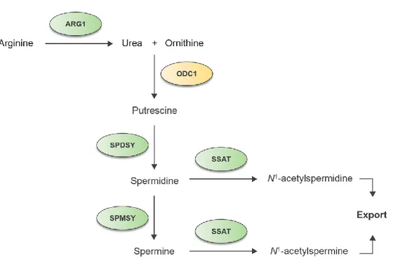
Figure 1. Polyamine biosynthesis pathway.
Arginase1 (ARG1) acts to make urea and ornithine from arginine. Ornithine decarboxylase 1 has a role in making putrescine from ornithine. Spermidine and spermine are produced by spermidine synthase (SPDSY) and spermine synthase (SPMSY), respectively. Spermidine/spermine N1-acetyltransferase 1 (SSAT) regulates the cellular content of polyamines by catalyzing acetylation of spermidine and spermine and exporting it out.23
8
II. MATERIALS AND METHODS
1. Cell culture
All cells used in the experiment were maintained in RPMI 1640 medium (Thermo Scientific, Waltham, MA USA) supplemented with 10% fetal bovine serum(FBS, Biowest, Kansas city, Missouri USA) and 1% antibiotic/antimycotic (GIBCO, Thermo Scientific, Waltham, MA USA). Cells were cultured at 37℃ in 5% CO2 incubator. For
siRNA transfection, cells were maintained in RPMI1640 supplement with 10% FBS, without antibiotics/ antimycotic for high efficiency
2. Western blot analysis
The western blot is a widely used analytical method in molecular biology, immunogenetics, and other disciplines to detect specific proteins in a sample of tissue homogenate or extract. The proteins are denaturated, followed by gel electrophoresis. Secondary antibodies are added and the primary antibody recognizes and binds to their target protein. Secondary antibodies can be visualized through staining or fluorescence to detect the desired target protein.
Whole cells were lysed in 1x Radioimmunoprecipitation assay (RIPA) lysis buffer (Rockland, Pottstown, PA USA) on ice for 15minutes. Lysates were collected and supernatant was obtained by centrifugation at 12,000 rpm, 4℃for 15minutes. To determine protein concentration, Pierce™ BCA Protein Assay (Thermo Scientific, Waltham, MA USA) was performed. Then protein was mixed with 4x protein sample buffer(BIORAD, Contra Costa County, CA USA) and 2-mercaptoethanol was boiled at 100℃ for 5minutes. 2-mercaptoethanol acts on the sample and breaks the SS bond and electrophoresis by converting the protein of dimer or more into monomer state.Sodium dodecyl sulfate - polyacrylamide gel electrophoresis (SDS-PAGE) was transferred to the polyvinylidene fluoride (PVDF) membrane. Unlike nitrocellulose and nylon membranes, PVDF is a hydrophobic material and does not adhere to a protein unless activated with methanol. Transferred membranes were treated with 5% skim milk in tris buffered saline with tween 20 (TBST) for 2hours at room temperature. Primary antibodies were reacted overnight at 4℃. The primary antibodies against ornithine decarboxylase (Catalog No. ab-126590), β-actin (Catalog No. ab- 8227) were purchased from abcam.
9
The primary antibodies to detect caspase-3 (Catalog No. 9662s), and caspase-9 (Catalog No. 9502s) were obtained from Cell Signaling Biotechnology and Bax (Catalog No. sc-7480), Bcl-2 (Catalog No. sc-7382), and cyclin A (Catalog No. sc-271682) antibodies were from santa cruz biotechnology. After washing five times with TBST, secondary antibodies were treated for 2hrs at room temperature. After chemoluminescence detection using the Electrochemiluminescence (ECL) solution (BIORAD, Contra Costa County, CA USA), blots were developed by image count LAS 4000.
3. Cell viability assay
Cells were plated in a 96-well plate (SPL LIFE SCIENCE) at a density of 1x10⁴
cells/well in triplicate and after 24hours, treated with different concentrations of (0mM to 5mM) difluoromethylornithine (Sigma, St.Louis, Missouri USA) in culture media. After incubation for 24, 48, and 72 hours, the supernatant was suctioned and the cells were treated with Cell Counting Kit-8 assay (Dojindo laboratories, Kumamoto, Japan), and incubated for 2 hours. Cell viability was determined at OD450nm with a plate reader
(VersaMax, Molecular Device, Sunnyvale, CA, USA). A Cell Counting Kit-8 (CCK-8) was used for cell proliferation and cytotoxicity assays. Water-soluble tetrazolium salt (WST-8), a major component of CCK-8, was reduced by dehydrogenase in living cells to produce an orange form (formazan). The amount of formazan dye produced was directly proportional to the number of living cells.
4. Colony formation assay
The colony formation assay is an in vitro cell survival assay which measures the colony forming ability of a single cell to grow into a colony. Colony formation is the ability to cell division indefinitely. Leaking colonies comprise of at least 50 cells. Unlike normal cells, cancer cells do not die even if they grow excessively and grow in multiple layers beyond the monolayer membrane. In soft agar, cancer cells grow well without attaching to the ground due to anchorage-independent growth characteristics. The colony formation assay can be used to determine the effectiveness of cytotoxic agents.43Cells were plated
in a 6-well plate (SPL LIFE SCIENCE) at a density of 1x10⁴cells/well and after 24hours,
treated with 0, 1.25, 2.5, and 5mM of DFMO in culture media. After one week, we changed the media containing DFMO and the cells were incubated for a total of 2 weeks
10
at 37℃. After 2 weeks, colony formation ability was photographed with a microscope mounted camera (ZIESS, Oberkochen, Germany), for observing the colony formation ability.
5. Wound healing assay
The wound healing assay is an in vitro experiment used to examine collective cell migration in two dimensions. Exposure to a cell-free area induces cells to migrate into the gap. Migration is showed by epithelial and endothelial monolayers that move in two dimensions while maintaining their intercellular junctions and migration occurs in diverse processes such as cancer metastasis, embryonic morphogenesis, and tissue injury.44
In this experiment, the monolayer was scratched with a pipette tip and migration into the gap was imaged over several hours using a microscope equipped for live-cell imaging. Cells were plated in a 6-well plate at a density of 1x10⁵cells/well and after 24hours, treated
with 0, 1.25, 2.5, and 5mM of DFMO. After 4days, DFMO-treated cells were harvested and seeded into culture-insert 2-well dish (ibidi, Nürnberg, Germany). When cell density reached 90~100%, the culture-insert 2-well was removed and the dish was filled with culture media containing either 0, 1.25, 2.5, and 5mM of DFMO. Wound recovery changes were photographed with a microscope mounted camera (ZIESS, Oberkochen, Germany).
6. siRNA transfection
siRNA binds to RNA-induced silencing complex (RISC), resulting in strands with a stable 5'-end. The single-stranded siRNA component then guides and combines the RISC on the target mRNA through the action of catalytic RISC protein.45
Lipofectamine is used to increase the transfection efficiency of RNA (including mRNA and siRNA) or plasmid DNA into the in vitro cell. Lipofectamine has a positively charged liposome formulation and forms a complex with a negatively charged nucleic acid molecule to penetrate the cell membrane. For expression of the transgene of a cell, the nucleic acid must extend to the nucleus of the cell to begin transcription. However, transfected genetic material is interrupted during the delivery process and cannot reach the nucleus from the transcription start. During cell division, the material may reach the nucleus by being trapped in the reassembling nuclear envelope following mitosis.46
11
BIONEER. We seeded SKBR3 cells by 5x10⁵ cells per 6-well plate and proceed with
reverse transfection. To improve transfection efficiency, Lipofectamine 2000 (Invitrogen, Thermo Scientific, Waltham, MA USA) was diluted in Opti-mem reduced serum medium (GIBCO, Thermo Scientific, Waltham, MA USA) and 200 pmol siRNA was also diluted in Opti-mem medium. Mix 1: 1 with lipofectamine 2000 and siRNA diluted in Opti-mem reduced serum medium. The mixture was then incubated for 20min at room temperature. siRNA with Lipofectamine 2000 was transfected onto SKBR3. ODC1 expression was measured by RT-PCR and western blot. Targeted siRNA sequence information is shown in Table 1.
7.qPCR
Total RNA was extracted from the cells with TRIzol™ Reagent (Thermo Scientific, Waltham, MA USA) according to the manufacturer’s instructions. In brief, growth media was removed and TRIzol™ Reagent was added directly to the culture dish by pipetting, to lyse the cells and homogenize the lysate. The lysate was centrifuged and the clear supernatant was transferred to a new tube. Chloroform was added to the mixture in order to separates into a lower red phenol-chloroform, and interphase, and a colorless upper aqueous phase. Isopropanol was added, so that total RNA precipitate formed a white gel-like pellet at the bottom of the tube. Finally, the pellet was resuspended in 75% ethanol for washing. cDNA was produced using a cDNA Synthesis Kit (Thermo scientific, Waltham, MA USA). It is capable of synthesizing cDNA up to 20 kb from a wide range of total RNA amounts (1 pg to 5 µg) at elevated temperatures (45-65 °C). The reaction product of the first strand cDNA synthesis can be used directly in qPCR.
The PCR process consists of a series of temperature changes that are repeated 25–50 times. These cycles normally consist of three stages. First, at a temperature of around 95 °C, the nucleic acids double chain is separated. Second, at a temperature of around 50–60 °C, the primers are bound with the DNA template. Third, at between 68–72 °C, DNA polymerase is carried out. 47,48 To process PCR, we need to DNA polymerase,
dNTPs, template, and primer, and anything else. For qPCR, we use SYBR Green mix containing dNTPs, MgCl2, and DNA polymerase. SYBR Green is a reagent that displays fluorescence by binding to double-stranded DNA. It binds to double-stranded DNA synthesized by a PCR reaction to emit fluorescence, and we can detect the fluorescence
12
sensitivity to measure the amount of amplified products produced.
The qPCR conditions were 95℃/5min for one cycle, 95℃/15sec and 60℃/ 30sec and 72℃/ 45sec for 30cycles, followed by 95℃/ 15sec, 60℃/1min, 95℃/ 15sec 60℃/ 15sec for one cycle. ODC1 expression levels were represented relative to Glyceraldehyde 3-phosphate dehydrogenase (GAPDH). The primer sequences used in the study are listed in Table 2.
8.Apoptosis analysis.
To identify apoptotic cells, cells were detected by flow cytometry using the FITC annexinⅤ apoptosis detection kit (BD Biosciences, Bergen County, NJ, USA) according to the manufacturer’s instruments. In apoptotic cells, membrane phospholipid phosphatidylserine (PS) is translocated from the inner to the outer leaflet of the plasma membrane. Annexin V is a Ca2+ dependent phospholipid-binding protein with a high affinity for PS, and binds to cells with exposed PS. Annexin V may be conjugated to FITC.This format retains its high affinity for PS and thus serves as a sensitive probe for flow cytometric analysis of cells that are undergoing apoptosis. Since externalization of PS occurs in the earlier stages of apoptosis, FITC Annexin V staining can identify apoptosis at an earlier stage than assays based on nuclear changes such as DNA fragmentation.
Thirty hours after siODC1 transfection, cells were harvested by scraper and washed with 1x phosphate-buffed saline (PBS) and resuspended in the 1x binding buffer. Annexin V and propidium iodide(PI) mix was added to the cells and mixed lightly. The mixture was incubated for 15minutes at room temperature in the dark, and apoptosis detection was performed using FACSDiva flow (BD Biosciences, Bergen County, NJ, USA).
9.Cell cycle analysis
For determine of cell cycle arrest cells, cells were recognized by flow cytometry using the PI solution in FITC annexin V apoptosis detection kit (BD Biosciences, Bergen County, NJ, USA). The fluorescence intensity of cells stained with PI is related to the amount of DNA the cells contain. DNA content in each cell cycle is often expressed as a histogram, which provides information about the relative frequency of cells.
13
Thirty hours after siODC1 transfection, cells were harvested by scraper and washed with 1x phosphate-buffed saline (PBS) and resuspended in the 1x binding buffer. Cells were stained with PI (50ug/Ml) for 15minutes at room temperature in the dark. Cell cycle analysis was performed using FACSDiva flow (BD Biosciences, Bergen County, NJ, USA).
Table1. siODC1 sequence
siODC1
number sense antisense
1 GAC UAG GAU AUG GGU CAC A UGU GAC CCA UAU CCU AGU C 2 GAG AUC ACC GGC GUA AUC A UGA UCA CGC CGG UCA UCU C
3 CGA CGA UCC ACC AUG UGA U AUC ACA UAG UAG AUC GUC G
4 CUG CCA CUU CCU CGA UGA A UCC AUC GAG GAA GUG GCA G
5 GAC UGU GCU AGC AAG ACU G CAG UCU UGC UAG CAC AGU C
Table2. Primer sequence
Name Primer sequence
ODC1 F : 5’-GAC GAG TTT GAC TGC CAC ATC-3’ R : 5’-CGC AAC ATA GAA CGC ATC CTT-3’ GAPDH F : 5’-GAG TCA ACG CAT TTG GTC GT-3’
14
III. RESULTS
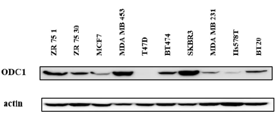
1.ODC1 expression level in breast cancer.
We used western blotting to qualify ODC1 protein in ten breast cancer cell lines. The results show that SKBR3 had the highest ODC1 expression level while MCF7, T47D, and Hs578T had lowest. (Figure 2) We selected SKBR3 for treatment with DFMO and ODC1 siRNA transfection. Due to their relatively high levels of ODC1 protein expression.
Figure 2. ODC1 expression level in breast cancer
Confluent monolayers of ZR-75-1, ZR-75-30, MCF-7, MDA-MB-453, T-47D, BT-474, SKBR3, MDA-MB-231, Hs578T, BT-20 were extracted with RIPA lysis buffer. 15 mg total protein from each cell extract was resolved by SDS-PAGE, transferred to PVDF, and blotted with antibodies against ODC1.
15
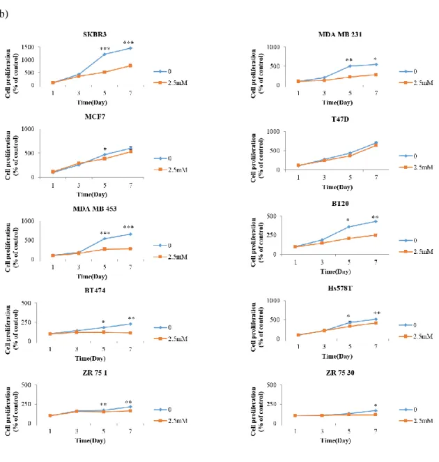
2.DFMO treatment highly reduced breast cancer cell growth.
We investigated whether DFMO inhibits cell growth by measuring the degree of ODC1 expression.SKBR3 and MDA MB 453, which had high ODC1 expression, was sensitive to low DFMO (1.25mM). MDA MB 231, T47D, and MCF7, which had low ODC1 expression, were less sensitive to high DFMO concentration. As an exception, BT474 and ZR 75 30 did not respond to the high concentration of DFMO (Figure 3a). Also, the growth of SKBR3 and MDA MB 453 treated with 2.5mM DFMO for 7 days decreased significantly after 3 days. Whereas the other cells showed slight differences or no difference at all (Figure 3b).
16
17
(b)
Figure 3. DFMO inhibits breast cancer cell growth.
(a) Cell viability was measured in cells treated with DFMO at 0, 1.25, 2.5, and 5mM by Cell Counting Kit-8 assay. (b) After treated with DFMO 2.5Mm, growth for 7 days was also measured by Cell Counting Kit-8 assay. *p<0.1,** p<0.01, *** p<0.001
18
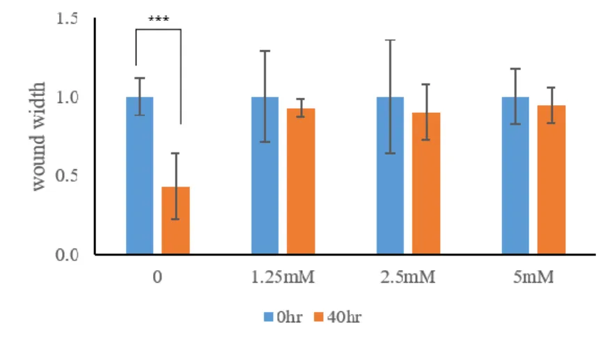
3. DFMO treatment decreased colony formation and cell migration ability.
Unlike normal cells, when cancer cells grow excessively, they grow in multiple layers beyond the monolayer membrane, and form a colony. Thus, we conducted colony formation assays to determine if DFMO exhibited these properties in breast cancer cell lines. Fifteen days after DFMO treatment on SKBR3, colony formation efficiency was reduced from the lowest concentration of 1.25mM DFMO and is further rapidly at higher concentrations of DFMO (Figure 4a). Compared to the ability of each cell to migrate by wound healing assay, cells treated with 1.25, 2, and 5mM concentrations of DFMO healed at a slower rate than untreated cells and did not move for 40 hours after wounding (Figure 4b, c).
(a)
19
(c)
Figure 4. DFMO inhibits cell colony formation and cell migration.
(a) Colony formation ability of SKBR3 decreased from the lowest concentration of 1.25mM DFMO, which showed greater reduction at higher concentrations. (b)&(c) Comparison of the migration ability of each cell by wound healing assay. The migration rate of treated SKBR3 from 1.25mM to 5mM was slower than that DFMO-untreated SKBR3. *p<0.1, ** p<0.01, *** p<0.001
20
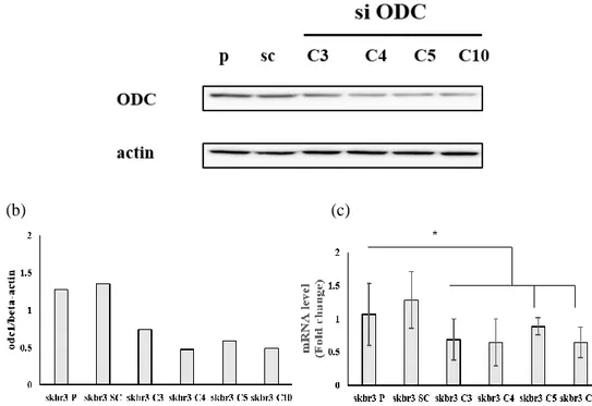
4. Decreased ODC1 expression through siRNA transfection
We transfected siODC1 into a breast cancer cell, SKBR3 by lipofectamine to investigate the role of ODC1 in apoptosis and cell cycle arrest in breast cancer. We used a combination of two siRNAs to select the most effective combination. Combinations are that C3 are siODC 1+ siODC 4, C4 are siODC 1+ siODC 5, C5 are siODC 2+ siODC 3, and C10 are siODC 4+ siODC 5 (total 200pmol). We confirmed the reduction of ODC1 expression by siRNA transfection with western blot (Figure 5a). Our data showed that the expression of ODC1 was reduced, especially for siODC C4,10 (Figure 5b). The graph shows that reduced mRNA levels of ODC1 in siRNA combinations (Figure 5c).
(a)
(b) (c)
Figure 5. Downregulation of ODC1 by siRNA (a) ODC expression was analyzed by
Western blot in SKBR3 cells expressing parental, scramble, and siODC. (b) ODC1 expression was reduced by half, especially si ODC1 C4 and 10, in a graph showing quantified data normalized with β-actin. (c) RT-PCR showed siODC1 transfection into SKBR3. The graph shows the reduced value compared to GAPDH by calculating the delta-delta CT value.
21
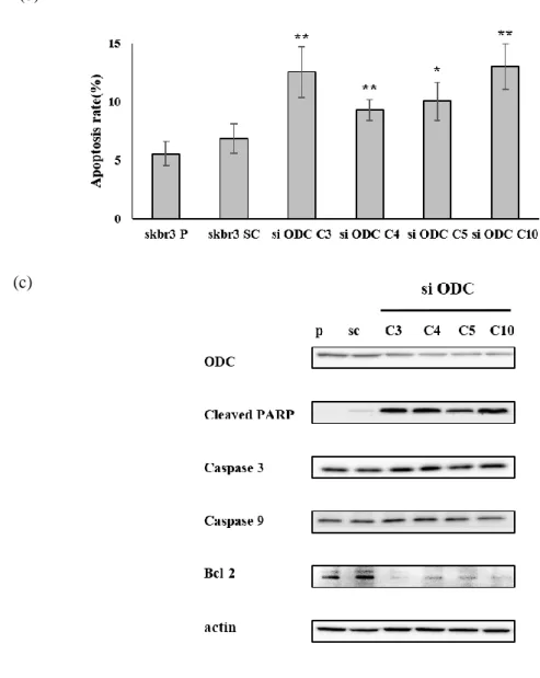
5. Reduced ODC1 expression in breast cancer cell induces apoptosis.
Apoptosis was measured by flow cytometry using Annexin V and PI staining of normal cells and cell with reduced ODC1 expression. Compared to parental and scramble cells, total apoptosis was more than doubled in ODC1 knocked down cells (Figure 6a). Normal cells with intact cell membranes were negative because PI and Annexin V + FITC were not stained. In early apoptosis, phosphatidylserine (PS) was released to outside, Annexin V + FITC was positive and PI was negative. In late apoptosis, dead cells were positive for PI and Annexin V + FITC. The apoptosis rate is the sum of early apoptosis corresponding to Q1 and late apoptosis or dead cells of Q2 (Figure 6b).If DNA damage does not recover above the repair level, not only caspase-3 and 9, but also cleaved PARP is activated and Bcl-2, which prevents apoptosis, is reduced, resulting in apoptosis. Western blot analysis showed that reduced ODC1 induced increased expression of cleaved PARP, caspase-3 and caspase-9, and suppressed expression of Bcl-2 (Figure 6c).
(a)
22
(b)
(c)
Figure 6. Silencing of ODC1 expression by siODC1 induces apoptosis.
(a) Apoptosis was induced when SKBR3 was transfected with siODC1. (b) The apoptosis rate, the sum of early apoptosis and late apoptosis or dead cells, was significantly increased after the siODC1 transfection. (c) Apoptosis induction was confirmed by the western blot. When ODC1 expression was decreased, expression of Cleaved PARP, Caspase-3, and Caspase- 9 was increased, whereas Bcl-2 was reduced.
23
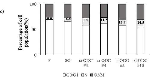
6.Downregulation of ODC1 in breast cancer cells induces cell cycle arrest at S phase. We observed cyclin A by western blot in order to confirm that reduced ODC1 expression results in cell cycle arrest. Decreased expression of cyclin A stops DNA synthesis and replication. Therefore, the cell is accumulated in the S stage and can't proceed to the next step. Western blot analysis showed that reduced ODC1 induced decreased expression of cyclin A (Figure 7a). We analyzed cells stained with PI solution through flow cytometry and observed that cell cycle arrest at S phase was more than doubled in ODC1 knocked down cells, compared to parental and scramble cells (Figure 7 b, c).
(a)
24
(c)
Figure 7. Silencing of ODC1 expression by siODC1 induces cell cycle arrest.
(a) SKBR3 was transfected with siODC1 and it was confirmed by Western blot that cyclin A expression was decreased. (b) Cell cycle was analyzed by flow cytometry and cell cycle arrest occurred in S phase due to the decrease in ODC1 expression. (c)It is a graph of (b). P1 is G0 / G1, P2 is S, and P3 is G2 / M phase.
25
IV. DISCUSSION
Breast cancer is the most common cancer in women in the world. Like all tumors, breast cancer is caused by a regulatory disorder of intracellular signaling pathways associated with self-sufficiency in growth signals, insensitivity to anti-growth signals, evading apoptosis, tissue invasion and metastasis, limitless replicative potential, sustained angiogenesis.34,4934,49 For example, The estrogen steroid hormone receptor
(ER) and human epidermal growth factor receptor 2 (HER2) are the mediators of two key pathways involved in breast cancer cell growth, metastasis, and invasion of other organs.ER is composed of six functional domains. Among them, activating function 1 (AF1) and activating function 2 (AF2) that regulate transcriptional activity are located in N-terminal A/B and hormone-binding domains, respectively.50When ER is activated
by estrogen stimulation, it moves from the cytoplasm to the nucleus and binds to cis-regulatory DNA regions of genes, regulating the expression of other genes. ER stimulates the proliferation of breast cancer cells and increases cell division and DNA replication. This leads to mutation and cell cycle disruption, which increases tumorigenesis.51 HER2 activates intracellular signals via PI3K/Akt/mTOR and
MAPK/ERK pathways. When HER2 is overexpressed in breast cancer cells, the expression of Bcl-2 and Bcl-XL, which play an anti-apoptotic role, is up-regulated.52
The mechanisms contributing to the development of breast cancer are complex, many studies are currently underway.
The intracellular concentration of polyamines, including putrescine, spermidine, and spermine, heavily influences cell growth and differentiation. Polyamines are present at high concentrations in exocrine and endocrine cells and exhibit the highest concentrations in insulin-producing beta cells. Inhibition of biosynthesis of polyamines by genetic engineering results in decreased levels of mass and differentiation of exocrine pancreas and beta cells.53 Polyamines and polyamine- related enzymes have a role in enhancing the
differentiation of stem cells and central transcription factors, and are involved in bone development. Polyamine, spermidine, and spermine are involved in the hypertrophy and terminal differentiation of chondrocytes.54 In general, as the concentration of polyamine
increases, cell growth rapidly increases. It is overexpressed in breast, colon, endometrial, and prostate cancers.30,32,55,56 Overexpression of polyamines, including ODC1, promotes
26
Overexpression of ODC1 has been shown to contribute to the formation of tumors in mouse skin.58
DFMO is the best-known polyamine biosynthesis inhibitor. The L-enantiomer of DFMO forms an enzyme-inhibitor complex. DFMO functions by binding to the active sites of lys 69 and lys 360 of ODC1 after enzymatic decarboxylation and fluorine loss.42 DFMO is a
specific inhibitor of ODC1 activity accompanied by growth inhibition.59 DFMO has a
great effect in reducing glioma cell growth and invasion.60 Depletion of cellular
polyamines by DFMO substantially decrease colorectal cell growth.61 DFMO attenuates
proliferation in vascular smooth muscle cells and accumulated cells in the S phase of the cell cycle and reduces cyclin A expression.62 DFMO also inhibits cell migration and
invasion in neuroblastoma cell.63 One clinical trial reported a combination of DFMO and
PCV (procarbazine / lomustine / vincristine) was given to anaplastic glioma patients after radiation therapy. The study results indicate that survival was increased with combination of DFMO and PCV, compared with PCV alone.64 In another clinical trial, two-year
administration of DFMO in high-risk neuroblastoma patients, which account for 15% of childhood cancer deaths, has been shown to be effective in preventing relapse.65
In our study, the cells with high ODC1 expression show that cell growth is reduced from 3 days after the reaction with a low concentration of DFMO. The colony formation ability of cells treated with DFMO was lower than that of the control, and markedly decreased at higher concentrations. Migration of cells not treated with DFMO was very rapid, compared to cells treated with various concentrations of DFMO. However, in clinical trials investigating small cell lung cancer treatment, the use of DFMO alone or in combination with other inhibitors was shown to be minimally effective, and the toxicity issue was not resolved. Other concentrations of DFMO are effective, depending on the cancer type. So many studies are being conducted the issue, including toxicity studies, to enable the safe use DFMO in clinical practice.34,64,66
The relationship between ODC1 expression and apoptosis is still unknown. Apoptosis is a form of cell death that removes unneeded and dangerous cells from an organism, allowing tissue remodeling and homeostasis. Apoptosis destroys problematic DNA and cellular proteins, which are absorbed and killed by phagocytosis.67 In some intestinal
epithelial cell lines, depletion of polyamines either increases or decreases sensitivity to apoptosis depending on the nature of the apoptosis. Decreasing cellular polyamines
27
induces cell cycle arrest but, does not induce apoptosis in NIH3T3 cells.68 Polyamine
depletion has been shown to block apoptosis in rat intestinal epithelial cells by decreasing cytochrome c release, and reducing the activities of caspase-3 and 9.69 Many reports
indicate that apoptosis is induced when polyamines are reduced.33 In the present study,
when apoptosis is measured by flow cytometry after staining cells with Annexin V and PI, apoptosis is increased in cells with ODC1 downregulated by siRNA compared to parental and scramble cells. In addition, siODC1 C3, 4 increased the expression of caspase-3, 9, and cleaved PARP. In contrast, anti-apoptotic Bcl-2, which induces anti-apoptosis signaling, was decreased. When DNA damage does not recover beyond the repair level, p53 activates Bax expression and Bax induces cytochrome c release, which in turn induces caspase-3, and 9 in the mitochondrial pathway.70-72 Anti-apoptotic Bcl-2 proteins tightly
bind all BH3 proteins to inhibit apoptosis, and Bax can heterodimerize with Bcl-2 to provoke apoptosis.73 Poly [ADP-ribose] polymerase-1 (PARP-1) is a nuclear enzyme that
has a role in protecting the genome from DNA damage. When PARP-1 is activated, apoptosis-inducing factor translocate from the mitochondria to the nucleus,74 Once PARP
is cleaved by caspases, apoptosis is induced.75 Our results confirmed that inhibiting ODC1
in breast cancer cell resulted in cell death due to apoptosis.
Polyamines are essential for cell growth and proliferation.The transcription of ODC1 is activated by c-MYC and linked to the cell cycle and expressed in the G1 / S phase.76
DFMO induces G1 phase arrest of human gastric cancer cell (MKN45) for up to 72 hours through the expression of p21 and phosphorylation of stat1.77 DFMO acts by blocking
ODC1 activity, and accumulates the cells in S phase of the cell cycle in vascular smooth muscle cells.62 Polyamine biosynthesis changes peak periodically as enzyme activity is
regulated during transcription, translation, and post-translation during the cell cycle. When polyamines do not increase normally during cell proliferation, DNA replication is negatively affected. The most sensitive cell cycle is the S phase.78 Decreased cyclin A
expression prevents DNA synthesis and slows the rate at which cells accumulate in the S phase and cross over to G2 / M.62 The results of the current study suggest that reduced
ODC1 expression arrests breast cancer cell cycle in the S phase by decreasing the cyclin A expression. When confirming the cell cycle by flow cytometry after staining with PI, cell cycle arrest also occurs in S phase at cells reduced ODC1 expression.
28
V. CONCLUSION
The purpose of this study was to characterize the role of ODC1 in breast cancer. We showed that low concentration of DFMO inhibits the growth of breast cancer cell with high ODC1 expression. But other cells that had low ODC1 expression were less sensitive to high DFMO concentration. Our study also demonstrated that ODC1 depletion not only increased apoptosis, but also induced cell cycle arrest of the breast cancer cell. These results suggest that ODC1 may be a target for the treatment of breast cancer. Developing new drugs that more effectively inhibit ODC1 would be helpful to treat breast cancer.
29
REFERENCES
1. Siegel RL, Miller KD, Jemal A. Cancer statistics, 2018. CA Cancer J Clin 2018;68:7-30.
2. Barone I, Giordano C, Bonofiglio D, Ando S, Catalano S. The weight of obesity in breast cancer progression and metastasis: Clinical and molecular perspectives. Semin Cancer Biol 2019; doi:10.1016/j.semcancer.2019.09.001.
3. Zolfaroli I, Tarin JJ, Cano A. Hormonal contraceptives and breast cancer: Clinical data. Eur J Obstet Gynecol Reprod Biol 2018;230:212-6.
4. De Cicco P, Catani MV, Gasperi V, Sibilano M, Quaglietta M, Savini I. Nutrition and Breast Cancer: A Literature Review on Prevention, Treatment and Recurrence. Nutrients 2019;11.
5. Coughlin SS. Epidemiology of Breast Cancer in Women. Adv Exp Med Biol 2019;1152:9-29.
6. Budny A, Staroslawska E, Budny B, Wojcik R, Hys M, Kozlowski P, et al. [Epidemiology and diagnosis of breast cancer]. Pol Merkur Lekarski 2019;46:195-204.
7. Brinton LA, Daling JR, Liff JM, Schoenberg JB, Malone KE, Stanford JL, et al. Oral contraceptives and breast cancer risk among younger women. J Natl Cancer Inst 1995;87:827-35.
8. Soroush A, Farshchian N, Komasi S, Izadi N, Amirifard N, Shahmohammadi A. The Role of Oral Contraceptive Pills on Increased Risk of Breast Cancer in Iranian Populations: A Meta-analysis. J Cancer Prev 2016;21:294-301.
9. Penniecook-Sawyers JA, Jaceldo-Siegl K, Fan J, Beeson L, Knutsen S, Herring P, et al. Vegetarian dietary patterns and the risk of breast cancer in a low-risk population. Br J Nutr 2016;115:1790-7.
10. Chang YJ, Hou YC, Chen LJ, Wu JH, Wu CC, Chang YJ, et al. Is vegetarian diet associated with a lower risk of breast cancer in Taiwanese women? BMC Public Health 2017;17:800.
11. Butler LM, Wu AH, Wang R, Koh WP, Yuan JM, Yu MC. A vegetable-fruit-soy dietary pattern protects against breast cancer among postmenopausal Singapore Chinese women. Am J Clin Nutr 2010;91:1013-9.
12. Brennan SF, Cantwell MM, Cardwell CR, Velentzis LS, Woodside JV. Dietary patterns and breast cancer risk: a systematic review and meta-analysis. Am J Clin Nutr 2010;91:1294-302.
13. Low SK, Zembutsu H, Nakamura Y. Breast cancer: The translation of big genomic data to cancer precision medicine. Cancer Sci 2018;109:497-506.
14. Neuhausen SL, Brummel S, Ding YC, Singer CF, Pfeiler G, Lynch HT, et al. Genetic variation in insulin-like growth factor signaling genes and breast cancer risk among BRCA1 and BRCA2 carriers. Breast Cancer Res 2009;11:R76. 15. Girardini JE, Napoli M, Piazza S, Rustighi A, Marotta C, Radaelli E, et al. A
Pin1/mutant p53 axis promotes aggressiveness in breast cancer. Cancer Cell 2011;20:79-91.
16. Carraro DM, Koike Folgueira MA, Garcia Lisboa BC, Ribeiro Olivieri EH, Vitorino Krepischi AC, de Carvalho AF, et al. Comprehensive analysis of BRCA1, BRCA2 and TP53 germline mutation and tumor characterization: a portrait of early-onset breast cancer in Brazil. PLoS One 2013;8:e57581.
30
17. Wang T, Xu Y, Sheng S, Yuan H, Ouyang T, Li J, et al. HER2 somatic mutations are associated with poor survival in HER2-negative breast cancers. Cancer Sci 2017;108:671-7.
18. Frost AR, Hurst DR, Shevde LA, Samant RS. The influence of the cancer microenvironment on the process of metastasis. Int J Breast Cancer 2012;2012:756257.
19. Haricharan S, Bainbridge MN, Scheet P, Brown PH. Somatic mutation load of estrogen receptor-positive breast tumors predicts overall survival: an analysis of genome sequence data. Breast Cancer Res Treat 2014;146:211-20.
20. Gray JM, Rasanayagam S, Engel C, Rizzo J. State of the evidence 2017: an update on the connection between breast cancer and the environment. Environ Health 2017;16:94.
21. Yip CH, Rhodes A. Estrogen and progesterone receptors in breast cancer. Future Oncol 2014;10:2293-301.
22. Li X, Yang J, Peng L, Sahin AA, Huo L, Ward KC, et al. Triple-negative breast cancer has worse overall survival and cause-specific survival than non-triple-negative breast cancer. Breast Cancer Res Treat 2017;161:279-87.
23. Casero RA, Jr., Murray Stewart T, Pegg AE. Polyamine metabolism and cancer: treatments, challenges and opportunities. Nat Rev Cancer 2018;18:681-95. 24. Shantz LM, Pegg AE. Translational regulation of ornithine decarboxylase and
other enzymes of the polyamine pathway. Int J Biochem Cell Biol 1999;31:107-22.
25. Ye Z, Zeng Z, Shen Y, Yang Q, Chen D, Chen Z, et al. ODC1 promotes proliferation and mobility via the AKT/GSK3beta/beta-catenin pathway and modulation of acidotic microenvironment in human hepatocellular carcinoma. Onco Targets Ther 2019;12:4081-92.
26. Pegg AE. Regulation of ornithine decarboxylase. J Biol Chem 2006;281:14529-32. 27. Pendeville H, Carpino N, Marine JC, Takahashi Y, Muller M, Martial JA, et al. The ornithine decarboxylase gene is essential for cell survival during early murine development. Mol Cell Biol 2001;21:6549-58.
28. Manni A, Grove R, Kunselman S, Aldaz CM. Involvement of the polyamine pathway in breast cancer progression. Cancer Lett 1995;92:49-57.
29. Scalabrino G, Pigatto P, Ferioli ME, Modena D, Puerari M, Caru A. Levels of activity of the polyamine biosynthetic decarboxylases as indicators of degree of malignancy of human cutaneous epitheliomas. J Invest Dermatol 1980;74:122-4. 30. Mohan RR, Challa A, Gupta S, Bostwick DG, Ahmad N, Agarwal R, et al.
Overexpression of ornithine decarboxylase in prostate cancer and prostatic fluid in humans. Clin Cancer Res 1999;5:143-7.
31. Canizares F, Salinas J, de las Heras M, Diaz J, Tovar I, Martinez P, et al. Prognostic value of ornithine decarboxylase and polyamines in human breast cancer: correlation with clinicopathologic parameters. Clin Cancer Res 1999;5:2035-41. 32. Kim HI, Schultz CR, Buras AL, Friedman E, Fedorko A, Seamon L, et al.
Ornithine decarboxylase as a therapeutic target for endometrial cancer. PLoS One 2017;12:e0189044.
31
33. He W, Roh E, Yao K, Liu K, Meng X, Liu F, et al. Targeting ornithine decarboxylase (ODC) inhibits esophageal squamous cell carcinoma progression. NPJ Precis Oncol 2017;1:13.
34. Gerner EW, Meyskens FL, Jr. Polyamines and cancer: old molecules, new understanding. Nat Rev Cancer 2004;4:781-92.
35. Auvinen M, Paasinen A, Andersson LC, Holtta E. Ornithine decarboxylase activity is critical for cell transformation. Nature 1992;360:355-8.
36. Tang X, Kim AL, Feith DJ, Pegg AE, Russo J, Zhang H, et al. Ornithine decarboxylase is a target for chemoprevention of basal and squamous cell carcinomas in Ptch1+/– mice. Journal of Clinical Investigation 2004;113:867-75. 37. Oredsson SM. Polyamine dependence of normal cell-cycle progression. Biochem
Soc Trans 2003;31:366-70.
38. Scorcioni F, Corti A, Davalli P, Astancolle S, Bettuzzi S. Manipulation of the expression of regulatory genes of polyamine metabolism results in specific alterations of the cell-cycle progression. Biochem J 2001;354:217-23.
39. Pietenpol JA, Stewart ZA. Cell cycle checkpoint signaling: cell cycle arrest versus apoptosis. Toxicology 2002;181-182:475-81.
40. Bachmann AS. The role of polyamines in human cancer: prospects for drug combination therapies. Hawaii Med J 2004;63:371-4.
41. Casero RA, Jr., Marton LJ. Targeting polyamine metabolism and function in cancer and other hyperproliferative diseases. Nat Rev Drug Discov 2007;6:373-90.
42. Alexiou GA, Lianos GD, Ragos V, Galani V, Kyritsis AP.
Difluoromethylornithine in cancer: new advances. Future Oncol 2017;13:809-19. 43. Bilibin AF, Il'inskii Iu A, Gracheva NM. [Modern antibiotics in the clinical picture
of infectious diseases]. Antibiotiki 1980;25:615-9.
44. Jonkman JE, Cathcart JA, Xu F, Bartolini ME, Amon JE, Stevens KM, et al. An introduction to the wound healing assay using live-cell microscopy. Cell Adh Migr 2014;8:440-51.
45. Dana H, Chalbatani GM, Mahmoodzadeh H, Karimloo R, Rezaiean O, Moradzadeh A, et al. Molecular Mechanisms and Biological Functions of siRNA. Int J Biomed Sci 2017;13:48-57.
46. Dalby B, Cates S, Harris A, Ohki EC, Tilkins ML, Price PJ, et al. Advanced transfection with Lipofectamine 2000 reagent: primary neurons, siRNA, and high-throughput applications. Methods 2004;33:95-103.
47. Clemmons DR. Quantitative measurement of IGF-I and its use in diagnosing and monitoring treatment of disorders of growth hormone secretion. Endocr Dev 2005;9:55-65.
48. Giri L, Subramanian AR. Hydrodynamic properties of protein S1 from Escherichia coli ribosome. FEBS Lett 1977;81:199-203.
49. Hanahan D, Weinberg RA. The hallmarks of cancer. Cell 2000;100:57-70. 50. Wada T, Tsuneki H, Sasaoka T. New insights into metabolic regulation via
bifurcated function of estrogen receptor alpha. Diabetes 2013;62:3996-8.
51. Cagnet S, Ataca D, Sflomos G, Aouad P, Schuepbach-Mallepell S, Hugues H, et al. Oestrogen receptor alpha AF-1 and AF-2 domains have cell population-specific functions in the mammary epithelium. Nat Commun 2018;9:4723.
32
52. Kumar R, Mandal M, Lipton A, Harvey H, Thompson CB. Overexpression of HER2 modulates bcl-2, bcl-XL, and tamoxifen-induced apoptosis in human MCF-7 breast cancer cells. Clin Cancer Res 1996;2:1215-9.
53. Mastracci TL, Robertson MA, Mirmira RG, Anderson RM. Polyamine biosynthesis is critical for growth and differentiation of the pancreas. Sci Rep 2015;5:13269.
54. Borzi RM, Guidotti S, Minguzzi M, Facchini A, Platano D, Trisolino G, et al. Polyamine delivery as a tool to modulate stem cell differentiation in skeletal tissue engineering. Amino Acids 2014;46:717-28.
55. Wallace HM, Duthie J, Evans DM, Lamond S, Nicoll KM, Heys SD. Alterations in polyamine catabolic enzymes in human breast cancer tissue. Clin Cancer Res 2000;6:3657-61.
56. Paz EA, LaFleur B, Gerner EW. Polyamines are oncometabolites that regulate the LIN28/let-7 pathway in colorectal cancer cells. Mol Carcinog 2014;53 Suppl 1:E96-106.
57. Dai F, Yu W, Song J, Li Q, Wang C, Xie S. Extracellular polyamines-induced proliferation and migration of cancer cells by ODC, SSAT, and Akt1-mediated pathway. Anticancer Drugs 2017;28:457-64.
58. O'Brien TG, Megosh LC, Gilliard G, Soler AP. Ornithine decarboxylase overexpression is a sufficient condition for tumor promotion in mouse skin. Cancer Res 1997;57:2630-7.
59. Pegg AE, Casero RA, Jr. Current status of the polyamine research field. Methods Mol Biol 2011;720:3-35.
60. Terzis AJ, Pedersen PH, Feuerstein BG, Arnold H, Bjerkvig R, Deen DF. Effects of DFMO on glioma cell proliferation, migration and invasion in vitro. J Neurooncol 1998;36:113-21.
61. Chi W, Song X, Jiang C, Liu X, Li W, Wang X. Lentiviral vector-mediated downregulation of ornithine decarboxylase inhibits tumor cell growth in vitro and in vivo. Tumour Biol 2006;27:243-51.
62. Odenlund M, Holmqvist B, Baldetorp B, Hellstrand P, Nilsson BO. Polyamine synthesis inhibition induces S phase cell cycle arrest in vascular smooth muscle cells. Amino Acids 2009;36:273-82.
63. Koomoa DL, Geerts D, Lange I, Koster J, Pegg AE, Feith DJ, et al. DFMO/eflornithine inhibits migration and invasion downstream of MYCN and involves p27Kip1 activity in neuroblastoma. Int J Oncol 2013;42:1219-28. 64. Levin VA, Hess KR, Choucair A, Flynn PJ, Jaeckle KA, Kyritsis AP, et al. Phase
III randomized study of postradiotherapy chemotherapy with combination alpha-difluoromethylornithine-PCV versus PCV for anaplastic gliomas. Clin Cancer Res 2003;9:981-90.
65. Sholler GLS, Ferguson W, Bergendahl G, Bond JP, Neville K, Eslin D, et al. Maintenance DFMO Increases Survival in High Risk Neuroblastoma. Sci Rep 2018;8:14445.
66. Raj KP, Zell JA, Rock CL, McLaren CE, Zoumas-Morse C, Gerner EW, et al. Role of dietary polyamines in a phase III clinical trial of difluoromethylornithine (DFMO) and sulindac for prevention of sporadic colorectal adenomas. Br J Cancer 2013;108:512-8.
33
67. Sprick MR, Walczak H. The interplay between the Bcl-2 family and death receptor-mediated apoptosis. Biochim Biophys Acta 2004;1644:125-32.
68. Landau G, Ran A, Bercovich Z, Feldmesser E, Horn-Saban S, Korkotian E, et al. Expression profiling and biochemical analysis suggest stress response as a potential mechanism inhibiting proliferation of polyamine-depleted cells. J Biol Chem 2012;287:35825-37.
69. Yuan Q, Ray RM, Johnson LR. Polyamine depletion prevents camptothecin-induced apoptosis by inhibiting the release of cytochrome c. Am J Physiol Cell Physiol 2002;282:C1290-7.
70. Jin Z, El-Deiry WS. Overview of cell death signaling pathways. Cancer Biol Ther 2005;4:139-63.
71. Speidel D. The role of DNA damage responses in p53 biology. Arch Toxicol 2015;89:501-17.
72. Jurgensmeier JM, Xie Z, Deveraux Q, Ellerby L, Bredesen D, Reed JC. Bax directly induces release of cytochrome c from isolated mitochondria. Proc Natl Acad Sci U S A 1998;95:4997-5002.
73. Zheng JH, Viacava Follis A, Kriwacki RW, Moldoveanu T. Discoveries and controversies in BCL-2 protein-mediated apoptosis. FEBS J 2016;283:2690-700. 74. Yu SW, Wang H, Poitras MF, Coombs C, Bowers WJ, Federoff HJ, et al.
Mediation of poly(ADP-ribose) polymerase-1-dependent cell death by apoptosis-inducing factor. Science 2002;297:259-63.
75. Ghavami S, Hashemi M, Ande SR, Yeganeh B, Xiao W, Eshraghi M, et al. Apoptosis and cancer: mutations within caspase genes. J Med Genet 2009;46:497-510.
76. Brooks WH. Polyamine involvement in the cell cycle, apoptosis, and autoimmunity. Med Hypotheses 1995;44:331-8.
77. Nemoto T, Kamei S, Seyama Y, Kubota S. p53 independent G(1) arrest induced by DL-alpha-difluoromethylornithine. Biochem Biophys Res Commun 2001;280:848-54.
78. Fredlund JO, Johansson MC, Dahlberg E, Oredsson SM. Ornithine decarboxylase and S-adenosylmethionine decarboxylase expression during the cell cycle of Chinese hamster ovary cells. Exp Cell Res 1995;216:86-92.
34