1527 www.eymj.org
INTRODUCTION
Dashboard injury is the most common mechanism of femoral head fractures, commonly known as Pipkin fractures, and asso-ciated hip dislocation; the type of Pipkin fracture is determined by the orientation of the leg when the accident occurred.1,2 Pip-kin type II fractures, in which the fracture line extends cephalad to the fovea into the weight-bearing surface of the femoral head, specifically require accurate alignment. Anatomical reduction of the fracture and restoration of injured cartilage generally yield good long-term results.3,4 All procedures should therefore focus on restoration of joint congruency and cartilage. Several
treatment options such as arthroscopic lavage and debride-ment, marrow stimulation, osteochondral allograft, autograft, and autogenous chondrocyte implantation have been pro-posed for restoration of damaged cartilage and chondral de-fects.3-7 Hangody and Karpati introduced osteochondral autolo-gous graft transplantation surgery (OATS) in the 1990s, and OATS remains a popular technique.7 We report a case of OATS for femoral head fracture with dislocation harvested from its own femoral articular surface through surgical hip dislocation. The osteochondral graft was harvested from the inferior non-weight-bearing articular surface and grafted to the osteochon-dral defect.
CASE REPORT
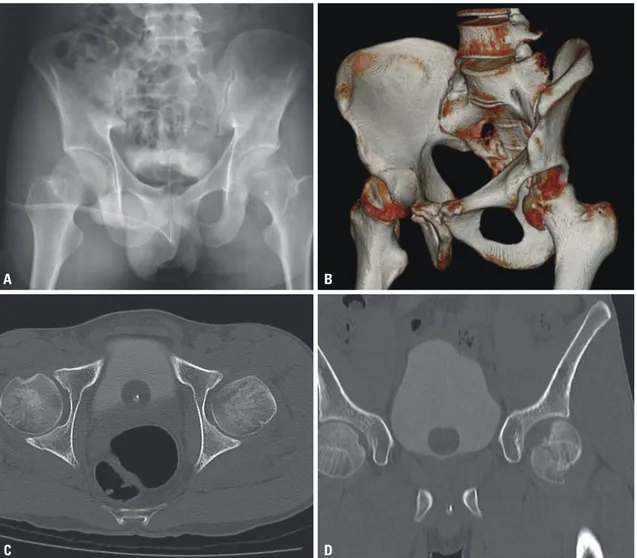
A 31-year-old healthy man presented after a fall during a soccer game that resulted in dislocated right posterior hip with frac-ture and acetabular wall fracfrac-ture. In the emergency depart-ment, he was diagnosed with right posterior hip dislocation and Pipkin type II femoral head fracture with intra-articular bony fragments. Plain radiographs and a computed tomogra-phy scan confirmed the cephalad direction of the femoral head
Osteochondral Autograft from the Ipsilateral Femoral
Head by Surgical Dislocation for Treatment of Femoral
Head Fracture Dislocation: A Case Report
Yougun Won
1, Gi-Soo Lee
1, Sang Bum Kim
1, Sun Joong Kim
1, and Kyu-Hyun Yang
21Department of Orthopedic Surgery, College of Medicine, Konyang University, Daejeon; 2Department of Orthopedic Surgery, Yonsei University College of Medicine, Seoul, Korea.
As anatomical reduction of the articular surface of femoral head fractures and restoration of damaged cartilage are essential for good long-term results, many treatment options have been suggested, including fixation of the fracture using various surgical ex-posures and implants, as well as arthroscopic irrigation and debridement, bone marrow stimulating techniques, osteochondral allograft, autograft, and autogenous chondrocyte implantation. We report a case of osteochondral autograft harvested from its own femoral articular surface through surgical hip dislocation. The osteochondral graft was harvested from the inferior non-weight-bearing articular surface and grafted to the osteochondral defect. One year later, the clinical and radiological results were good, without the collapse of the femoral head or arthritic change. This procedure introduced in our case is considered conve-nient and able to lessen surgical time without morbidity of the donor site associated with the harvest.
Key Words: Femoral head, hip dislocation, autograft transplant, fractures cartilage
Yonsei Med J 2016 Nov;57(6):1527-1530 http://dx.doi.org/10.3349/ymj.2016.57.6.1527
Case Report
pISSN: 0513-5796 · eISSN: 1976-2437
Received: October 5, 2015 Revised: March 8, 2016 Accepted: April 28, 2016
Corresponding author: Dr. Gi-Soo Lee, Department of Orthopedic Surgery, College
of Medicine, Konyang University, 158 Gwanjeodong-ro, Seo-gu, Daejeon 35365, Korea.
Tel: 82-42-600-9862, Fax: 82-42-545-2373, E-mail: gs1899@hanmail.net •The authors have no financial conflicts of interest.
© Copyright: Yonsei University College of Medicine 2016
This is an Open Access article distributed under the terms of the Creative Com-mons Attribution Non-Commercial License (http://creativecomCom-mons.org/licenses/ by-nc/3.0) which permits unrestricted non-commercial use, distribution, and repro-duction in any medium, provided the original work is properly cited.
http://dx.doi.org/10.3349/ymj.2016.57.6.1527 1528
OATS for Femoral Head Fracture Dislocation
fracture extending around the fovea in the coronal plane, with bony fragments in the postero-superior and postero-inferior aspects of the joint (Fig. 1). One day after the accident, he un-derwent open reduction and internal fixation and OATS on his right hip and pelvis (Fig. 2).
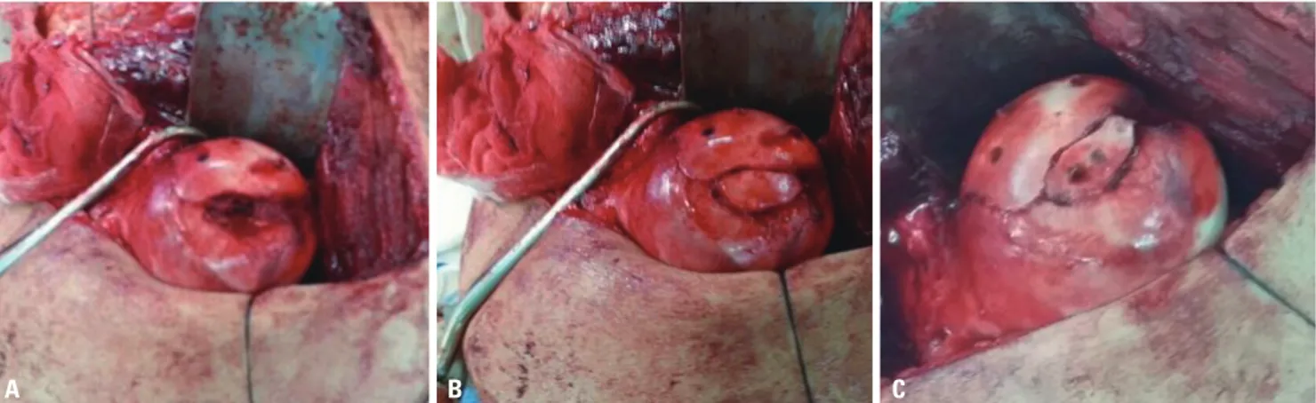
A longitudinal incision was made distally from 5 cm above the tip of the greater trochanter, and the fascia lata was incised in line with the skin incision. An osteotomy that was introduced by Ganz, et al.8 was made on the lateral border of the femur af-ter distal-to-proximal palpation of the vastus laaf-teralis ridge. By gently externally rotating and flexing the leg, the vastus lateralis and intermedius were sharply dissected from the femur. A Z-type capsulotomy was made. In order to dislocate the hip, the leg was brought into further external rotation and flexed. By manipulating the leg, the femoral head and the acetabulum were exposed. The major fragment was reduced and fixed with three 3.5-mm cannulated screws (Fig. 2). Small fragments of the femoral head without capsular attachment could not be fixed. The cartilage defect was 2.5×1 cm and located vertically from the fovea to the proximal weight-bearing zone. The
non-weight-bearing portion of cartilage was harvested. OATS was performed on the cartilage defect, and the harvested fragment was fixed with two 3.5-mm cannulated screws. The trochanteric osteotomy was reduced and fixed with three 3.5-mm screws. Weight bearing was prohibited for four weeks, followed by crutch ambulation for eight weeks.
One year later, the surgical, clinical, and radiological results were good, without the collapse of the femoral head or arthritic change (Modified Harris Hip Score: 82 points). However, the patient had a decreased range of external hip rotation with 90 degrees of flexion. Adhesive capsulitis was suspected; thus, we performed arthroscopic capsulotomy (mainly posterior cap-sule), and the coexistent pathology was identified and ad-dressed via labral debridement, synovectomy, and excision of fibrotic tissue from the acetabular fossa. The joint was accessed through a standard anterolateral portal and a mid-anterior por-tal, with direct visualization. The OATS site showed mild soften-ing and fraysoften-ing (Fig. 3). The overall surface of the OATS site was regenerated with fibrocartilage tissue and was otherwise nor-mal. Before completing the operation, we performed brisement,
Fig. 1. (A) Plain radiograph showing femoral head fracture. (B and C) CT scan showing Pipkin type II femoral head fracture. (D) CT sagittal scan show femoral defect of weight bearing portion.
A
C
B
1529 http://dx.doi.org/10.3349/ymj.2016.57.6.1527
Yougun Won, et al.
resulting in 130 degrees of hip flexion on full external rotation.
DISCUSSION
The good results of mosaicplasty when applied to the knee joint have led to its application to other joints, including the talus, tibial plateau, and humeral capitellum. Good clinical results of OATS for the knee joint were reported at 2-year follow-ups us-ing clinical scores.9,10 Nam, et al.11 previously implemented OATS harvested from the non-weight-bearing knee joint for repair of a femoral head defect via arthrotomy in a 15-year-old boy. They fixed this portion with three bio-absorbable screws. Another case of OATS harvested from the lateral border of the lateral femoral condyle was performed on a 28-year-old man. Hart, et al.12 treated this case with posterior mosaicplasty of the osteo-chondral defect of the femoral head. In this report, we intro-duced and presented the initial clinical results of the use of an intact non-weight-bearing portion of the ipsilateral femoral ar-ticular cartilage to restore the osteochondral defect of the weight-bearing antero-superior articular surface of the femoral head. This procedure is considered to be convenient and is able to lessen surgical time without morbidity of the donor site
asso-ciated with the harvest. However, compared to our case, harvest from other joints is likely to require more time and cost, and it may also increase the risk of surgical co-morbidity.
Utilization of the non-weight-bearing intact femoral surface was first reported in 1978 by Sugioka,13 and favorable results were reported. While scientific evidence to explain the relation-ship between transplantation and Sugioka’s osteotomy is lack-ing, utilization of less-invasive methods such as OATS, com-pared to standard osteotomy and fixation, is expected to increase.
As Ganz, et al.8 described in 2001, surgical dislocation of the hip joint with osteotomy of the great trochanter of the femur can expose the acetabulum without the risk of avascular necro-sis and avoid injury to the medial femoral circumflex artery. With this technique, we can access and handle the whole as-pect of the femoral head in order to harvest the proper articular surface.
Among the indications for OATS, the recommended location and size of the graft differ among authors. In cases of osteone-crosis of a femoral head treated with OATS, outcomes of the large-sized pre-collapse stage are compared to those of the small- and medium-sized pre-collapse stages among cases with unsuccessful results.14 Under certain conditions, mosaicplasty
Fig. 2. (A) 2.5×1 cm femoral head defect is visible after fixation of the main fragment. (B) OATS was performed with a bone plug harvested from the inferior non-weight bearing portion of the affected femoral head. (C) OATS was performed using a headless 3.5 mm compression screw. OATS, osteochondral autologous graft transplantation surgery.
A B C
Fig. 3. (A and B) One year after the operation, plain radiograph and CT scan shows good fixation and articulation. (C) Arthroscopic findings one year after the operation show mild fraying and softening, but the overall surface was regenerated with fibrocartilage tissue.
http://dx.doi.org/10.3349/ymj.2016.57.6.1527 1530
OATS for Femoral Head Fracture Dislocation
can be used as a salvage procedure for defects as large as 8 to 9 cm2, although such extension of the indication can result in a higher rate of donor-site morbidity.15 Autogenous osteochon-dral mosaicplasty is an innovative and promising treatment for focal chondral and osteochondral articular surface defects that are between 1 and 4 cm2 in size. The success of the procedure de-pends largely on adherence to proper indications and caution regarding technical details.15
We believe that surgical dislocation combined with OATS harvested from the ipsilateral femoral head is a good option for osteochondral defects in femoral head fractures, as it offers suf-ficient exposure of the femoral head while preserving its vascu-lature and minimizes the damage to other joints and other complications, which enables proper restoration of the articular cartilage surface.
REFERENCES
1. Pipkin G. Treatment of grade IV fracture-dislocation of the hip. J Bone Joint Surg Am 1957;39-A:1027-42 passim.
2. Davis JB. Simultaneous femoral head fracture and traumatic hip dislocation. Am J Surg 1950;80:893-5.
3. Butler JE. Pipkin Type-II fractures of the femoral head. J Bone Joint Surg Am 1981;63:1292-6.
4. Henle P, Kloen P, Siebenrock KA. Femoral head injuries: which treatment strategy can be recommended? Injury 2007;38:478-88. 5. Hougaard K, Thomsen PB. Traumatic posterior
fracture-disloca-tion of the hip with fracture of the femoral head or neck, or both. J Bone Joint Surg Am 1988;70:233-9.
6. Mowery C, Gershuni DH. Fracture dislocation of the femoral head treated by open reduction and internal fixation. J Trauma 1986;26:
1041-4.
7. Kish G, Módis L, Hangody L. Osteochondral mosaicplasty for the treatment of focal chondral and osteochondral lesions of the knee and talus in the athlete. Rationale, indications, techniques, and re-sults. Clin Sports Med 1999;18:45-66, vi.
8. Ganz R, Gill TJ, Gautier E, Ganz K, Krügel N, Berlemann U. Surgi-cal dislocation of the adult hip a technique with full access to the femoral head and acetabulum without the risk of avascular necro-sis. J Bone Joint Surg Br 2001;83:1119-24.
9. Horas U, Pelinkovic D, Herr G, Aigner T, Schnettler R. Autologous chondrocyte implantation and osteochondral cylinder transplan-tation in cartilage repair of the knee joint. A prospective, compara-tive trial. J Bone Joint Surg Am 2003;85-A:185-92.
10. Smith GD, Richardson JB, Brittberg M, Erggelet C, Verdonk R, Knutsen G, et al. Autologous chondrocyte implantation and osteo-chondral cylinder transplantation in cartilage repair of the knee joint. J Bone Joint Surg Am 2003;85-A:2487-8.
11. Nam D, Shindle MK, Buly RL, Kelly BT, Lorich DG. Traumatic os-teochondral injury of the femoral head treated by mosaicplasty: a report of two cases. HSS J 2010;6:228-34.
12. Hart R, Janecek M, Visna P, Bucek P, Kocis J. Mosaicplasty for the treatment of femoral head defect after incorrect resorbable screw insertion. Arthroscopy 2003;19:E1-5.
13. Sugioka Y. Transtrochanteric anterior rotational osteotomy of the femoral head in the treatment of osteonecrosis affecting the hip: a new osteotomy operation. Clin Orthop Relat Res 1978:191-201. 14. Gagala J, Tarczyn´ska M, Gawe˛da K. Clinical and radiological
out-comes of treatment of avascular necrosis of the femoral head us-ing autologous osteochondral transfer (mosaicplasty): preliminary report. Int Orthop 2013;37:1239-44.
15. Hangody L, Kish G, Kárpáti Z, Udvarhelyi I, Szigeti I, Bély M. Mo-saicplasty for the treatment of articular cartilage defects: applica-tion in clinical practice. Orthopedics 1998;21:751-6.