BJP
R E S E A R C H P A P E R
Neurotrophic interactions between neurons and astrocytes
following AAV1
‐Rheb(S16H) transduction in the hippocampus
in vivo
Min
‐Tae Jeon
1,2|
Gyeong Joon Moon
1,2|
Sehwan Kim
1,2|
Minji Choi
4|
Yong
‐Seok Oh
5|
Dong Woon Kim
6|
Hyung
‐Jun Kim
7|
Kea Joo Lee
7|
Youngshik Choe
7|
Chang Man Ha
7|
Il
‐Sung Jang
8,9|
Michiko Nakamura
9|
Catriona McLean
10,11|
Won
‐Suk Chung
12|
Won
‐Ho Shin
13|
Seok
‐Geun Lee
4,14|
Sang Ryong Kim
1,2,3,91
School of Life Sciences, Kyungpook National University, Daegu, Korea 2
BK21 plus KNU Creative BioResearch Group, Kyungpook National University, Daegu, Korea 3
Institute of Life Science and Biotechnology, Kyungpook National University, Daegu, Korea 4
Department of Science in Korean Medicine, Graduate School, Kyung Hee University, Seoul, Korea 5
Department of Brain‐Cognitive Science, Daegu‐Gyeongbuk Institute of Science and Technology, Daegu, Korea 6
Department of Anatomy, Brain Research Institute, Chungnam National University School of Medicine, Daejeon, Korea 7
Department of Neural Development and Disease, Department of Structure and Function of Neural Network, Korea Brain Research Institute, Daegu, Korea 8
Department of Pharmacology, School of Dentistry, Kyungpook National University, Daegu, Korea 9
Brain Science and Engineering Institute, Kyungpook National University, Daegu, Korea 10
Victorian Brain Bank Network, Florey Institute of Neuroscience and Mental Health, Parkville, Victoria, Australia 11
Department of Anatomical Pathology, Alfred Hospital, Melbourne, Victoria, Australia 12
Department of Biological Sciences, Korea Advanced Institute of Science and Technology (KAIST), Daejeon, Korea 13
Predictive Model Research Center, Korea Institute of Toxicology, Daejeon, Korea 14
KHU‐KIST Department of Converging Science and Technology, Kyung Hee University, Seoul, Korea Correspondence
Sang Ryong Kim, School of Life Sciences, Kyungpook National University, Daegu 41566, Korea.
Email: [email protected]
Seok‐Geun Lee, Department of Science in Korean Medicine, Graduate School, Kyung Hee University, Seoul 02447, Korea.
Email: [email protected]
Background
and
Purpose:
We
recently
reported
that
AAV1
‐Rheb(S16H)
transduction could protect hippocampal neurons through the induction of
brain
‐derived neurotrophic factor (BDNF) in the rat hippocampus in vivo. It is still
unclear how neuronal BDNF produced by AAV1
‐Rheb(S16H) transduction induces
neuroprotective effects in the hippocampus and whether its up
‐regulation contributes
to the enhance of a neuroprotective system in the adult brain.
-This is an open access article under the terms of the Creative Commons Attribution‐NonCommercial License, which permits use, distribution and reproduction in any medium, provided the original work is properly cited and is not used for commercial purposes.
© 2019 The Authors. British Journal of Pharmacology published by John Wiley & Sons Ltd on behalf of British Pharmacological Society
Abbreviations: AAV, adeno‐associated virus; AD, Alzheimer's disease; AP, anterior–posterior; BDNF, brain‐derived neurotrophic factor; CA1, cornu ammonis 1; CM, conditioned medium; CNTF, ciliary neurotrophic factor; CNTFR, ciliary neurotrophic factor receptor; CNTFRα, ciliary neurotrophic factor receptor subunit α; DV, dorsal–ventral; GDNF, glial cell line‐derived neurotrophic factor; GFAP, glial fibrillary acidic protein; Iba1, ionized calcium‐binding adapter molecule 1; MAP 2, microtubule‐associated protein 2; ML, medial–lateral; mTORC1, mammalian target of rapamycin complex 1; MTT, 3‐(4,5‐dimethylthiazol‐2‐yl)‐2,5‐diphenyltetrazolium bromide; NeuN, neuronal nuclei; p‐4E‐BP1, phosphorylation of the Thr37/46 residues of 4E‐BP1; PBST, Tween‐20 in PBS; PD, Parkinson's disease; Rheb(S16H), Ras homolog enriched in brain containing a serine‐to‐histidine mutation at position 16; RT, room temperature; SD, Sprague–Dawley; SN, substantia nigra; TrkB, tropomyosin receptor kinase B
Min‐Tae Jeon, Gyeong Joon Moon, Sehwan Kim, and Minji Choi contributed equally to this work.
Funding information
KBRI Research Program of the Ministry of Science, ICT & Future Planning, Grant/Award Numbers: 18‐BR‐03‐01, 18‐RB‐03‐03 and 18‐ BR‐02‐06; Ministry of Health & Welfare, Grant/Award Numbers: HI15C1928 and HI14C1135; National Research Foundation of Korea, Grant/Award Numbers: NRF‐ 2017R1D1A1B03032218 and NRF‐ 2017R1A2B4002675
Experimental Approach:
To determine the presence of a neuroprotective system in
the hippocampus of patients with Alzheimer's disease (AD), we examined the levels of
glial fibrillary acidic protein, BDNF and ciliary neurotrophic factor (CNTF) and their
receptors, tropomyocin receptor kinase B (TrkB) and CNTF receptor
α(CNTFRα), in
the hippocampus of AD patients. We also determined whether AAV1
‐Rheb(S16H)
transduction stimulates astroglial activation and whether reactive astrocytes
contrib-ute to neuroprotection in models of hippocampal neurotoxicity in vivo and in vitro.
Key
Results:
AD patients may have a potential neuroprotective system,
demonstrated by increased levels of full
‐lengthTrkB and CNTFRα in the hippocampus.
Further AAV1
‐Rheb(S16H) transduction induced sustained increases in the levels of
full
‐length TrkB and CNTFRα in reactive astrocytes and hippocampal neurons.
More-over, neuronal BDNF produced by Rheb(S16H) transduction of hippocampal neurons
induced reactive astrocytes, resulting in CNTF production through the activation of
astrocytic TrkB and the up
‐regulation of neuronal BDNF and astrocytic CNTF which
had synergistic effects on the survival of hippocampal neurons in vivo.
Conclusions and Implications:
The results demonstrated that Rheb(S16H)
transduc-tion of hippocampal neurons could strengthen the neuroprotective system and this
inten-sified system may have a therapeutic value against neurodegeneration in the adult brain.
1
|I N T R O D U C T I O N
Neurotrophic factors play important roles in enhancing neuronal survival, function and plasticity in the adult brain (Huang & Reichardt, 2001; Jeon et al., 2015; Jeong, Nam, Jin, & Kim, 2015; Siegel & Chauhan, 2000). The effects of neurotrophic factors are mediated by specific receptors, such as ciliary neurotrophic factor receptor (CNTFR), which is composed of its ligand‐specific α subunit (CNTFRα),
leukaemia inhibitory factor receptorβandgp130(Siegel & Chauhan, 2000; Leibinger & Andreadaki et al., 2012) for ciliary neurotrophic factor (CNTF), as well as tropomyosin receptor kinase B (TrkB; Jeon et al., 2015; Jeong & Nam et al., 2015) for brain‐derived neurotrophic factor (BDNF). The loss of neurotrophic factors such BDNF and CNTF may be associated with the pathogenesis of brain diseases (Chauhan, Siegel, & Lee, 2001; Jeon et al., 2015; Jeong et al., 2015; Phillips et al., 1991; Sopova, Gatsiou, Stellos, & Laske, 2014). On the other hand, the up‐regulation of these factors induces neuroprotective effects through the activation of mammalian target of rapamycin complex 1 (mTORC1), which stimulates the neuronal survival signalling pathway against neurodegeneration in animal models of neurodegen-erative diseases (Jeon et al., 2015; Leibinger, Andreadaki, & Fischer, 2012; Smith et al., 2014). These observations suggest that the sustained expression of neurotrophic factors may be useful for protecting neurons in the adult brain, and the regulatory system of a key molecule that can produce neurotrophic factors is a potential therapeutic target against neurodegenerative diseases.
We recently reported an increase in reactive astrocytes, which protect dopaminergic neurons, in the substantia nigra (SN) of patients with Parkinson's disease (PD; Nam et al., 2015), suggesting
the presence of an endogenous neuroprotective system in lesioned human brains. It is unclear whether reactive astrocytes in the brains of patients with Alzheimer's disease (AD) have a similar potential to protect hippocampal neurons. However, if a target molecule activated by cell‐type‐specific transduction consistently constructs and intensifies the neuroprotective system through neuron–glia interactions in the lesioned hippocampus of the adult brain, it may
What is already known
• AAV1‐Rheb(S16H) transduction could protect
hippocampal neurons by inducing brain‐derived
neurotrophic factor in the rat hippocampus.
What does this study add
• AAV1‐Rheb(S16H) transduction of hippocampal neurons increased astrocytic ciliary neurotrophic factor expression.
• AAV1‐Rheb(S16H) transduction strengthened the
neuroprotective system through neuron–glia
neurotrophic interactions in the rat hippocampus.
What is the clinical significance
• The AAV1‐Rheb(S16H) transduction approach may be useful for protecting hippocampal neurons against neurodegenerative diseases.
be important to develop a novel therapeutic strategy against neurodegeneration.
Ras homolog enriched in brain (Rheb), which is a member of the
Ras family of small GTP‐binding proteins, is a key regulator of mTORC1 activation associated with neuronal survival and functional maintenance (Jeon et al., 2015; Jeong et al., 2015; Kim, Kareva, Yarygina, Kholodilov, & Burke, 2012). Neuronal transduction with Rheb(S16H), the constitutively active form of Rheb containing a serine‐to‐histidine mutation at position 16, sustains the activation of mTORC1 (Jeon et al., 2015; Jeong et al., 2015; Kim et al., 2011; Kim et al., 2012). This activation can protect hippocampal neurons by inducing neurotrophic effects, such as the production of neuronal BDNF against thrombin‐induced neurotoxicity in vivo (Jeon et al., 2015). However, it is still unknown whether the production of BDNF
following Rheb(S16H) transduction of hippocampal neurons
contributes to glial activation, which can further produce neuro-trophic factors and promote neuroprotective effects (Nam et al., 2015) via paracrine stimulation in the adult brain. In the present study in order to determine the presence of a neuroprotective sys-tem, which has been observed in the SN of patients with PD (Nam et al., 2015), we examined the protein levels of glial fibrillary acidic protein (GFAP), a marker of astrocytes, and neurotrophic factors such as BDNF and CNTF with their respective receptors. TrkB and CNTFRα. in the post‐mortem hippocampus of patients with AD and age‐matched controls. In addition, to verify the possibility of the development of a neuroprotective system following cell‐type‐specific gene transduction in the hippocampus of the adult brain, we exam-ined whether adeno‐associated virus 1 (AAV1)–Rheb(S16H) trans-duction of hippocampal neurons stimulates the activation of astrocytes in the hippocampus of the adult brain and whether the induction of reactive astrocytes following Rheb(S16H) transduction contributes to neuroprotection in models of hippocampal neurotoxic-ity in vivo and in vitro.
2
|M E T H O D S
2.1
|Animals
Female Sprague–Dawley (SD) rats (200–220 g, 8‐week‐old; RRID: RGD_1566440) and pregnant SD rats were obtained from Daehan Biolink (Chungbuk, Korea). For hippocampal neuronal cultures, embryonic (embryonic days 18–19) SD rats were used. For astrocyte cultures, newborn (postnatal day 1) SD rats were used.
2.2
|Human brain tissues
Frozen brain tissues were obtained from the Victorian Brain Bank Network, supported by the Florey Institute of Neuroscience and Mental Health, The Alfred and the Victorian Forensic Institute of Medicine, and funded by Australia's National Health & Medical Research Council and Parkinson's Victoria. The information of the human post‐mortem brain samples is shown in Table 1.
2.3
|Ethics approval and consent to participate
All animal experimental procedures were carried out in accordance with the Guidelines for Animal Care and Use of Kyungpook National University, approved by the Animal Care and Use Committee of Kyungpook National University (No. KNU 2012‐37 and 2016‐42). All animals were maintained with a 12‐h light–dark cycle in a temperature‐controlled room and had free access to food and water. The animals were randomly divided into different groups. Human tissue experiments were approved by the Bioethics Committee, Institutional Review Board of Kyungpook National University Industry Foundation (IRB No. 2013‐0016 and 2016‐0011). Animal studies are reported in compliance with the ARRIVE guidelines (Kilkenny & Browne et al., 2010) and with the recommendations by the British Journal of Pharmacology.
2.4
|Production of AAV viral vectors
All vectors used were the AAV1 serotype as previously described (Kim et al., 2011; Kim et al., 2012). A plasmid carrying Rheb was purchased from OriGene Technologies (Rockville, MD, USA). Rheb DNA was amplified and modified to incorporate a FLAG‐encoding sequence at the 3′‐end by expand long‐template PCR (Roche, Basel, Switzerland). Constitutively active Rheb [Rheb(S16H)] was generated with the Phusion Site‐Directed Mutagenesis Kit (Cat No. E0554S, New England Biolabs, Ipswich, MA, USA) in the pGEM‐T vector (Cat No. A1360, Promega, Madison, WI, USA) and cloned into an AAV packaging construct that utilizes the chicken β‐actin promoter and contains a TABLE 1 Information of human post‐mortem brain samples
AD patients Healthy controls
P value Number of patients 5 5 Region of brain Hippocampus Hippocampus Age (years) 76.4 ± 2.6 76.6 ± 2.5 >.05 Male gender, n (%) 4 (80) 4 (80) >.05 PMI (hr) 18.5 ± 3.5 40.4 ± 2.4 <.05 Braak stage 6 ± 0 0.4 ± 0.4 <.05 Medication (n) AChE inhibitor (3), antihypertensive (2), antidiabetic (1), antiplatelet (1), antipsychotic (2), antidepressant (2) and hypnotic (3) agents PPI (1), NSAID (1), antigout agent (1), antiplatelet agent (1) and antihypertensive agent (2)
Abbreviations: NSAID, nonsteroidal anti‐inflammatory drug; PMI, post‐ mortem interval; PPI, proton‐pump inhibitor.
3′ WPRE (pBL). AAVs were produced by the University of North Caro-lina Vector Core, and the genomic titre of Rheb(S16H) was 2 × 10¹²viral
genomes·ml−1. eGFP, used as a control, was subcloned into the same viral backbone, and viral stocks were produced at titres of 1 × 10¹²viral
genomes·ml−1.
2.5
|Intra
‐hippocampal injection
Animals were anaesthetized with 360 mg·kg−1chloral hydrate (Cat No. C8383; Sigma, St. Louis, MO, USA) by intraperitoneal injection and placed in a stereotaxic frame (David Kopf Instrument, Tujunga, CA, USA). AAV1‐GFP or AAV1‐Rheb(S16H) was infused unilaterally into the hippocampal cornu ammonis 1 (CA1) area of SD rats (anterior– posterior [AP]:−3.8 mm; medial–lateral [ML]: −2.4 mm; dorsal–ventral [DV]:−3.0 mm, relative to the bregma) according to the brain atlas (Paxinos & Watson, 2007) using 30‐gauge injection needles connected to a 10‐μl Hamilton syringe. With an automated syringe pump, 2.0 μl of viral vector suspension was infused at a rate of 0.1μl·min−1over 20 min and the injection needle was left in place for an additional 5 min to allow diffusion into the tissue and minimize dragging back along the injection track.
To determine the contribution of various signalling pathways, we used neutralizing antibodies and a recombinant protein. Three weeks after viral injection, 200 ng of neutralizing antibodies alone or in combination with 20 U of thrombin (Cat No. T4648; Sigma) dissolved in 4‐μl PBS were injected into the right hippocampal CA1 region as described above. Recombinant BDNF protein (200 ng in 2‐μl PBS; Cat No. 450‐02; PeproTech, Rocky Hill, NJ, USA) was injected into the right hippocampal CA1 region of normal rats as described above. The follow-ing neutralizfollow-ing antibodies were used: anti‐BDNF (Cat No. SC‐20981; RRID:AB_2064213; Santa Cruz Biotechnology, Dallas, TX, USA), anti‐ TrkB (Cat No. AF1494; RRID:AB_2155264; R&D Systems, Minneapolis, MN, USA), anti‐CNTF (Cat No. AB‐557‐NA; RRID:AB_354368; R&D Systems) and anti‐CNTFRα (Cat No. AF‐303‐NA; RRID:AB_2083208; R&D Systems).
2.6
|Astrocyte cultures and collection of
conditioned medium
Primary astrocytes from the cerebral hemispheres of newborn SD rats were cultured as described previously (Kaech & Banker, 2006). In brief, cortices were triturated into single cells in DMEM (Cat No. SH30243.01; Hyclone, Logan, UT, USA) containing 10% horse serum (Cat No. 16050130; Invitrogen, Carlsbad, CA, USA) and 1% antibiotics–antimycotics (Cat No. 30‐004‐CI; Corning, Inc., Corning, NY, USA) and plated on 75‐cm2poly‐D‐lysine‐coated culture flasks (0.5 hemisphere/flask; BD Biosciences, Franklin Lakes, NJ, USA). After 2 days, unattached cells and debris were removed by changing the medium. Cultures were fed every 2–3 days with fresh medium until they reached 70–80% confluence.
To determine whether BDNF‐treated astrocyte‐conditioned medium (CM) contributes to hippocampal neuronal survival after
exposure to thrombin, secondary astrocyte cultures were washed and treated with 80 ng·mL−1BDNF for 24 hr. The cells were washed three times with PBS and incubated in serum‐free medium for 24 hr. The CM was then collected and centrifuged at 2,000× g for 10 min at 4°C and filtered through a 0.2‐μm filter to remove cell debris. The untreated astrocyte‐CM served as the control. All media were stored at−70°C until use.
2.7
|Treatment of astrocyte cultures
To determine whether exogenous BDNF leads to the activation of mTORC1 and release of CNTF in astrocytes, the primary astrocyte cultures were treated with recombinant BDNF (10, 40, and 80 ng·ml−1) for 24 hr in serum‐free medium. We further examined whether the BDNF/TrkB signalling pathway is critical for increasing CNTF release by activating mTORC1 in astrocytes. The primary astrocyte cultures
were exposed to 80 ng·ml−1 recombinant BDNF alone or in
combination with anti‐TrkB neutralizing antibody, 10‐μM GNF5837 (Cat No. 4559; Tocris Bioscience, Bristol, UK), or 80‐nM rapamycin (Cat No. 9904; Cell Signaling Technology, Danvers, MA, USA) in serum‐free medium for 24 hr.
2.8
|Hippocampal neuronal cultures and treatment
Primary hippocampal neurons were obtained from fetal SD rats as described previously (Kaech & Banker, 2006). In brief, hippocampal tissues were dissociated by mild mechanical trituration, plated at 4 × 10⁵ cells·ml−1 in 24‐well culture plates previously coated with
1 mg·ml−1 poly‐D‐lysine (Cat No. P6407; Sigma), and maintained in DMEM supplemented with 5% FBS (Cat No. SH30048.03; Hyclone).
Three days after plating, the medium was replaced with
Neurobasal/B27 medium (Cat No. 21103049; Invitrogen) containing cytosine arabinoside (final concentration 5μM; Cat No. C6645; Sigma) to halt the proliferation of non‐neuronal cells. One third of the medium was replaced with fresh medium every 3 days. The cultures were maintained at 37°C in a humidified 5% CO2 atmosphere. To
determine the effects of BDNF‐treated astrocyte‐CM on hippocampal neuronal survival after thrombin treatment, hippocampal neuronal cul-tures were exposed to 50 U·ml−1thrombin alone or in combination with BDNF‐treated astrocyte‐CM or anti‐CNTFRα neutralizing anti-body for 48 hr.
2.9
|MTT assay
Hippocampal neuronal cell viability was evaluated by 3 ‐(4,5‐dimethyl-thiazol‐2‐yl)‐2,5‐diphenyltetrazolium bromide (MTT; Cat No. M5655; Sigma) assay. In brief, hippocampal neuronal cells were incubated with 0.5 mg·ml−1MTT in serum‐free medium at 37°C under 5% CO2for
2 hr. After incubation, the medium was removed, and an equal volume of dimethylformamide (Cat No. 227056; Sigma) was added. The plate was gently mixed on an orbital shaker for 10 min. The absorbance was
measured at 570 nm using the VersaMax Microplate Reader (Molecular Devices, Sunnyvale, CA, USA).
2.10
|Immunohistochemical staining
The immuno‐related procedures used comply with the recommenda-tions made by the British Journal of Pharmacology (Alexander et al., 2018). Animals were transcardially perfused and their brains were fixed and brain sections (30μm thick) were processed for immunohis-tochemical staining as previously described with some modifications (Kim et al., 2018). In brief, brain sections were rinsed with PBS and incubated at 4°C with primary antibodies for 48 hr. Then the sections were rinsed with PBS‐0.5% BSA, incubated at room temperature (RT) with the appropriate biotinylated secondary antibodies, which included goat anti‐mouse IgG (1:400; Cat No. 5450‐0011; RRID: AB_2687537; SeraCare Life Sciences, Milford, MA, USA), anti‐rabbit IgG (1:400; Cat No. BA‐1000; RRID:AB_2313606; Vector Laborato-ries, Burlingame, CA, USA), and rabbit anti‐goat IgG (1:400; Cat No. BA‐5000; RRID:AB_2336126; Vector Laboratories), and processed
with an avidin–biotin complex kit (Cat No. PK6100; RRID:
AB_2336819; Vector Laboratories). The signal was detected by incu-bating the sections with 0.5 mg·ml−13,3′‐diaminobenzidine (Cat No. D5637; Sigma) in 0.1‐M PBS containing 0.003% H2O2. The stained
samples were analysed under a bright‐field microscope (Axio Imager; RRID:SCR_016980; Carl Zeiss, Jena, Germany). The primary
antibod-ies were goat anti‐CNTF (1:100; Cat No. AB‐557‐NA; RRID:
AB_354368; R&D Systems), goat anti‐TrkB (1:100; Cat No. AF1494; RRID:AB_2155264; R&D Systems), goat anti‐CNTFRα (1:100; Cat No. AF‐303‐NA; RRID:AB_2083208; R&D Systems), rabbit anti‐GFAP (1:2,000; Cat No. AB5804; RRID:AB_2109645; Millipore, Billerica, MA, USA), rabbit anti‐ionized calcium‐binding adapter molecule 1 (Iba1; 1:2,000; Cat No. 019‐19741; RRID:AB_839504; Wako Pure Chemical Industries, Osaka, Japan), rabbit anti‐phospho‐4E‐BP1 (1:1,000; Cat No. 2855; RRID:AB_560835; Cell Signaling Technology) and mouse anti‐neuronal nuclei (NeuN; 1:500; Cat No. MAB377; RRID:AB_2298772; Millipore). For Nissl staining, brain sections were mounted on gelatin‐coated slides, stained with 0.5% cresyl violet (Cat No. C5042; Sigma) and analysed using a bright‐field microscope (RRID:SCR_016980; Axio Imager; Carl Zeiss).
For immunofluorescence labelling, brain sections were rinsed and incubated for 48 hr with one of the following pairs: rabbit anti‐FLAG (1:3,000; Cat No. F7425; RRID:AB_439687; Sigma) and mouse anti‐ NeuN (1:500; Millipore), rabbit anti‐FLAG (1:3,000; Sigma) and mouse anti‐GFAP (1:2,000; Millipore), mouse anti‐FLAG (1:2,000; Cat No. F3165; RRID:AB_259529; Sigma) and rabbit anti‐Iba1 (1:2,000; Wako Pure Chemical Industries), mouse anti‐NeuN (1:500; Millipore) and rabbit anti‐BDNF (1:200; Santa Cruz Biotechnology), rabbit anti‐GFAP (1:2,000; Millipore) and goat anti‐TrkB (1:100; R&D Systems), mouse anti‐OX‐42 (1:500; Cat No. MCA275GA; RRID:AB_566455; Serotec, Oxford, UK) and goat anti‐TrkB (1:100; R&D Systems), rabbit anti‐p‐ 4E‐BP1 (1:1,000; Cell Signaling Technology) and mouse anti‐GFAP (1:2,000; Millipore), mouse anti‐GFAP (1:2,000; Millipore) and goat
anti‐CNTF (1:100; R&D Systems), mouse anti‐NeuN (1:500; Millipore) and goat anti‐CNTFRα (1:100; R&D Systems), mouse anti‐GFAP (1:2,000; Millipore) and goat anti‐CNTFRα (1:100; R&D Systems), mouse anti‐OX‐42 (1:500; Serotec) and goat anti‐CNTFRα (1:100; R&D Systems), mouse anti‐OX‐42 (1:500; Serotec) and rabbit anti‐p‐ 4E‐BP1 (1:1,000; Cell Signaling Technology), rabbit anti‐Iba1 (1:2,000; Wako Pure Chemical Industries) and goat anti‐CNTF (1:100; R&D Systems), or mouse anti‐microtubule‐associated protein 2 (MAP 2; 1:500; Cat No. MAB3418; RRID:AB_94856; Millipore) and goat anti‐TrkB (1:100; R&D Systems). The sections were then rinsed, incubated with Texas Red‐conjugated second antibodies: goat anti‐rabbit IgG (1:400; Cat No. TI‐1000; RRID:AB_2336199; Vector Laboratories), rabbit anti‐goat IgG (1:400; Cat No. TI‐5000; RRID: AB_2336129; Vector Laboratories), horse anti‐mouse IgG (1:400; Cat No. TI‐2000; RRID:AB_2336178; Vector Laboratories), fluorescein isothiocyanate‐conjugated donkey anti‐rabbit IgG (1:200; Cat No. 711‐095‐152; RRID:AB_2315776; Jackson Lab, West Grove, PA, USA), or horse anti‐mouse IgG (1:200; Cat No. FI‐2000; RRID: AB_2336176; Vector Laboratories) for 1 hr, washed and mounted
with Vectashield mounting medium (Cat No. H‐1000; RRID:
AB_2336789; Vector Laboratories). The stained sections were imaged using a fluorescence microscope (Axio Imager; Carl Zeiss). Negative controls for BDNF and TrkB were prepared by omitting the primary antibodies. The sections were incubated with 1.5μg·ml−1diamidino‐
2‐phenylindole solution (Cat No. D1306; RRID:AB_2629482;
Invitrogen) for 5 min, washed, and mounted with Vectashield mount-ing medium (Vector Laboratories).
2.11
|Western blot analysis
Western blot analysis was performed as described previously (Kim et al., 2018). In brief, the lysates obtained from astrocyte cultures and hippocBPHampal tissues were homogenized and centrifuged at 4°C for 15 min at 14,000× g. The supernatant was transferred to a
fresh tube, and the concentration was determined using a
bicinchoninic acid assay kit (Cat No. 5000116; Bio‐Rad Laboratories, Hercules, CA, USA). The samples were boiled at 100°C for 5 min before gel loading and equal amounts of protein (20μg) were loaded into each lane with loading buffer. Proteins analysed by gel electro-phoresis were transferred to polyvinylidene difluoride membranes (Millipore) using an electrophoretic transfer system (Bio‐Rad Laborato-ries) and the membranes were incubated overnight at 4°C with specific primary antibodies. The following primary antibodies were used: mouse anti‐β‐actin (1:1,000; Cat No. SC‐47778; RRID: AB_626632; Santa Cruz Biotechnology), rabbit anti‐BDNF (1:500; Santa Cruz Biotechnology), goat anti‐CNTF (1:1,000; R&D Systems), goat anti‐TrkB (1:1,000; R&D Systems), goat anti‐CNTFRα (1:1,000; R&D Systems), rabbit anti‐IL‐1 β (1:1,000; Cat No. SC‐7884; RRID:
AB_2124476; Santa Cruz Biotechnology), mouse anti‐TNF‐α
(1:1,000; Cat No. SC‐52746; RRID:AB_630341; Santa Cruz
Biotechnology), rabbit anti‐p‐4E‐BP1 (1:1,000; Cell Signaling Technology), rabbit anti‐4E‐BP1 (1:1,000; Cell Signaling Technology),
mouse anti‐NeuN (1:500; Millipore), mouse anti‐GFAP (1:500; Millipore) and rabbit anti‐Iba1 (1:1,000; Wako Pure Chemical Industries). After washing, the membranes were incubated with
HRP‐conjugated secondary antibodies (1:5,000; Santa Cruz
Biotechnology), which included goat anti‐mouse IgG (Cat No. SC‐ 2005; RRID:AB_631736), anti‐rabbit IgG (Cat No. SC‐2004; RRID: AB_631746) and mouse anti‐goat IgG (Cat No. SC‐2354; RRID: AB_628490) for 1 hr at RT and the blots were developed using enhanced chemiluminescence western blot detection reagents (GE Healthcare Life Sciences, Little Chalfont, UK) with an X‐ray film (Agfa, Mortsel, Belgium) or the LAS‐500 image analyser (GE Healthcare Life Sciences). The signal was analysed with Multi Gauge version 3.0 (RRID:SCR_014299; Fuji Photo Film, Tokyo, Japan). All histograms were quantitatively analysed based on the density of target proteins normalized to theβ‐actin band for each sample.
2.12
|Materials
The chemicals used in the present study were chloralhydrate, throm-bin, cytosine arabinoside, MTT, and dimethylformamide (Sigma, St. Louis, MO, USA); GNF5837( Tocris Bioscience, Bristol, UK); rapamycin (CellSignaling Technology, Danvers, MA, USA).
2.13
|Quantification of CNTF
To quantify the CNTF released in the BDNF‐treated astrocyte culture medium, we used commercially availableELISAkits according to the manufacturer's protocol (Cat No. DY557; R&D Systems). In brief, a mouse‐anti‐rat CNTF capture antibody was coated at 2 μg·ml−1 in 96‐well immunoassay plates (Corning) overnight at RT. The plates were blocked with 1% BSA for at least 1 hr at RT and washed with 0.01% Tween‐20 in PBS (PBST). Next, 100 μl of the CM of astrocyte cultures was added to each well, incubated for 2 hr at RT and washed with PBST. Biotinylated goat anti‐rat CNTF detection antibody, diluted to 200 ng·ml−1with blocking buffer, was added to the wells for 1 hr at RT. The assay was developed with substrate solution for 20 min at RT followed by the addition of stop solution. The absor-bance was measured at 450 nm using the VersaMax Microplate Reader (Molecular Devices).
2.14
|Quantification of NeuN
‐positive neurons and
p
‐4E‐BP‐1‐positive cells in the hippocampus
As previously described (Jeon et al., 2015), alternate sections were obtained at 3.3‐, 3.6‐, 4.16‐ and 4.3‐mm posterior to the bregma.
NeuN‐positive neurons were counted in a rectangular box
(1 × 0.05 mm) for every selected section. The rectangular box was located over the CA1 cell layer beginning 1.5 mm lateral to the midline and only neurons with normal visible nuclei were counted for two sections in each level (eight regions for each animal). The number of p‐4E‐BP‐1‐positive cells in the hippocampus was quantified in six rectangular areas of 0.4 × 0.3 mm for each animal. The number of cells
was expressed as a percentage of the contralateral control using a light microscope (Carl Zeiss) at a magnification of 200×.
2.15
|Statistical analysis
All experiments were independently conducted five times, and all measurements were performed by an investigator who was blinded to the experimental groups. The precise number of animals used are given in the figure legend. The data and statistical analysis comply with the recommendations of the British Journal of Pharmacology on experimental design and analysis in pharmacology (Curtis et al., 2015). All values are expressed as the mean ± SEM. Data normality was assessed using the Shapiro‐Wilk test, and subsequent testing was performed using a Student'sunpaired t‐test, or one‐way ANOVA with Tukey's post‐hoc test. If the data is not satisfying the condition of normality then the non‐parametric test was performed using the Kruskal‐Wallis test. P values < 0.05 were considered statistically sig-nificant. All statistical analyses and graph generation were performed using SigmaPlot software (RRID:SCR_003210; Systat Software, San Jose, CA, USA).
2.16
|Nomenclature of targets and ligands
Key protein targets and ligands in this article are hyperlinked to corresponding entries in http://www.guidetopharmacology.org, the common portal for data from the IUPHAR/BPS Guide to PHARMA-COLOGY (Harding et al., 2018) and are permanently archived in the Concise Guide to PHARMACOLOGY 2019/2020 (Alexander et al., 2019).
3
|R E S U L T S
3.1
|Elevation of the protein levels of GFAP,
full
‐length TrkB and CNTFRα in the post‐mortem
hippocampus of patients with AD
To investigate the presence of an endogenous neuroprotective system in the hippocampus of patients with AD, which may be similar to the increase in reactive astrocytes exerting neuroprotection in the SN of patients with PD (Nam et al., 2015), we measured the protein levels of GFAP and neurotrophic factors such as BDNF and CNTF with their respective receptors TrkB and CNTFRα in the post‐mortem hippocam-pus of AD patients and age‐matched controls by western blotting (Figure 1). Western blot analysis of NeuN (a marker of neurons) showed significant neuronal loss in the hippocampus of AD patients compared with age‐matched controls (Figure 1a). However, the pro-tein levels of GFAP were significantly higher in the hippocampus of AD patients compared with age‐matched controls (Figure 1a;
*P < .05 vs. CON). In addition, we observed a significant decrease in
BDNF and a tendency for CNTF expression to decrease in the hippocam-pus of AD patients compared with age‐matched controls (Figure 1b). In contrast, the levels of both full‐length TrkB (TrkB‐FL), which has a high
affinity for BDNF (Ferrer et al., 1999; Vidaurre et al., 2012) and CNTFRα, were significantly increased in the hippocampus of AD patients (Figure 1c). Therefore, similar to PD patients (Nam et al., 2015), a neuroprotective system involving an increase in reactive astrocytes may be present in the hippocampus of AD patients. However this neuroprotective system may not be sufficient to control neurodegeneration in the brain of AD patients.
3.2
|Astroglial activation following Rheb(S16H)
transduction of hippocampal neurons in the adult brain
We previously reported that neuronal transduction with AAV1 ‐Rheb(-S16H) could induce neuroprotective effects against thrombin‐induced neurotoxicity by increasing BDNF expression in hippocampal neurons in vivo (Jeon et al., 2015). Although the expression of a target protein following Rheb(S16H) transduction was limited to within neurons (Figures 2a,d and S1A), we recently found that its administration induced reactive astrocytes in the hippocampus of the rat brain.Double immunofluorescence staining for GFP and NeuN or FLAG epi-tope and NeuN demonstrated that the target proteins GFP and FLAG were expressed in hippocampal neurons (Figure 2a) but not glial cells such as astrocytes (Figure 2d) and microglia (Figure S1A). These were stained with anti‐GFAP and anti‐Iba1, respectively, at 4 weeks after intra‐hippocampal injection of AAV1‐GFP or AAV1‐Rheb(S16H). Consistent with our previous study (Jeon et al., 2015), the increased expression of neuronal BDNF was observed in the Rheb(S16H)‐ treated hippocampus, as demonstrated by immunohistochemical staining and western blotting (Figure 2b,c). However, there were apparent morphological changes in astrocytes (Figure 2d,e) and microglia (Figures S1A–C and S2A) following Rheb(S16H) transduction of hippocampal neurons. The protein levels of GFAP (Figure 2f;*) and Iba1 (Figure S2B) were significantly increased in the Rheb(S16H)‐ treated hippocampus compared with intact and GFP‐transduced controls. There was no staining signal for BDNF in negative control tissues (Figure S3A). Moreover, western blot analysis revealed that the protein levels ofIL‐1βandTNF‐α, which are pro‐inflammatory cytokines associated with neurodegeneration in the adult brain (Jang FIGURE 1 Comparison of protein expression profiles in the hippocampus of AD patients and age‐matched controls. (a) Patient and tissue information. A total of 10 post‐mortem brains were used: five from individuals with a clinical diagnosis of probable AD and five from
individuals without neurological disorders (CON). (b–d) Western blot analysis of the levels of NeuN (neuronal marker), GFAP (astrocyte marker), BDNF, CNTF, TrkB, CNTFRα (trophic factors and their receptors) and β‐actin in the hippocampus of CON (n = 5) and AD patients (n = 5). Differences between groups were evaluated by Student's unpaired t‐test at a significance level of *P < .05
et al., 2013; Nam et al., 2014; Shin et al., 2015), were not increased in the Rheb(S16H)‐transduced hippocampus (Figure S2B). Therefore, these results suggest that Rheb(S16H) transduction of hippocampal neurons could induce glial activation in the hippocampus of the adult brain, which may not be related to neurotoxic inflammatory responses. TrkB is a specific receptor involved in BDNF‐mediated neuro-trophic effects (Jeon et al., 2015; Jeong et al., 2015). The protein level of TrkB‐FL, which contains a catalytic kinase domain that can be acti-vated (Ferrer et al., 1999; Vidaurre et al., 2012), was significantly lower in the frontal cortex of AD patients (Ferrer et al., 1999) but not the
hippocampus of AD patients (Connor et al., 1996; Ferrer et al.,
1999), suggesting a selective decrease in the BDNF/TrkB
neurotrophic signalling pathway in AD. Our immunohistochemical staining results revealed increased TrkB expression in glia‐like cells (marked with black arrows) in the Rheb(S16H)‐treated hippocampus of the rat brain at 4 weeks after intra‐hippocampal viral injection (Figure 2g). To identify the glial cell type overexpressing TrkB in the Rheb(S16H)‐treated hippocampus, we further performed double immunofluorescence staining for GFAP and TrkB or OX‐42, another marker of microglia (Shin et al., 2015) and TrkB. The results showed FIGURE 2 Induction of astrocytic activation and TrkB expression by transduction with Rheb(S16H) in hippocampal neurons. Hippocampal tissue sections and protein lysates were obtained from the AAV1‐GFP‐treated and AAV1‐Rheb(S16H)‐treated rat brain at 4 weeks after injection. (a) Double immunofluorescence staining against GFP (green) and NeuN (red) or FLAG epitope (green) and NeuN (red) showing the co‐localization of these two markers in the CA1 region of the hippocampus. Scale bar, 20μm. (b) Double immunofluorescence staining against NeuN (green) and BDNF (red) showing the co‐localization of these two markers in the CA1 region of the hippocampus of CON and AAV1‐Rheb(S16H)‐injected rats. Scale bar, 50μm. (c) Western blot analysis showing the levels of BDNF and β‐actin in the hippocampus of CON, AAV1‐GFP‐injected, and AAV1‐ Rheb(S16H)‐injected rats. Differences among groups were evaluated by one‐way ANOVA and Tukey's post hoc analysis. *P < .05 versus CON and GFP (n = 5 for each group). (d) FLAG was not expressed in astrocytes as determined by anti‐GFAP antibody staining in the CA1 region of the hippocampus of CON and AAV1‐Rheb(S16H)‐injected rats. Scale bar, 50 μm. (e) Immunohistochemical staining showing the morphological changes in astrocytes in the CA1 region of the hippocampus of CON, AAV1‐GFP‐injected and AAV1‐Rheb(S16H)‐injected rats. Scale bars, 500 μm (inset 20μm). (f) Western blot analysis showing the levels of GFAP and β‐actin in the hippocampus of CON, AAV1‐GFP‐injected and AAV1‐Rheb (S16H)‐injected rats. Differences among groups were evaluated by one‐way ANOVA and Tukey's post hoc analysis. *P < .01 versus CON and GFP (n = 5 for each group). (g) Immunohistochemical staining showing increased TrkB expression in glia‐like cells of the hippocampal CA1 region of AAV1‐Rheb(S16H)‐injected rats (black arrows). Scale bars, 500 μm (inset 20 μm). (h) Double immunofluorescence staining against GFAP (green) and TrkB (red) or OX‐42 (green) and TrkB (red) showing the co‐localization of these two markers in the CA1 region of the hippocampus of CON and AAV1‐Rheb(S16H)‐injected rats (marked with a yellow arrow). Scale bar, 20 μm. (i) Western blot analysis showing the levels of TrkB‐FL (140 kDa), TrkB‐T (100 kDa) and β‐actin in the hippocampus of CON, AAV1‐GFP‐injected and AAV1‐Rheb(S16H)‐injected rats (n = 5 for each group). (j) Western blot analysis showing the levels of TrkB‐FL, TrkB‐T, and β‐actin in the hippocampus after treatment with AAV1‐Rheb(S16H) alone, with or without BDNF neutralizing antibody (B.NA). Differences among groups were evaluated by one‐way ANOVA and Tukey's post hoc analysis. *P < .05 versus CON and#P < .05 versus Rheb(S16H) (n = 5 for each group)
the up‐regulation of TrkB in GFAP‐positive astrocytes but not microg-lia (Figure 2h). There was no TrkB‐positive signal in negative control sections (Figure S3B). In addition, western blotting demonstrated a significant increase in TrkB‐FL expression in the Rheb(S16H)‐treated hippocampus compared with the controls (Figure 2i; *P < . 05 vs. CON); however, there was no significant change in truncated TrkB (TrkB‐T) expression (Figure 2i). Moreover, the inhibition of BDNF activity induced by neutralizing antibodies against BDNF (Jeon et al., 2015; Jeong et al., 2015) significantly attenuated the increase in TrkB‐FL following Rheb(S16H) administration as demonstrated by western blotting at 1 day after treatment with neutralizing antibodies (Figure 2j) and there was no change in TrkB‐FL expression in the hippocampus treated with neutralizing antibodies alone (Figure S4A). These results were further confirmed by the expression of GFAP and TrkB‐FL in astrocyte cultures after recombinant BDNF treatment in vitro. The results of preliminary western blot analysis showed that the expression of GFAP and TrkB‐FL were increased in astrocytes exposed to recombinant BDNF (80 ng·ml−1) for 24 hr (Figure S5). Therefore, these results suggest that the production of neuronal BDNF following Rheb(S16H) transduction could induce an increase in TrkB‐FL expression in reactive astrocytes in the hippocampus.
3.3
|Activation of astrocytic mTORC1 by Rheb
(S16H)
‐induced neuronal BDNF through the
TrkB
‐dependent signalling pathway
In addition to the activation of mTORC1 in hippocampal neurons fol-lowing Rheb(S16H) transduction (Jeon et al., 2015) the phosphorylation of the Thr37/46 residues of 4E‐BP1 (p‐4E‐BP1), which is a representa-tive substrate of mTORC1 (Jeon et al., 2015; Jeong et al., 2015), was increased in reactive astrocytes (yellow arrows) in the Rheb(S16H)‐ treated hippocampus (Figure 3a). However, it was not increased in the microglia (Figure S1B). In addition, p‐4E‐BP‐1‐positive cells were significantly increased in the Rheb(S16H)‐treated hippocampus com-pared with the control (Figure 3a). The inhibition of BDNF and TrkB activities by treatment with specific neutralizing antibodies significantly attenuated the up‐regulation of p‐4E‐BP1 in the Rheb(S16H)‐treated hippocampus of the rat brain (Figure 3b) and there was no significant change in p‐4E‐BP1 expression in the hippocampus treated with neu-tralizing antibodies alone (Figure S4B). To verify that neuronal BDNF produced by Rheb(S16H) transduction could induce mTORC1 activa-tion in astrocytes, we further evaluated p‐4E‐BP1 expression following treatment with recombinant BDNF in rat astrocyte‐enriched cultures. Treatment with recombinant BDNF induced a significant increase in p‐4E‐BP1 expression in astrocyte‐enriched cultures compared with untreated controls (Figure 3c). Further the activation of astrocytic mTORC1 was significantly inhibited by co‐treatment with recombinant BDNF and neutralizing antibodies against TrkB (Figure 3c), suggesting that Rheb(S16H)‐induced neuronal BDNF could stimulate astrocytic mTORC1 activation through the TrkB‐dependent signalling pathway in the hippocampus of the adult brain.
3.4
|Production of astrocytic CNTF through a
BDNF/TrkB
‐dependent signalling pathway in the
AAV1
‐Rheb(S16H)‐treated hippocampus
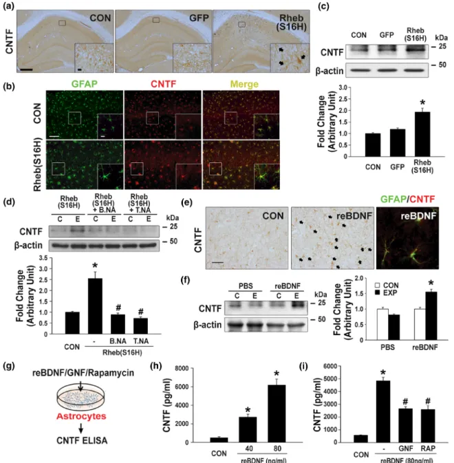
Reactive astrocytes are involved in producing CNTF in the area sur-rounding the lesioned brain in vivo (Albrecht et al., 2003; Nam et al., 2015), suggesting that the induction of astroglial activation, which results in the production of neurotrophic factors such as CNTF. This may be a useful strategy for protecting neurons in the adult brain. In the present study, immunohistochemical staining for CNTF showed that Rheb(S16H) transduction of hippocampal neurons induced an increase in CNTF expression in glial cells in the hippocampus (marked with black arrows) at 4 weeks post intra‐hippocampal viral injection (Figure 4a). Moreover, increased CNTF expression was observed in reactive astrocytes, as demonstrated by double immunofluorescence staining for GFAP and CNTF (Figure 4b). However, it was not observed in the microglia (Figure S1C). Consistent with these results of immunohistochemical staining for CNTF, western blotting also showed the up‐regulation of CNTF following Rheb(S16H) transduc-tion in the hippocampus (Figure 4c). To further clarify the mechanism of astrocytic CNTF production in vivo we examined whether the inhi-bition of BDNF or TrkB activity by treatment with specific neutralizing antibodies, attenuates the production of CNTF following Rheb(S16H) transduction in the hippocampus. Western blot analysis showed no significant change in CNTF expression in the hippocampus treated with neutralizing antibodies alone (Figure S4C). However, the neutral-ization of BDNF and TrkB significantly attenuated the increase in CNTF expression in the Rheb(S16H)‐treated hippocampus (Figure 4d). In addition, treatment with recombinant BDNF by intra‐hippocampal injection significantly increased the level of CNTF expression in reac-tive astrocytes in the hippocampus, as demonstrated by immunohis-tochemical staining (Figure 4e) and western blotting (Figure 4f). The increase in astrocytic CNTF following Rheb(S16H) transduction of hippocampal neurons in vivo, which may be controlled by inhibition of the BDNF/TrkB signalling pathway, was further confirmed by treatment with recombinant BDNF and co‐treatment with GNF‐ 5837 (a Trk inhibitor; Albaugh et al., 2012) and BDNF for 24 hr followed byELISA(Figure 4g–i). Moreover, treatment with rapamycin as a specific inhibitor of mTORC1 (Jeon et al., 2015; Jeong et al., 2015) induced a significant decrease in CNTF expression in BDNF‐ treated astrocyte‐enriched cultures (Figure 4i). Therefore, these in vivo and in vitro results suggest that the activation of astrocytic TrkB following an increase in neuronal BDNF in the Rheb(S16H)‐ treated hippocampus could promote the production of CNTF through the mTORC1‐dependent signalling pathway.
3.5
|Contribution of Rheb(S16H)
‐induced astrocytic
CNTF production to neuroprotection against
thrombin
‐induced neurotoxicity in the hippocampus
The results of immunohistochemical staining for CNTFRα at 4 weeks after intra‐hippocampal viral injection showed the expression of
CNTFRα in hippocampal neurons in vivo and AAV1‐Rheb(S16H) trans-duction increased its expression in hippocampal neurons (Figure 5a). Consistent with the results of immunohistochemical staining, western blotting for CNTFRα showed a significant increase in CNTFRα expres-sion in the Rheb(S16H)‐treated hippocampus compared with control and GFP‐transduced controls (Figure 5b). Double immunofluorescence staining for NeuN and CNTFRα also showed that increased CNTFRα
following Rheb(S16H) transduction was localized in hippocampal neu-rons (Figure 5c) but not GFAP‐positive astrocytes and OX‐42‐positive microglia (Figure 5d). These results suggest that AAV1‐Rheb(S16H) transduction could intensify the CNTF/CNTFRα neurotrophic signalling pathway in hippocampal neurons in vivo.
To verify that the increase in astrocytic CNTF following Rheb
(S16H) transduction of hippocampal neurons contributes to
FIGURE 3 Induction of mTORC1 activation in hippocampal astrocytes via increased BDNF following Rheb(S16H) transduction of hippocampal neurons. Hippocampal tissue sections and protein lysates were obtained from the control (CON) or AAV1‐Rheb(S16H)‐treated rat brain at 4 weeks after injection. (a) Immunohistochemical staining showing increased p‐4E‐BP1 expression in glia‐like cells of the hippocampal CA1 region of AAV1‐Rheb(S16H)‐injected rats (black arrows). Double immunofluorescence staining against GFAP (green) and p‐4E‐BP1 (red) showing the co‐ localization of these two markers in the CA1 region of the hippocampus of AAV1‐Rheb(S16H)‐injected rats (marked with a yellow arrow). Scale bar, 50μm. The bar graph represents the number of p‐4E‐BP‐1‐positive cells in the CA1 region of the hippocampus. Differences between groups were evaluated by independent samples t test.#P < .05 versus CON. (b) Western blot analysis showing the levels of p
‐4E‐BP1, 4E‐BP1, and β‐ actin in the hippocampus of AAV1‐Rheb(S16H)‐injected rats, with or without BDNF neutralizing antibody (B.NA) or TrkB neutralizing antibody (T. NA). Differences among groups were evaluated by one‐way ANOVA and Tukey's post hoc analysis. *P < .05 versus CON and#P < .05 versus Rheb
(S16H) (n = 5 for each group). (c) Schematic of the experimental design for western blot analysis of mTORC1 activity after the treatment of hippocampal astrocyte cultures with recombinant BDNF (80 ng·ml−1), with or without T.NA. Differences among groups were evaluated by Kruskal–Wallis test and Tukey's post hoc analysis. *P < .05 versus CON and#P < .05 versus recombinant BDNF alone (n = 5 for each group)
FIGURE 4 Elevation of CNTF production in hippocampal astrocytes via activation of the BDNF/TrkB/mTORC1 signalling pathway. (a) Immunohistochemical staining showing increased CNTF expression in glia‐like cells of the hippocampal CA1 region of CON, AAV1‐GFP‐ injected, and AAV1‐Rheb(S16H)‐injected rats (black arrows). Scale bars, 500 μm (inset 20 μm). (b) Double immunofluorescence staining against GFAP (green) and CNTF (red) showing the co‐localization of these two markers in the CA1 region of the hippocampus of CON and AAV1‐Rheb (S16H)‐injected rats. Scale bars, 100 μm (inset 10 μm). (c, d) Western blot analysis of the levels of CNTF expression in the hippocampus of AAV1‐ GFP‐injected or AAV1‐Rheb(S16H)‐injected rats, with or without BDNF neutralizing antibody (B.NA) or TrkB neutralizing antibody (T.NA). Differences among groups were evaluated by Kruskal–Wallis test (c) or one‐way ANOVA (d) and Tukey's post hoc analysis. *P < .05 versus CON and#P < .05 versus Rheb(S16H) alone (n = 5 for each group). (e) Immunohistochemical staining showing increased CNTF expression in glia
‐like cells of the hippocampal CA1 region of recombinant BDNF‐injected rats (black arrows). Double immunofluorescence staining against GFAP (green) and CNTF (red) showing the co‐localization of these two markers in the CA1 region of the hippocampus of recombinant BDNF‐injected rats. Scale bar, 50μm. (f) Western blot analysis of CNTF. Differences between groups were evaluated by Student's unpaired t test. *P < .05 versus CON (n = 5 for each group). (g–i) Measurement of CNTF concentration in the conditioned medium (CM) of recombinant BDNF‐treated astrocyte cultures usingELISAkits. (g) Schematic of the experimental design for measuring the levels of CNTF in hippocampal astrocyte cultures. (h) Histogram showing the dose‐dependent effects of recombinant BDNF on CNTF release in astrocyte cultures. Differences among groups were evaluated by Kruskal–Wallis test and Tukey's post hoc analysis. *P < .05 versus CON (n = 5 for each group). (i) Quantitative results showing the CNTF concentration after the treatment of astrocyte cultures with recombinant BDNF (80 ng·ml−1), with or without GNF‐5837 (10 μM) or rapamycin (80 nM). Differences among groups were evaluated by one‐way ANOVA and Tukey's post hoc analysis. *P < .05 versus CON and
neuroprotection in the hippocampus of the adult brain, we deter-mined whether the inhibition of CNTF, CNTFRα and TrkB activities by treatment with specific neutralizing antibodies diminished the Rheb(S16H)‐induced neuroprotective effects against thrombin‐ induced neurotoxicity in the hippocampus of the rat brain (Jeong
et al., 2015). The results of immunohistochemical staining for NeuN showed that the administration of AAV1‐Rheb(S16H) in the hippo-campus of the rat brain protected NeuN‐positive neurons from thrombin‐induced neurotoxicity in the counting area of the hippo-campal CA1 region by 95% compared with that of the contralateral FIGURE 5 Contribution of increased neuronal CNTFRα and astrocytic CNTF to the neuroprotective effects of AAV1‐Rheb(S16H) transduction in the hippocampus. (a, b) The expression and localization of CNTFRα in the hippocampus of the rat brain were determined by
immunohistochemical staining and western blot analysis. (a) Immunohistochemical staining showing the differences in CNTFRα expression in neuronal cells of the hippocampal CA1 region of CON, AAV1‐GFP‐injected and AAV1‐Rheb(S16H)‐injected rats. Scale bars, 500 μm (inset 20 μm). (b) Western blot analysis showing the levels of CNTFRα and β‐actin in the hippocampus of CON, AAV1‐GFP‐injected and AAV1‐Rheb(S16H)‐ injected rats. Differences among groups were evaluated by one‐way ANOVA and Tukey's post hoc analysis. *P < .01 versus CON and GFP (n = 5 for each group). (c, d) The expression and localization of NeuN (green) and CNTFRα (red), GFAP (green) and CNTFRα (red) or OX‐42 (green) and CNTFRα (red) in the hippocampus of the rat brain were determined by double immunofluorescence staining. (c) Double immunofluorescence staining against NeuN and CNTFRα showing the co‐localization of these two markers in the CA1 region of the hippocampus of AAV1‐Rheb (S16H)‐injected rats. Scale bar, 40 μm. (d) CNTFRα was not expressed in GFAP‐positive astrocytes and OX‐42‐positive microglia in the CA1 region of the hippocampus of AAV1‐Rheb(S16H)‐injected rats. Scale bar, 10 μm. (e) To assess the neuroprotective effects of AAV1‐Rheb(S16H) transduction, NeuN staining was performed to quantify the number of hippocampal neurons. Surviving neurons in the CA1 region of the hippocampus were counted after treatment with AAV1‐Rheb(S16H) and thrombin (20 U), with or without neutralizing antibodies (NA; 200 ng) against anti‐CNTF, anti‐TrkB, or anti‐CNTFRα. Scale bars, 500 μm. The number of NeuN‐positive hippocampal neurons in the CA1 region was expressed quantitatively as a percentage of the contralateral control. Differences among groups were evaluated by one‐way ANOVA and Tukey's post hoc analysis. *P < .05 versus CON,#P < .05 versus thrombin alone and†P < .05 versus Rheb(S16H) + thrombin (n = 5 for each group). (f) Neuronal cell viability was assessed by MTT assay after 48‐hr treatment with thrombin (50 U·ml−1), with or without recombinant BDNF‐treated astrocyte‐conditioned media (A‐CM) or anti‐CNTFRα‐neutralizing antibody (CNTFRα NA) in primary hippocampal neuron cultures. The histogram shows the MTT assay results expressed as a percentage of the control. Differences among groups were evaluated by one‐way ANOVA and Tukey's post hoc analysis. *P < .05 versus CON,#P < .05 versus thrombin alone, and†P < .05 versus A‐CM + thrombin (n = 5 for each group)
controls at 7 days post‐lesion (Figure 5e). On the other hand, the inhibition of CNTF, TrkB and CNTFRα activities by intra‐hippocampal injection of a mixture of specific neutralizing antibodies and thrombin significantly attenuated the preservation of NeuN‐positive neurons by 15%, 33% and 34% respectively, compared with the preserved neurons with Rheb(S16H) treatment alone (Figure 5e;). Treatment with neutralizing antibodies alone did not result in significant changes in the number of NeuN‐positive neurons compared with contralateral controls (Figure S6). In addition to the in vivo results, we observed that treatment with CM obtained from astrocyte‐ enriched cultures at 24 hr after recombinant BDNF treatment protected hippocampal neurons against thrombin‐induced neurotox-icity in vitro, as demonstrated by cell viability analysis at 48 hr post‐lesion (Figure 5f). CM‐induced neuroprotection was significantly attenuated by co‐treatment with neutralizing antibodies against CNTFRα (Figure 5f). Therefore, these results demonstrated that increased astrocytic CNTF following AAV1‐Rheb(S16H) administra-tion could contribute to the protecadministra-tion of hippocampal neurons against thrombin‐induced neurotoxicity in the hippocampus.
In our previous study (Jeon et al., 2015), we found that Rheb (S16H)‐induced neuronal BDNF could contribute to neuroprotection against thrombin‐induced neurotoxicity in the hippocampus of the adult brain. However, it was unclear whether the activation of TrkB, expressed in hippocampal neurons is associated with neuroprotection
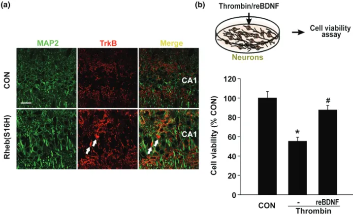
following Rheb(S16H) transduction. Similar to the results showing TrkB up‐regulation in astrocytes (Figure 2h) double immunofluores-cence staining for MAP 2, a marker of soma and dendrites (Chung, Jan, & Jan, 2006) and TrkB revealed an increase in TrkB expression in non‐neuronal cells (white arrows; Figure 6a). However, TrkB expres-sion was also observed in hippocampal dendrites in vivo (Figure 6a). Moreover, in comparison with treatment with thrombin alone, co‐ treatment with recombinant BDNF and thrombin significantly attenu-ated thrombin‐induced neurotoxicity in hippocampal neuron‐enriched cultures (Figure 6b). Taken together, in addition to the neuroprotec-tive effects following activation of the paracrine signalling pathway between hippocampal neurons and astrocytes, Rheb(S16H)‐induced neuronal BDNF could protect hippocampal neurons by activating neu-ronal TrkB, suggesting the activation of an autocrine signalling path-way in the hippocampus of the adult brain (Figure 7).
4
|D I S C U S S I O N
Various studies have demonstrated the therapeutic potential of AAV vectors for treating neurodegeneration in the adult brain with the cell‐type‐specific transduction of AAVs in the CNS (Chtarto et al., 2013; Fitzsimons & During, 2006; Kim et al., 2011). The beneficial effects of neuronal transduction with AAV in the adult brain were first
FIGURE 6 Neuroprotective effects of BDNF against thrombin‐induced neurotoxicity in vitro. (a) Double immunofluorescence staining showing the expression pattern of MAP 2 and TrkB in the hippocampus of AAV1‐Rheb(S16H)‐injected rats. Scale bar, 40 μm. White arrows indicate TrkB expression in MAP 2‐negative cells. (b) Schematic of the experimental design. Neuronal cell viability was assessed by MTT assay 48 hr after treatment with thrombin (50 U·ml−1) and BDNF. BDNF treatment significantly reduced thrombin‐induced neuronal cell death. The histogram shows the results expressed as a percentage of the control (n = 5 for each group). Differences among groups were evaluated by one‐way ANOVA and Tukey's post hoc analysis. *P < .05 versus CON and#P < .05 versus thrombin alone
reported by Kaplitt et al., 1994, who showed that AAV transduction with a gene encoding TH in the nigral dopaminergic system induced functional recovery in a 6‐hydroxydopamine‐lesioned animal model of PD (Kaplitt et al., 1994). Transduction with AAV1 in the CNS, such as the hippocampus, SN and striatum, has shown that the expression of a transduced gene is limited to neurons without affecting glial cells such as astrocytes and microglia (Burger et al., 2004; Jeon et al., 2015; Kim et al., 2012), indicating that AAV1 is suitable for the neuronal transduction of a specific gene in the adult brain. In addition, specific gene delivery by AAV1 has been reported to have beneficial effects such as neuroprotection and functional recovery in animal models of neurodegenerative disorders such as PD and AD (Jeon et al., 2015; Jeong et al., 2015; Kim et al., 2011; Kim et al., 2012; Kiyota et al., 2015; Ries et al., 2006). However, although many studies have used AAV1 constructs for therapeutic interventions in the adult brain, it remains unclear whether the production of biomolecules following neuronal transduction with a specific gene using AAV1 stimulates glial activation, which simultaneously supports neuroprotection in the adult brain.
We previously reported that the induction of Rheb(S16H) expression in hippocampal neurons by AAV1 transduction could acti-vate the mTORC1 signalling pathway, resulting in the production of BDNF as a neuroprotective biomolecule against thrombin‐induced neurotoxicity in the hippocampus of the rat brain (Jeon et al., 2015). Similar to the beneficial effects in the hippocampus (Jeon et al., 2015), AAV1‐Rheb(S16H) transduction in the SN of the murine brain could induce an increase in the protein levels of BDNF, CNTF and glial cell line‐derived neurotrophic factor (GDNF) through the activation of mTORC1 in nigral dopaminergic neurons, resulting in neuroprotection in the nigrostriatal dopaminergic system in vivo (Jeong et al., 2015;
Kim et al., 2011; Kim et al., 2012). The expression patterns of neurotropic factors are different between the Rheb(S16H)‐treated SN and hippocampus and the mechanism involved remains unclear. However, the findings suggest that the sustained activation of the Rheb/mTORC1 signalling pathway in adult neurons can produce neurotrophic factors in the adult brain in vivo. In addition, we found that Rheb(S16H) transduction of hippocampal neurons increased astrocytic CNTF expression in the hippocampus but not hippocampal neurons of the rat brain (Figure 4a,b), suggesting the beneficial activa-tion of astrocytes following the transducactiva-tion of hippocampal neurons with a specific gene in vivo. To further investigate how Rheb(S16H) transduction of hippocampal neurons could induce glial activation in the hippocampus despite the limited expression of a target protein within neurons, we examined whether Rheb(S16H)‐induced neuronal BDNF, which was observed previously (Jeon & Nam et al., 2015), stimulates glial activation in the hippocampus of the adult brain and secondly whether reactive astrocytes contribute to Rheb(S16H)‐ induced neuroprotection in the lesioned hippocampus through interactions between neurons and astrocytes.
Astrocytes are the most abundant cells in the brain and play an important role in the homeostatic maintenance of physiological environments, such as the ion concentration and pH in the CNS (Mulligan & MacVicar, 2004; Sofroniew, 2005). Although previous studies indicated that astrocyte activation is involved in neurodegen-eration through inflammatory responses in the adult brain (Jang et al., 2013; Nam et al., 2014), reactive astrocytes can induce benefi-cial effects such as increasing neuronal survival, growth and activity through barrier function to restrict tissue damage and neuroinflamma-tion (Cekanaviciute et al., 2014; Cekanaviciute et al., 2014; Sofroniew, 2005), also they produce neurotrophic factors such as GDNF and FIGURE 7 Schematic representation of the enhancing neuroprotective system following AAV1‐Rheb(S16H) administration. The results suggest that AAV1‐Rheb(S16H) transduction of hippocampal neurons could intensify a potential neuroprotective system through functional interactions between neurons and astrocytes triggered by an increase in neuronal BDNF. The intensified system could contribute to neuroprotection via both autocrine and paracrine signalling activation in the hippocampus in vivo
CNTF in animal models of neurodegenerative diseases (Harada et al., 2002; Harada et al., 2011; Nam et al., 2015). Moreover, a study reported that the loss of astrocytes in the brain of GFAP‐knockout mice resulted in a larger infarct area induced by ischaemic brain injury following middle cerebral artery occlusion (Nawashiro, Brenner, Fukui, Shima, & Hallenbeck, 2000). The loss of astrocytic density in the hippocampus may be associated with abnormal phenotypes in an ani-mal model of post‐traumatic stress disorder (Saur et al., 2016). Overall, these findings suggest that astrocytes are important for maintaining a normal neural system in the adult brain. Furthermore, the induction of reactive astrocytes, which can protect against neurodegeneration in patients with neurodegenerative diseases such as AD and PD, can support neuronal survival and functional maintenance in vivo. In the present study, morphological changes in astrocytes (Figures 2–4) and microglia (Figures 2, S1, and S2) were clearly observed in the Rheb (S16H)‐treated hippocampus of the rat brain. However, there was no significant change in the levels of pro‐inflammatory cytokines such as IL‐1β and TNF‐α, which could induce neurodegeneration in the adult brain (Jang et al., 2013; Nam et al., 2014; Shin et al., 2015) and the Rheb(S16H)‐treated hippocampus (Figure S2). Although the role of microglia, which are not involved in the production of BDNF and CNTF in the Rheb(S16H)‐treated hippocampus, is unclear, the results suggest that astroglial activation following Rheb(S16H) transduction of hippocampal neurons is important in neuroprotection, but not for inducing neurotoxic inflammation in the adult brain.
The cellular effects of BDNF (a neurotrophin that mediates neuronal survival and differentiation) are initiated by its binding to the specific receptor TrkB expressed in hippocampal neurons resulting in the activation of mTORC1 (Bhave, Ghoda, & Hoffman, 1999; Jeon et al., 2015; Lambert et al., 2013). Increased BDNF expression induces neuroprotective effects, such as anti‐neurodegeneration and improved cognitive ability in models of AD in vivo and in vitro (Jeon et al., 2015; Jeronimo‐Santos et al., 2015; Massa et al., 2010; Nagahara et al., 2009). A recent study found that the primary recipient cells of neuronal BDNF are astrocytes in the visual system (Stahlberg, Kügler, & Dean, 2018). Several studies have shown that TrkB‐FL and TrkB‐T1 are highly expressed on astrocytes (Cheng et al., 2007; Climent, Sancho‐Tello, Minana, Barettino, & Guerri, 2000; Cragnolini, Huang, Gokina, & Friedman, 2009; Gabryel & Bernacki, 2009). However, little is known about the role of BDNF in astrocytes. A previous study reported that BDNF delivery stimulated astrocytes to release neuropro-tective factors, contributing to neuronal survival in the CNS (Saba et al., 2018), and the production of these neurotrophic factors was attenuated in GFAP‐specific TrkB knockout mice (Harada et al., 2011), indicating that the induction of neurotrophic factors in astrocytes could be trig-gered by activation of the BDNF/TrkB signalling pathway.
CNTF, a neurotrophic cytokine belonging to the IL‐6 family, is an important regulator of neuronal survival, neurogenesis and neuronal plasticity in the CNS (Blanchard et al., 2010; Chohan et al., 2011; Jeong et al., 2015; Lipinski et al., 2015; Nam et al., 2015). Its delivery has been found to alleviate cognitive impairments in models of AD in vivo and in vitro (Garcia et al., 2010; Wang et al., 2015). The effects of CNTF are mediated by the tripartite receptor complex including
leukemia inhibitory factor receptorβ, gp130, and CNTFRα (Leibinger & Andreadaki et al., 2012; Jeong & Nam et al., 2015). Especially, CNTFRα is highly expressed in hippocampal neurons (MacLennan & Vinson et al., 1996; Lee & Hofmann et al., 1997). In animal models of PD, we recently found that the induction of astrocytic CNTF prevented the degeneration of dopaminergic neurons in vivo (Nam et al., 2015). In addition, the expression of CNTFRα and CNTF was sig-nificantly higher in dopaminergic neurons and reactive astrocytes respectively in the post‐mortem SN of PD patients compared with age‐matched controls (Nam et al., 2015), suggesting that there might be an endogenous neuroprotective system following injury in the SN. BDNF expression levels have been reported to be decreased in the hippocampus and cortex of patients with AD (Peng, Wuu, Mufson, & Fahnestock, 2005; Phillips et al., 1991). However, the increased expression of TrkB‐FL, which contains a catalytic kinase domain that can be activated (Armanini, McMahon, Sutherland, Shelton, & Phillips, 1995), was detected in glial cells in the hippocampus of AD patients (Connor et al., 1996; Ferrer et al., 1999), suggesting the presence of a neuroprotective system in the lesioned hippocampus of AD patients, similar to the SN of PD patients. In the present study, the expression of CNTFRα, GFAP and TrkB‐FL was significantly higher in the post‐ mortem hippocampus of AD patients compared with age‐matched controls (Figure 1c). However, a decrease in CNTF and BDNF was observed in the hippocampus of AD patients (Figure 1b). These obser-vations suggest that there may also be a compensatory system for neuroprotection in the lesioned hippocampus of AD patients. Although the compensatory system is not sufficient to control neurodegeneration, the induction of astrocytic CNTF and neuronal CNTFRα may be useful approach for strengthening the neuroprotec-tive system, consequently resulting in the protection of hippocampal neurons in the adult brain. However, the current study has some lim-itations in defining the clinical relevance because measurements were restricted to a limited number of post‐mortem human brain samples.
As demonstrated in Figures 2 and 3, Rheb(S16H) transduction of hippocampal neurons induced mTORC1 activation (Figure 3a) and increased the expression levels of both TrkB (Figure 2g–i) and CNTF (Figure 4a–c) in reactive astrocytes. In addition, BDNF was produced in hippocampal neurons in vivo (Figure 2b,c) even though transduction with Rheb(S16H) was limited in neurons (Figures 2a,d and S1A; Jeon et al., 2015). To clarify the mechanism of astrocytic CNTF production following AAV1‐Rheb(S16H) transduction in the hippocampus, we investigated the effects of neutralizing antibodies against BDNF or TrkB on the production of astrocytic CNTF in the Rheb(S16H)‐treated hippocampus in vivo. Our results demonstrated that increased astro-cytic CNTF following Rheb(S16H) transduction was significantly attenuated by the neutralization of BDNF and TrkB (Figure 4d). Similar to the effects in vivo, the production of CNTF induced by treatment
with recombinant BDNF was suppressed by inhibiting the
TrkB/mTORC1 signalling pathway in rat astrocyte‐enriched cultures in vitro (Figure 4g–i). In addition to increasing astrocytic CNTF, AAV1‐Rheb(S16H) transduction induced a significant increase in CNTFRα expression in hippocampal neurons in vivo (Figure 5a–d). Moreover, Rheb(S16H)‐induced neuroprotection against thrombin‐