저작자표시-비영리-변경금지 2.0 대한민국 이용자는 아래의 조건을 따르는 경우에 한하여 자유롭게 l 이 저작물을 복제, 배포, 전송, 전시, 공연 및 방송할 수 있습니다. 다음과 같은 조건을 따라야 합니다: l 귀하는, 이 저작물의 재이용이나 배포의 경우, 이 저작물에 적용된 이용허락조건 을 명확하게 나타내어야 합니다. l 저작권자로부터 별도의 허가를 받으면 이러한 조건들은 적용되지 않습니다. 저작권법에 따른 이용자의 권리는 위의 내용에 의하여 영향을 받지 않습니다. 이것은 이용허락규약(Legal Code)을 이해하기 쉽게 요약한 것입니다. Disclaimer 저작자표시. 귀하는 원저작자를 표시하여야 합니다. 비영리. 귀하는 이 저작물을 영리 목적으로 이용할 수 없습니다. 변경금지. 귀하는 이 저작물을 개작, 변형 또는 가공할 수 없습니다.
Master’s Thesis of Science in Biomodulation
Identification of Inhibitory effects of Gomisin N from
Omija (Schisandra chinensis) on Early Stage of
Atherosclerosis
오미자 유래 고미신 N의 초기 동맥경화 억제 효능 규명
August 2018
The Graduate School
Seoul National University
Biomodulation Major
i
ABSTRACT
Omija (Schisandra chinensis) has long been used as a food material for its unique color and taste, and also as herbal medicine for its abundant bioactive substances. Omija is known to have the protective effect against cardiovascular diseases, but its bioactive compounds and underlying mechanisms remain unclear. In this study, I compared the preventive effect of the dibenzocyclooctadiene lignans which are specific compounds in Omija on early atherosclerosis. Among the lignans rich in Omija, gomisin N most effectively attenuated adhesion of THP-1 monocytes to Human umbilical vein endothelial cells (HUVECs). Western blot, ELISA and RT-qPCR results showed that gomisin N suppressed tumor necrosis factor-alpha (TNF-α)-induced expression of vascular adhesion molecule (VCAM)-1 and monocyte chemoattractant protein (MCP)-1 related to monocyte adhesion. In addition, gomisin N downregulated the phosphorylation of proteins involved in Nuclear Factor-Kappa B (NF-κB) pathway in a concentration-dependent manner.
ii
Using a kinase profiling and IC50 assays, we elucidated that gomisin N significantly inhibited the activity of mammalian sterile-20 3 (MST3/STK24). Gomisin N also suppressed TNF-α-induced phosphorylation of ezrin/radixin/moesin (ERM), the substrate of MST3/STK24, in HUVECs. Taken together, gomisin N markedly inhibited THP-1 monocyte adherence to vascular endothelial cells by targeting MST3/STK24. Our findings suggest that gomisin N is a potential functional material for preventing early atherosclerosis.
Keywords:
Gomisin N, Schisandra chinensis, dibenzocycoloctadiene lignan, atherosclerosis prevention, monocyte-endothelial adhesion;
iii
CONTENTS
ABSTRACT………i
CONTENTS……….……… i i i Ⅰ. INTRODUCTION………...………1
Ⅱ. MATERIALS AND METHODS………...4
1. Chemicals and reagents………4
2. Cell culture………...………5
3. Cell viability assay……….………6
4. THP-1 monocyte adhesion assay……….…………...6
5. Western blot assay...7
6. Enzyme-linked immunosorbent assay...8
7. Real-time quantitative PCR...8
8. Luciferase assay...9
9. Kinase assay...10
10. Statistical analysis...11
Ⅲ. RESULTS………...….….12
1. Gomisin N most effectively inhibits TNF-α-induced THP-1 monocyte-endothelial adhesion compared to lignans rich in Omija (Schisandra chinensis) ...12
iv
2. Gomisin N attenuates TNF-α-induced THP-1 monocyte-endothelial adhesion in a concentration-dependent
manner ...15
3. Gomisin N suppresses TNF-α-induced VCAM-1 expression in HUVECs ...17
4. Gomisin N suppresses TNF-α-induced MCP-1 expression in HUVECs ...19
5. Gomisin N downregulates TNF-α-induced transactivation of NF-κB in HUVECs ...23
6. Gomisin N reduces MST3/STK24 activity ...24
7. Gomisin N suppresses TNF-α-induced phosphorylation of ERM in HUVECs ...28
Ⅳ. DISCUSSION ...30
Ⅴ. REFERENCES ...35
1
Ⅰ. INTRODUCTION
Atherosclerosis is a typical aspect of cardiovascular diseases that has
been a major cause of death[1, 2] all over the world. It can develop for
many years without symptoms and lead to sudden death or disability[3].
Therefore, it is recommended to maintain vascular health through
prevention. Atherosclerosis is a chronic inflammatory disease with a
condition in which the elasticity of the arteries decreases and the blood
vessel width narrows. Circulating leukocytes, such as monocytes,
transmigrate into the subendothelial layer due to the effect of
overexpressed chemokines and adhesion molecules from endothelial
cells, and differentiate into macrophages[4, 5]. Uptake of oxidized
low-density lipoprotein (LDL) forms a lipid plaque[6] and abnormal
proliferation of vascular smooth muscle cells thickens the arterial wall.
As a result, the above procedures narrow the blood vessels and block
blood flow. In order to prevent early stage of atherosclerosis, it is
2
cells.
Omija (Schisandra chinensis) is a small red fruit that grows in Asian
countries. In Korea, Omija was named after five tastes like sweetness,
saltiness, bitterness, sourness and pungency. Omija has long been used
as not only a food for its unique color and taste but also an herbal
medicine because it contains abundant bioactive substances such as
organic acids, vitamins and essential oils[7]. In a Korean traditional
medical book, Donguibogam, it is said that Omija has the effect of
improving circulation. Previous studies presented that extract of Omija
has protective effects against cardiovascular diseases such as myocardial
infraction and hypertension[8, 9]. However, such studies have not
investigated its bioactive components and the underlying mechanism.
The specific bioactive ingredients of Omija are lignans with a
dibenzocyclooctadiene structure. Although they have similar structures,
each lignan has been reported to have different therapeutic effects of
cardiovascular disease such as vasorelaxation, anti-oxidative stress and
3
A are used as marker components for herbal medicine standards because
of their high contents in Omija. However, the preventive effects of those
lignans on monocyte-endothelial cell adhesion have not been reported.
In this study, I identified the most effective lignan rich in Omija for
preventing THP-1 monocyte-endothelial adhesion in HUVECs at first.
In addition, I elucidated the molecular mechanism and target of its
4
Ⅱ. MATERIALS AND METHODS
1. Chemicals and reagents
Gomisin N and gomisin A were purchased from ALB Technology (Hong Kong, China, ≥98%, HPLC). Schisandrin was purchased from Chemfaces (Hubei, China, ≥98.9%, HPLC). Medium199 (M199), hydrocortisone, 2-mercaptoethanol, puromycin, fetal bovine serum (FBS) and calcein AM dye were purchased from Sigma-Aldrich (St. Louis, MO, USA). RPMI 1640 medium was purchased from Welgene (Daegu, Republic of Korea). L-glutamine, fetal bovine serum (FBS), basic fibroblast growth factor (bFGF), and recombinant human epidermal growth factor (hEGF) were purchased from Gibco (Grand Island, NY, USA). Penicillin (10,000 units/ml)-streptomycin (10,000 µg/ml) (P/S) was purchased from Corning (Corning, NY, USA). Recombinant human tumor necrosis factor-alpha (TNF-α) was purchased from PeproTech Korea (Seoul, Republic of Korea). 3-(4,5-dimethylthiazol-2-yl)-2,5-diphenyltetrazolium bromide tetrazolium salt (MTT) solution was
5
purchased from USB Corporation (Cleveland, OH, USA). Dimethylsulfoxide (DMSO) was purchased from Duksan Pure Chemicals (Ansan, Republic of Korea). Antibodies against vascular cell adhesion molecule-1 (VCAM-1), β-actin and basal IκB kinase (IKK)α/β were purchased from Santa Cruz Biotechnology Inc. (Santa Cruz, CA, USA). Phosphorylated IKKα/β and IκBα, basal IκBα, phosphorylated ERM and basal ERM were purchased from Cell Signaling Biotechnology (Danvers, MA, USA).
2. Cell culture
Human umbilical vein endothelial cells (HUVECs) were purchased from Lonza (Walkersville, MD, USA) and cultured in M199 with 25 mM HEPES containing 3 growth factors (1 ng/ml hydrocortisone, 1 ng/ml hEGF and 2 ng/ml bFGF), 1% (v/v) P/S, 2 mM L-glutamine and 10% (v/v) FBS (Gibco). Usage of HUVECs was limited between passages 7 and 14. To culture THP-1 monocyte-like leukemia derived cells purchased from the Korean Cell Line Bank, RPMI1640 supplemented with 50 µM 2-mercaptoethanol, 1% (v/v) P/S, and 10% (v/v) FBS (Sigma-Aldrich) were
6
used. Density of THP-1 was maintained between 2 x 10⁵ and 1 x 10⁶ cells/ml.
3. Cell viability assay
Confluent HUVECs in 96-well plates were serum-starved with M199 containing 1% (v/v) P/S and 2 mM L-glutamine for 4 h and incubated with various concentrations of gomisin N and lignan standards for 22 h. MTT solution was added to each well to 0.5 mg/ml of medium. After 2 h of incubation, the medium was removed and 200 µl DMSO was added. After 30min of incubation, the absorbance was measured at wavelength of 570 nm.
4. THP-1 monocyte adhesion assay
Confluent HUVECs in 96-well plates were serum-starved for 4 h and
treated with gomisin N and lignan standards for 1 h. After stimulation
with 10 ng/ml TNF-α for 5 h, stained THP-1 cells with calcein AM were
added to HUVECs at density of 5 x 10⁵ cells/well in M199 and incubated
for 1h. The plates were washed off with phosphate-buffered saline (PBS)
7
measured with Infinite 200 PRO system (Tecan group Ltd., Männedorf,
Switzerland) at excitation and emission wavelengths of 485 nm and 538
nm, respectively.
5. Western blot assay
Confluent HUVECs in 6-well plates or 6 cm dishes were
serum-starved for 4h and treated with 10, 20 or 40 µM gomisin N for 1 h. The
cells were stimulated with 10 ng/ml TNF-α for 5-15 min (NF-κB
signaling and ERM) or 5 h (VCAM-1). To harvest cell lysates, the cells
were washed with cold PBS and scraped with lysis buffer. The extracts
containing equal amount of proteins were separated by 10% sodium
dodecyl sulfate-polyacrylamide gel electrophoresis and transferred to
polyvinylidene difluoride membranes. The membranes were blocked
with 5% fat-free dry milk and incubated with specific primary antibodies
at 4 °C overnight. After incubation with horseradish
peroxidase-conjugated secondary antibodies for 1h, protein bands were detected
using an enhanced chemiluminescence detection kit (GE Healthcare,
8
6. Enzyme-linked immunosorbent assay
Levels of MCP-1 in culture supernatant were determined using
Human MCP-1/CCL2 ELISA MAX Deluxe Sets (BioLegend, San Diego, CA, USA) according to the manufacturer’s protocol. Briefly, 100µl of diluted standards and culture supernatants were added to each well of
96-well plates pre-coated with diluted capture antibody and incubated for 2
h at room temperature. After each well was washed 4 times, incubated
with detection antibody and Avidin-HRP solution for 1 h and 30 min,
respectively. After washing the plates 5 times, TMB substrate solution
was added and incubated 20 min in the dark. Stop solution was added
and optical density of each well was measured using a microplate reader
at wavelengths of 450 and 570 nm. A standard curve was generated and
linear regression analysis was performed.
7. Real-time quantitative PCR
Total RNA was extracted from HUVECs using Trizol, RNA iso Plus (Takara Bio Inc., Shiga, Japan). RNA was quantified using a NanoDrop ND-2000 spectrophotometer (Thermo Fisher Scientific, Waltham, MA,
9
USA). Reverse transcription was conducted with 1 μg/μl RNA using a
PrimeScriptTM 1st strand cDNA Synthesis Kit (Takara Bio, Inc., Shiga,
Japan). Primers of human VCAM-1 forward (5’-CCC TCC CAG GCA CAC ACA-3’), human VCAM-1 reverse (5’-GAT CAC GAC CAT CTT CCC AGG-3’), human MCP-1 forward (5’-TCG CCT CCA GCA TGA AAG TC-3’), human MCP-1 reverse (5’-GGC ATT GAT TGC ATC TGG CT-3’), human GAPDH forward (5’-CAG GGC TGC TTT TAA CTC TGG TAA A-3’), and human GAPDH reverse (5’-GGG TGG AAT CAT ATT GGA ACA TGT AA-3’) were purchased from Bioneer (Daejeon,
Republic of Korea). For quantitative real-time PCR, iQTM SYBR
Green® Supermix and CFX ConnectTM Real-time PCR Detection
System (Bio-Rad Laboratories, Hercules, CA, USA) were used. The amount of target gene expression was calculated as a ratio of target gene relative to GAPDH in each sample.
8. Luciferase assay
The lentiviral expression vector pGF1-NF-kB-EF1-Puro (System
10
with the packaging vectors pMD2.G and psPAX2 (Addgene, Cambridge,
MA, USA). For transfection, JetPEI DNA transfection reagent
(Polyplus-transfection, New York, NY, USA) was used. After the
HEK293T cell culture was harvested and filtered, HUVECs were
transfected using the medium and 10 μg/ml polybrene (EMD Millipore,
Billerica, MA, USA). Stably-transfected HUVECs selected by 1 μg/ml
puromycin (InvivoGen, San Diego, CA, USA) were seeded at density of
1 x 10⁴ cells/well in 96-well plates. HUVECs were serum-starved for 4
h and treated with gomisin N 10, 20, 40 μM for 1 h before stimulation with 10 ng/ml TNF-α. After 14h of incubation, the cells were disrupted
using lysis buffer (0.1 M pH 7.8 PBS, 1% Triton X-100, 1 mM DTT and
2 mM EDTA) and luciferase activity was measured by a Luminoskan
Ascent (Thermo Electron, Helsinki, Finland).
9. Kinase assay
Kinase profiling was conducted by kinase assay service (Reaction
Biology Corporation, Malvern, PA). Kinases were incubated with
11
addition of the compound in dimethyl sulfoxide (DMSO) and 33P-ATP
(specific activity 10 µCi/µ l). After incubation for 120 min at room
temperature, reaction was spotted onto P81 ion exchange paper (GE
healthcare) and washed extensively in 0.75% phosphoric acid. Kinase
activity data was expressed as the percent remaining kinase activity in
test samples compared to vehicle (DMSO) reactions.
10. Statistical analysis
Statistical analyses were performed using SPSS (Statistical Analysis System Institute, 2017). All data were expressed as mean ± standard error of the mean (SEM) and analyzed using one-way analysis of variance (ANOVA) followed by Tukey’s Honest Significant Difference test. P < 0.05 was used for statistical significance.
12
Ⅲ. RESULTS
1. Gomisin N most effectively inhibits TNF-α-induced THP-1 monocyte-endothelial adhesion compared to other lignans rich in Omija (Schisandra chinensis)
To compare preventive effect of lignans rich in Omija (schisandra chinensis) on THP-1 monocyte-endothelial adhesion, HUVECs were pretreated with 40 μM of three compounds (gomisin N, gomisin A and schisandrin) for 1 h and induced with 10 ng/ml of TNF-α. Those compounds are abundant in Omija and have been used as marker components for herbal medicine standards. Among them, gomisin N showed superior inhibitory effect against TNF-α-induced adhesion of THP-1 to HUVECs compared to gomisin A and schisandrin (Fig. 1B). There is no effect on cell viability up to 40 μM within 24 h (Fig. 1C). Considering the contents in Omija and inhibitory effects on monocyte adhesion, I carried out this research with gomisin N.
13
Figure 1
B
A
C
14
Figure 1. Effect of dibenzocyclooctadiene lignans rich in Omija on THP-1 monocyte-endothelial adhesion
A. Structure of gomisin N, gomsin A and schisandrin. B. Representing images of TNF-α -stimulated THP-1 adhesion to HUVECs in treatment with 40 μM of gomisin N, gomisin A and schisandrin. Quantification of adhered THP-1 cells was conducted as described in the Materials and Methods. C. Cell cytotoxicity of all above compounds was tested by MTT assay. Data (n = 3) represent the means ± SD. Means with different letters are significantly different from each other at P < 0.05. GN, gomisin N; GA, gomisin A; Sch, schisandrin.
15
2. Gomisin N attenuates TNF-α-induced THP-1 monocyte-endothelial adhesion in a concentration-dependent manner To identify the inhibitory effect of gomisin N, which was the most effective lignan, on interaction between THP-1 monocytes and endothelial cells, HUVECs were treated with various concentrations of gomisin N for 1h and induced with 10 ng/ml of TNF-α. From 20 μM treated group, the interaction was significantly attenuated. In addition, 40 μM of gomisin N showed about 50% of inhibitory effects on monocyte adhesion compared to inducer group. Therefore, gomisin N inhibited TNF-α-induced THP-1 monocyte adhesion to HUVECs in a concentration-dependent manner (2A and B).
16
Figure 2
Figure 2. Effect of gomisin N on THP-1 monocyte-endothelial adhesion
A. Representing images of TNF-α-stimulated THP-1 adhesion to HUVECs in treatment with 10, 20, 40 μM of gomisin N. B. Quantification of adhered THP-1 cells was conducted as described in the Materials and Methods. Data (n = 3) represent the means ± SD. Means with different letters are significantly different from each other at P < 0.05.
A
17
3. Gomisin N suppresses TNF-α-induced VCAM-1 expression in HUVECs
I next investigated the effect of gomisin N on the expression of VCAM-1 which is a key adhesion molecule to regulate vascular inflammation and binding of monocytes to endothelial cells[13]. As shown in western blot analysis results, 10 – 40 μM of gomisin N significantly attenuated TNF-α-induced protein expression levels of VCAM-1 in HUVECs. In particular, the group treated with 20, 40 μM of gomisin N inhibited VCAM-1 expression to a level similar to that of the control group. (Fig. 3A). Similarly, RT-qPCR results showed that gomisin N suppressed TNF-α-induced mRNA expression levels of VCAM-1 in a concentration-dependent manner (Fig. 3B).
18
Figure 3
Figure 3. Effect of gomisin N on TNF-α-stimulated VCAM-1 expression levels in HUVECs
A. Western blot analysis shows inhibitory effect of gomisin N on TNF-α-stimulated VCAM-1 protein expression. B. VCAM-1 mRNA expression was investigated using RT-qPCR as described in the Materials and Methods. Data (n = 3) represent the means ± SD. Means with different letters are significantly different from each other at P < 0.05.
19
4. Gomisin N suppresses TNF-α-induced MCP-1 expression in HUVECs
We assessed the effect of gomisin N on expression of MCP-1 which is a chemokine that plays an important role in transmigration of monocytes into vascular intima[14]. As shown in ELISA results, gomisin N reduced TNF-α-induced secretion of MCP-1 protein in HUVECs (Fig. 4A). Similarly, RT-qPCR results showed that gomisin N suppressed TNF-α-induced mRNA expression levels of MCP-1 (Fig. 4B) in a concentration-dependent manner.
20
Figure 4
Figure 4. Effect of gomisin N on TNF-α-stimulated MCP-1 expression levels in HUVECs
A. ELISA analysis shows preventive effect of gomisin N on TNF-α-stimulated MCP-1 protein expression B. MCP-1 mRNA expression was investigated using RT-qPCR as described in the Materials and Methods. Data (n = 3) represent the means ± SD. Means with different letters are significantly different from each other at P < 0.05.
21
5. Gomisin N downregulates Tα-induced transactivation of NF-κB in HUVECs
‘I examined the preventive effect of gomisin N on NF-κB transactivation
in HUVECs transfected with luciferase reporter plasmids. NF-κB is a transcription factor that regulates production of VCAM-1 and MCP-1[15]. Luciferase assay results showed gomisin N inhibited TNF-α-induced transactivation of NF-κB in HUVECs (Fig. 5A). Western blot analysis results showed gomisin N drastically suppressed TNF-α-induced phosphorylation of IκBα that is an upstream protein of NF-κB signaling pathway (Fig. 5B) and the degradation of basal IκBα (Fig. 5C). In addition, gomisin N inhibited phosphorylation of IKK that is an upstream protein of IκBα in a concentration-dependent manner (Fig. 5D).
22
Figure 5
A
B
23
Figure 5. Effect of gomisin N on TNF-α-stimulated NF-κB signaling pathway in HUVECs
A. A luciferase reporter gene assay shows effect of gomisin N on TNF-α-stimulated NF-κB transcriptional activity. Western blot analyses show the effect of gomisin N on phosphorylated and total protein expressions related to NF-κB signaling pathway. B. β-actin protein expression levels was used as a control of phosphorylated IκBα and C. total IκBα. D. Total protein expression levels of IKK was used as a control of phosphorylated IKK. Data (n = 3) represent the means ± SD. Means with different letters are significantly different from each other at P < 0.05.
24
6. Gomisin N reduces MST3/STK24 activity
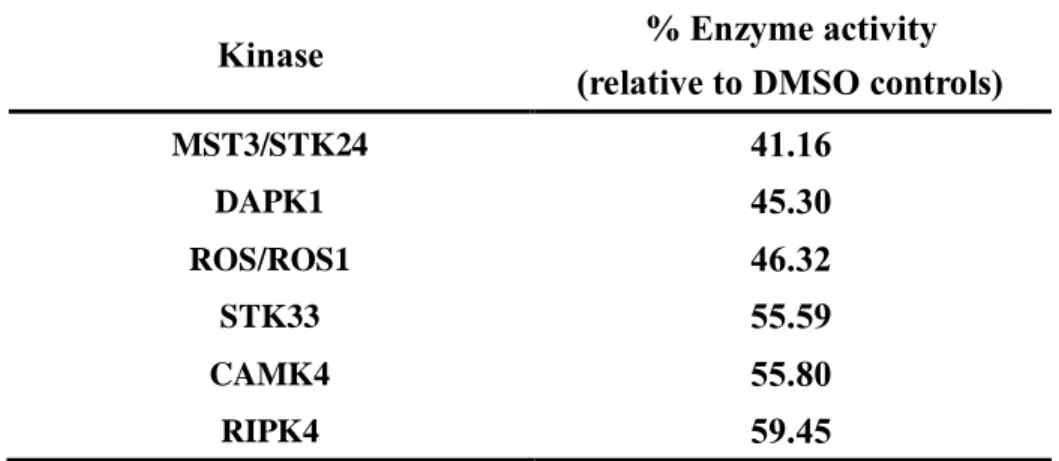
To find a direct target protein of gomisin N in suppressing TNF-α-induced signaling pathway, a kinase profiling analysis was conducted by kinase assay service (Reaction Biology Corporation). Activities of 369 human wild type kinases were analyzed with 20 μM gomisin N. From the results, I found 6 kinases which have less than 60% of enzyme activities (Table 1). In order to select the target, I excluded 4 kinases which are not expressed in HUVECs and 1 kinase which is less associated with monocyte adhesion. Finally, I selected MST3/STK24 as the target of gomisin N. Gomisin N also significantly attenuated the activity of MST3/STK24 with 8.1 μM of the half maximal inhibitory concentration (IC50) (Fig. 6A).
25
Table 1
Table 1. Kinase profiling of gomisin N (20 μM)Kinase profiling analysis of gomisin N was performed in duplicate mode at a concentration of 20 μM. 369 whole human type kinases were tested to find the target of gomisin N by kinase assay service (Reaction Biology Corporation). We selected 6 kinases whose activity was reduced by more than 40% by gomisin N.
Kinase % Enzyme activity
(relative to DMSO controls)
MST3/STK24 41.16 DAPK1 45.30 ROS/ROS1 46.32 STK33 55.59 CAMK4 55.80 RIPK4 59.45
26
Figure 6
Figure 6. Effect of gomisin N on MST3/STK24 activity
Gomisin N was tested for MST3/STK24 inhibitory activity from a 10-dose IC50 assay with 3-fold serial dilution starting at 20 μM.
27
7. Gomisin N suppresses TNF-α-induced phosphorylation of ERM in HUVECs
‘ Next, I investigated the effect of gomisin N on phosphorylation of
ERM family protein, a substrate of MST3/STK24, which is known as making docking system by binding VCAM-1 and up-regulating NF-κB signaling pathway indirectly[16, 17]. Western blot analysis results showed gomisin N markedly inhibited TNF-α-induced phosphorylation of ERM in HUVECs (Fig. 7A).
28
Figure 7
Figure 7. Effect of gomisin N on TNF-α-stimulated ERM phosphorylation in HUVECs
Western blot analysis shows inhibitory effect of gomisin N on TNF-α-stimulated phosphorylation of ERM. Data (n = 3) represent the means ± SD. Means with different letters are significantly different from each other at P < 0.05.
2 9 F igu re 8 . P rop os ed m ec h an is m o f gom is in N
F
ig
ur
e
8
30
Ⅳ. DISCUSSION
Dibenzocyclooctadiene lignans have been isolated to more than 300 species so far, and new compounds and their bioactivities are still being discovered and studied. Most of them are known to be abundant in Schisandra[18] and Kadsura[19] genera belonging to Schisandraceae family. Omija (Schisandra chinensis), a Korean medicinal plant, has also various kinds of lignans. Recent studies show that Omija contains about 5-9 mg/g of schisandrin, 2-3 mg/g of gomisin N and 1-2 mg/g of gomisin A[20-22]. Because these three lignans are highly concentrated in Omija, we asked if one of these is a bioactive compound of Omija which can exert the preventive effects of early stage of atherosclerosis.
In the present study, the preventive effect of gomisin N on atherosclerosis was determined by monocyte-endothelial adhesion assay. This is the first time to compare the three major components of Omija on monocyte-endothelial adhesion. Among the three lignans contained in Omija, we found that gomisin N is the most effective. Previous studies reported that gomisin N has the effects of improving sleep-behavior[23],
31
anti-obesity[24], anti-melanogenic[25] and so on. Especially, gomisin N exhibits inhibitory effects on nitric oxide production in lipopolysaccharide-stimulated RAW 264.7 macrophages[26]. Because of its anti-inflammatory properties, it is thought to have effectively worked against atherosclerosis which is aggravated by chronic inflammatory responses.
The monocytes that move around the blood vessels usually do not stick strongly to vascular endothelial cells[27]. However, when the endothelial cells are stimulated by a key pro-inflammatory cytokine such as TNF-α or oxidized LDL, they increase the secretion of MCP-1 and expression of VCAM-1. MCP-1 attracts monocytes into vascular intima and VCAM-1 causes the monocytes to adhere firmly to endothelial cells[28, 29]. We identified that gomisin N, which inhibited monocyte adhesion most effectively, also suppressed TNF-α-induced protein and mRNA expression levels of VCAM-1 and MCP-1 in a concentration-dependent manner at 10 to 40 μM. NF-κB signaling pathway is known to promote inflammatory reactions in atherosclerosis via regulating the
32
expression of VCAM-1 and MCP-1[30]. IκB proteins inhibit NF-κB signaling by tightly binding to NF-κB dimers until N-terminal phosphorylation by stimulated IKKs. Upon stimulation, IKKs phosphorylate IκBs leading to subsequent degradation of these proteins[31]. We demonstrated that gomisin N significantly inhibited the TNF-α-stimulated activity of the NF-κB transcription factor and also inhibited TNF-α-induced phosphorylation IκBα and IKK at 20 and 40 μM respectively. Taken together, these results indicate that gomisin N inhibits monocyte-endothelial adhesion, at least in part, via the inhibition
of NF-κB signaling. However, these results these results did not show
how gomisin N inhibits the phosphorylation of IKKs.
In this study, we also found that MST3/STK24 is a molecular target of gomisin N. MST3/STK24 plays a role in regulating cytoskeleton and mitogen-activated protein kinase (MAPK) signaling[32]. In particular, it is expressed in HUVECs and known to phosphorylate ERM protein involved in monocyte adhesion[33]. The ERM family which is known as substrate of MST3/STK24 is a key organizer of specialized membrane
33
domains. As ERM is activated by phosphorylation in the threonine residue, its folded structure expands and it can bind to other proteins[34]. Recent study reported that ERM indirectly promotes the NF-κB pathway by binding to ERM binding protein 50 (EBP50) with positive feedback[17]. Furthermore, it builds an endothelial docking structure to facilitate monocytes adherence by making a complex with VCAM-1[16]. We showed that gomisin N significantly inhibited the activity of MST3/STK24 and also suppressed TNF-α-stimulated phosphorylation of ERM in HUVECs. However, it remains to be determined how gomisin N structurally binds MST3/STK24 and regulates its downstream signaling.
In conclusion, the present study demonstrates that gomisin N significantly inhibited THP-1 monocyte-endothelial adhesion in vitro. We have shown that its efficacy is based on a mechanism that down-regulates NF-κB signaling pathway involved in VCAM-1 and MCP-1 expression by targeting MST3/STK24. Therefore, gomisin N is expected an active compound of Omija for preventing early stage of
34
atherosclerosis. However, it is necessary to develop processing techniques of Omija to increase contents of gomisin N and validate its efficacy in vivo and clinical studies.
35
Ⅴ. REFERENCES
1. Townsend, N., et al., Cardiovascular disease in Europe — epidemiological update 2015. European Heart Journal, 2015. 36(40): 2696-2705.
2. Mozaffarian, D., et al., Heart Disease and Stroke Statistics—2016 Update. A Report From the American Heart Association, 2016. 133(4): e38-e360.
3. Herrington, W., et al., Epidemiology of Atherosclerosis and the Potential to Reduce the Global Burden of Atherothrombotic Disease. Circ Res, 2016. 118(4): 535-46.
4. Rollins, B.J., et al., Cytokine-activated human endothelial cells synthesize and secrete a monocyte chemoattractant, MCP-1/JE. The American Journal of Pathology, 1990. 136(6): 1229-1233. 5. J., D.M., et al., The expression of the adhesion molecules ICAM‐
1, VCAM‐1, PECAM, and E‐selectin in human atherosclerosis. The Journal of Pathology, 1993. 171(3): 223-229.
6. Goldstein, J.L., et al., Binding site on macrophages that mediates uptake and degradation of acetylated low density lipoprotein, producing massive cholesterol deposition. Proceedings of the National Academy of Sciences of the United States of America, 1979. 76(1): 333-337.
7. Panossian, A. and G. Wikman, Pharmacology of Schisandra chinensis Bail.: an overview of Russian research and uses in medicine. J Ethnopharmacol, 2008. 118(2): 183-212.
8. Kim, E.Y., I.-H. Baek, and M.R. Rhyu, Cardioprotective effects of aqueous Schizandra chinensis fruit extract on ovariectomized and balloon-induced carotid artery injury rat models: Effects on serum lipid profiles and blood pressure. Journal of Ethnopharmacology, 2011. 134(3): 668-675.
9. Rhyu, M.R., et al., Aqueous extract of Schizandra chinensis fruit causes endothelium-dependent and -independent relaxation of isolated rat thoracic aorta. Phytomedicine, 2006. 13(9): 651-657.
36
10. Park, J.Y., et al., Gomisin A from Schisandra chinensis induces endothelium-dependent and direct relaxation in rat thoracic aorta. Planta Med, 2007. 73(15): 1537-42.
11. Chen, N. and M. Ko, Schisandrin B-Induced Glutathione Antioxidant Response and Cardioprotection Are Mediated by Reactive Oxidant Species Production in Rat Hearts. Biological and Pharmaceutical Bulletin, 2010. 33(5): 825-829.
12. Chiu, P.Y., et al., (–)Schisandrin B is more potent than its enantiomer in enhancing cellular glutathione and heat shock protein production as well as protecting against oxidant injury in H9c2 cardiomyocytes. Molecular and Cellular Biochemistry, 2006. 289(1): 185-191.
13. Cybulsky, M.I., et al., A major role for VCAM-1, but not ICAM-1, in early atherosclerosis. J Clin Invest, 2001. 107(10): 1255-62. 14. Tucci, M., et al., Deregulated expression of monocyte
chemoattractant protein-1 (MCP-1) in arterial hypertension: role in endothelial inflammation and atheromasia. Journal of Hypertension, 2006. 24(7): 1307-1318.
15. Brand, K., et al., Role of nuclear factor-kappa B in atherogenesis. Exp Physiol, 1997. 82(2): 297-304.
16. Barreiro, O., et al., Dynamic interaction of VCAM-1 and ICAM-1 with moesin and ezrin in a novel endothelial docking structure for adherent leukocytes. J Cell Biol, 2002. 157(7): 1233-45.
17. Leslie, K.L., et al., Ezrin-radixin-moesin-binding phosphoprotein 50 (EBP50) and nuclear factor-kappaB (NF-kappaB): a feed-forward loop for systemic and vascular inflammation. J Biol Chem, 2013. 288(51): 36426-36.
18. Opletal, L., H. Sovová, and M. Bártlová, Dibenzo[a,c]cyclooctadiene lignans of the genus Schisandra: importance, isolation and determination. Journal of Chromatography B, 2004. 812(1): 357-371.
19. Chen, Y.-G., et al., Compounds from Kadsura ananosma. Phytochemistry, 2001. 58(8): 1277-1280.
37
Different Parts of Fruit in Schisandra chinensis Baillon. Journal of the Korean Society of Food Science and Nutrition, 2016. 45(6): 851-858.
21. Lee, K.Y., et al., Micelle-Mediated Extraction of Dibenzocyclooctadiene Lignans from Schisandra chinensis with Analysis by High-Performance Liquid Chromatography. Journal of Chromatographic Science, 2014. 52(7): 745-750.
22. Nakajima, K., et al., Constituents of Schizandra chinensis Baill. XIII. Quantitative analysis of lignans in the fruits of Schizandra chinensis Baill. by high performance liquid chromatography. Yakugaku zasshi : Journal of the Pharmaceutical Society of Japan, 1983. 103(7): 743-749.
23. Zhang, C., et al., Gomisin N isolated from Schisandra chinensis augments pentobarbital-induced sleep behaviors through the modification of the serotonergic and GABAergic system. Fitoterapia, 2014. 96: 123-30.
24. Jang, M.-K., et al., Gomisin N inhibits adipogenesis and prevents high-fat diet-induced obesity. Scientific Reports, 2017. 7: 40345. 25. Lee, J., et al., Anti-melanogenic effect of gomisin N from Schisandra chinensis (Turcz.) Baillon (Schisandraceae) in melanoma cells. Arch Pharm Res, 2017. 40(7): 807-817.
26. Oh, S.Y., et al., Anti-inflammatory effects of gomisin N, gomisin J, and schisandrin C isolated from the fruit of Schisandra chinensis. Biosci Biotechnol Biochem, 2010. 74(2): 285-91. 27. Tellez Gil, L., et al., Modulation of soluble phases of
endothelial/leukocyte adhesion molecule 1, intercellular adhesion molecule 1, and vascular cell adhesion molecule 1 with interleukin-1beta after experimental endotoxic challenge. Crit Care Med, 2001. 29(4): 776-81.
28. Haraldsen, G., et al., Cytokine-regulated expression of E-selectin, intercellular adhesion molecule-1 (ICAM-1), and vascular cell adhesion molecule-1 (VCAM-1) in human microvascular endothelial cells. The Journal of Immunology, 1996. 156(7): 2558-2565.
38
29. Murao, K., et al., TNF-α Stimulation of MCP-1 Expression Is Mediated by the Akt/PKB Signal Transduction Pathway in Vascular Endothelial Cells. Biochemical and Biophysical Research Communications, 2000. 276(2): 791-796.
30. Suja, R., et al., NF‐κB, but not p38 MAP Kinase, is required for TNF‐α‐induced expression of cell adhesion molecules in endothelial cells. Journal of Cellular Biochemistry, 2008. 105(2): 477-486.
31. DiDonato, J.A., et al., A cytokine-responsive IκB kinase that activates the transcription factor NF-κB. Nature, 1997. 388: 548. 32. Thompson, B.J. and E. Sahai, MST kinases in development and
disease. The Journal of Cell Biology, 2015. 210(6): 871-882. 33. Zheng, X., et al., CCM3 signaling through sterile 20-like kinases
plays an essential role during zebrafish cardiovascular development and cerebral cavernous malformations. J Clin Invest, 2010. 120(8): 2795-804.
34. Fehon, R.G., A.I. McClatchey, and A. Bretscher, Organizing the cell cortex: the role of ERM proteins. Nat Rev Mol Cell Biol, 2010. 11(4): 276-87.
39
국문 초록
오미자는 오래 전부터 독특한 색과 맛 식품으로 사용되었지 만, 풍부한 생리 활성 물질을 가지고 있어 약초로도 사용되었 다. 한국 전통 의학서인 동의보감에는 오미자의 혈액순환 촉진 효능에 대하여 기록되어 있으며, 최근 연구들은 오미자의 심혈 관 질환 보호 효과에 대해 보고하고 있다. 그러나 오미자의 유 효 성분과 그 작용 기전에 대해서는 아직 밝혀져 있지 않다. 따라서 본 연구에서는 사람 혈관 내피 세포인 HUVEC과 단 핵구인 THP-1 세포주를 이용하여 오미자의 유효성분을 밝히 고, 그 성분이 염증으로 유도된 혈관 내피 세포에서 단핵구의 부착에 미치는 영향과 그 메커니즘을 규명하고자 하였다. 오미자에 다량 함유된 3가지 리그난들의 효능을 비교한 결 과, 고미신 N이 가장 효과적으로 TNF-α로 유도된 단핵구의 혈관 내피 세포에 대한 부착을 가장 효과적으로 억제 시켰다. 고미신 N은 단핵구의 부착에 관여하는 VCAM-1과 MCP-1 의 발현을 단백질과 mRNA 수준에서 농도 의존적으로 저해하40 였으며, NF-κB 합성 경로의 IKK와 IκBα 단백질의 인산화 또한 하향 조절하는 것으로 나타났다. 타겟 스터디를 위해 키 나아제 프로파일링 및 IC50 분석을 진행하였고, 고미신 N이 MST3/STK24의 활성을 유의하게 저해하였으며, 혈관 내피 세포에서 그 기질인 ERM의 TNF-α 유도된 인산화를 억제 하는 것을 밝혔다. 결론적으로, 고미신 N은 MST3/STK24를 타겟으로 하여, NF-κB 합성 경로를 하향 조절하였고 염증으로 유도된 혈관 내피 세포에 단핵구 부착을 현저히 저해하였다. 이러한 결과는 고미신 N이 심혈관 질환의 근본적인 병태인 동맥경화의 초기 단계를 예방할 수 있는 기능성 물질임을 시사한다.