저작자표시-비영리-변경금지 2.0 대한민국 이용자는 아래의 조건을 따르는 경우에 한하여 자유롭게 l 이 저작물을 복제, 배포, 전송, 전시, 공연 및 방송할 수 있습니다. 다음과 같은 조건을 따라야 합니다: l 귀하는, 이 저작물의 재이용이나 배포의 경우, 이 저작물에 적용된 이용허락조건 을 명확하게 나타내어야 합니다. l 저작권자로부터 별도의 허가를 받으면 이러한 조건들은 적용되지 않습니다. 저작권법에 따른 이용자의 권리는 위의 내용에 의하여 영향을 받지 않습니다. 이것은 이용허락규약(Legal Code)을 이해하기 쉽게 요약한 것입니다. Disclaimer 저작자표시. 귀하는 원저작자를 표시하여야 합니다. 비영리. 귀하는 이 저작물을 영리 목적으로 이용할 수 없습니다. 변경금지. 귀하는 이 저작물을 개작, 변형 또는 가공할 수 없습니다.
A Master’s Thesis
The Chloroform Fraction of Lemon (Citrus Limon)
Leaves Inhibits Human Gastric Cancer Cell
Proliferation via Induction of Apoptosis
Ahmed Abdelhamid Kamel Osman
Interdisciplinary Graduate Program in Advanced Convergence Technology & Science
Jeju National University
The
Chloroform
Fraction
of
Lemon
(Citrus
Limon)
Leaves
Inhibits
Human Gastric
Cancer
Cell prolif'eration
via
Induction
ofApoptosis
Ahmed
Abdelhamid
Kamel Osman lSupervisccl b.r [)rolcssor Sorli Kirn ('lro)A thesis strbrnittccl irr partial firllillrncnt lbr the degrcc o1'Mast"cr of'lliotcchlolosy
20 | 6. 0(r.
I'his clissertatiun lras bccn crantirrccl arrcl approvccl by
Prol-essor Dong Kcc .lcong. Vicc (lhairrlan. l)epaltnrurt oI' IJiotcchnologr. .lc.iLr National IJniversitv
-&p; q1''>
I)ro1'cssor Sorni Kirl (lho. I)cpartrncnl of Biotcchnoiogr'. .lc.iLr Natiorral IJpivcrsitr
Interdisciplinarl, (lraduatc Progranr in
Advanced Convergence'l'echnology & Sciencc
.fcju National Universitv
i Contents Contents ... i List of Figures ... ii List of Tables ... ii Abstract ... iii 1. Introduction ... 1
2. Materials and Methods ... 4
3. Results 3.1. Effect of CFLL on cell viability ... 8
3.2. CFLL induces nuclear fragmentation and condensation in AGS cells ... 12
3.3. CFLL increases the percentage of AGS cells in the sub-G1 fraction ... 15
3.4. Western blot analysis of apoptosis-related proteins ... 17
3.5. Compositional analysis by GC–MS ... 22
4. Conclusion ... 24
References ... 25
ii List of Figures
Figure 1. Cell growth inhibitions ··· 9 Figure 2. Chloroform fraction of lemon leaves (CFLL) induces nuclear fragmentation and condensation on AGS ··· 13 Figure 3. Effect of the chloroform fraction of lemon leaves (CFLL) on AGS cell cycle progression ··· 16 Figure 4. Chloroform fraction of lemon leaves (CFLL) induces apoptosis on AGS by altering the Bax/Bcl-2 ratio in favor of apoptosis cleavage of PARP and degradation of procaspases. ··· 19
List of Tables
Table 1. Compounds identified from the chloroform fraction of lemon leaves (CFLL) by GC/MS analysis ··· 23
iii Abstract
Little information about the biological activities of Citrus limon (lemon) leaves has been reported, whereas the fruit of Citrus limon (lemon) has been well-documented to contain various pro-health bio-functional compounds. In the present study, the antiproliferative activities of the lemon leaves were evaluated using several cancer cell lines. From the n-hexane, chloroform, ethyl acetate, n-butanol, and water fractions of methanolic extract of the leaves, the chloroform fraction of lemon leaves (CFLL) showed the most potent antiproliferative activity in the AGS human gastric cancer cells. The current study demonstrates that CFLL induces apoptosis in AGS cells, as evidenced by an increase in apoptotic bodies, cell population in the sub-G1 phase, Bax/Bcl-2 ratio, and cleavage of poly (ADP-ribose) polymerase (PARP), caspase-3 and caspase-9. Compositional analysis of the CFLL using gas chromatography mass spectrometry (GC-MS) resulted in the identification of 27 compounds including trans,trans-farnesol (3.19%), farnesol (3.26%), vanillic acid (1.45%), (-)-loliolide (5.24%) and palmitic acid (6.96%). Understanding the modes of action of these compounds individually and/or synergistically would provide useful information about their applications in cancer prevention and therapy.
1 1. Introduction
According to the World Health Organization (WHO), cancer is the uncontrolled growth and spread of cells that can affect almost any part of the body. Growths often invade surrounding tissue and can metastasize to distant sites (Wogan et al., 2004). In the year 2000, gastric cancer was the second most frequent cause of cancer death worldwide. Compared with other cancer types, gastric cancer was the fourth most common cancer with an estimated 650,000 deaths and 880,000 new cases per year. Almost two-thirds of these new cancer cases occurred in developing countries (Stewart and Kleihues, 2003).
Apoptosis (programmed cell death) is initially described by its morphological characteristics, including cell shrinkage, membrane blebbing, chromatin condensation and nuclear fragmentation (Kerr et al., 1972; Wyllie et al., 1980; Kerr et al., 1994). In the last two decades, basic cancer research has produced remarkable advances in our understanding of cancer biology and cancer genetics. Among the most important of these advances is the realization that mutations disrupt apoptosis genes leading to tumor initiation, progression or metastasis (Lowe and Lin, 2000). Activation of a caspase cascade occurs in apoptosis (Green, 2000). The induction of mitochondrial permeability transition and subsequent release of cytochrome c. Procaspase-9, cytochrome c, and oligomerised Apaf-1 form massive complexes, which known as the apoptosomes, resulting in the activation of the initiator procaspase-9. Active
2
caspase-9 activates the downstream executioners procaspase-3. Active caspase-3 cleaves the 116 kDa PARP proteins into an 89 kDa fragment a characteristic marker of apoptosis. As cancer is characterized by proliferation disorders and hindered apoptosis, many efforts have been made to isolate apoptosis-inducing agents from natural products for their potential use in new anti-tumor drugs (Kelloff et al., 2000; Sporn and Suh, 2000).
Since ancient times, the role of natural products has been recognized as a source for remedies (Farnsworth et al., 1985; Cragg et al, 1997). Natural products are known as rich source of agents valuable to medicine (Newman and Cragg, 2007). More than half of currently-available drugs are natural compounds or are related to natural products. In the case of cancer pharmaceuticals, this proportion surpasses 60%. This results in high interest from pharmaceutical companies and institutions in developing new natural based anticancer drugs (Wilson and Danishefsky, 2006; Wilson and Danishefsky, 2007). Experimental agents derived from natural products offer a great opportunity to evaluate not only totally new chemical classes of anticancer agents, but also novel and potentially-relevant mechanisms of action (da Rocha et al., 2001).
Citrus plants are the most important fruit tree crop in the world, with an annual production of approximately 1.02 hundred million tons (Hwang et al., 2012). While the fruit was described to have antioxidant activity and its flavonoids were described to have anticancer effect (Miyake et al., 1997; Ogata et al., 2000), leaves are major pruning waste that may be reused (Torres et al., 2015). However, in Tunisia, Citrus
3
leaves are used in traditional cuisine due to positive health effects (Hosni et al., 2013). Most aerial Citrus tissues and organs have oil glands that contain and emit a wide diversity of volatile terpenoids, such as hemiterpenes, monoterpenes, and sesquiterpenes, including their alcohol, ester, and acetate derivatives (Sawamura, 2000; Vekiari et al., 2002). Among the many kinds of citrus fruits, lemon (Citrus limon) fruit has been known as a healthy food for ages as it contains various bio-functional compounds (González-Molina et al., 2010). However, there is not much known about biological activity of lemon leaves and the potentially beneficial natural compounds contained within.
In this study, the antiproliferative activities of lemon leaves were evaluated using several cancer cell lines. The chloroform fraction of lemon leaves (CFLL) strongly inhibited the viability of AGS human gastric cancer cells among the tested cell lines. Cell death mechanism caused by CFLL on AGS cells was investigated by Hoechst 33342 nuclear staining, cell cycle analysis and western blotting. The CFLL induced formation of apoptotic bodies, increase in sub-G1 phase, change in the ratio of Bax/Bcl-2 in favor of apoptosis, and cleavage of Bax, poly (ADP-ribose) polymerase (PARP), caspase-3 and -9. Analyses of the CFLL by gas chromatograpy-mass spectrometry (GC-MS) tentatively identified 27 compounds, including trans,trans-farnesol (3.19%), trans,trans-farnesol (3.26%), vanillic acid (1.45%), (-)-loliolide (5.24%) and palmitic acid (6.96%). Our results provide the first evidence that chloroform fraction of a lemon leaf extract can induce the apoptosis in the AGS cells.
4 2. Materials and Methods
2.1. Lemon leaves extraction and fractionation
Nine kg of air dried lemon leaves were pulverized using electric mixer machine then extracted using 80% methanol by stirring for three days at room temperature. The extract was filtered and concentrated with a vacuum rotary evaporator under reduced pressure at 40ºC, giving (1,375 g) of methanolic extract. Then, 344 g of the methanolic extract dried powder was suspended in 4 liters of distilled water and fractionated individually by n-hexane, chloroform, ethyl acetate and n-butanol (1:1) 3 times each respectively in stepwise manner. Each fraction was dried by rotary evaporator at 40°C under vacuum, lyophilized and dissolved in dimethyl sulfoxide (DMSO) at a concentration of 100 mg/mL, which was used for subsequent experiments.
2.2. Reagents
DMEM medium, RPMI 1640 medium, fetal bovine serum (FBS), Hoechst 33342 dye and trypsin/EDTA were purchased from Invitrogen (Grand Island, NY). 3-(4,5-Dimethylthiazol-2-yl)-2,5-diphenyl-tetrazolium bromide (MTT) were purchased from AMRESCO (Cleveland, OH). Propidium iodide (PI) and RNase A were purchased from Sigma Chemical Co. (St. Louis, MO).
2.3. Cell lines and culture mediums
5
Bank (Seoul, Korea) and A549 human lung adenocarcinoma cell line was kindly provided by Professor Min Young Kim of the Department of Biotechnology, Jeju National University, Korea. AGS and SNU-1 were cultured in RPMI-1640, MDA-MB-231 cells were cultured in DMEM medium and F12K medium for the A549 cell line. Cultured media were supplemented with 1% antibiotic and 10% heat-inactivated fetal bovine serum (FBS). All cultures were maintained in a humidified incubator with 5% CO2 at 37ºC.
2.4. Cell viability assay
Cells were plated in 96-well plates at an initial density of (5 × 10⁴ cells/mL) cells/mL per well in a 100 µL medium containing 10% heat-inactivated FBS. The cells were left to be attached overnight then treated with various concentrations of lemon leaves fractions or quercetin for 48 h. After treatment, 20 µL MTT reagent (5 mg/mL) was added, and cells were incubated for 4 h. The medium was carefully removed and 150 µL DMSO was added. All experiments were conducted in quadruplicate. Cell viability was determined by measuring absorbance at 570 nm using a Sunrise microplate reader (Sunrise, Tecan, Salzburg, Austria). Cell viability is represented as the percentage of control cell viability (mean ± standard deviation [SD]).
2.5. Morphology
AGS (5 × 10⁴ cells/mL) were plated in 6-well plates and treated with CFLL for 24 h. After 24 h, cells were stained with 10 µM Hoechst33342 and observed under a florescence microscope (Olympus, Essex, UK).
6 2.6. Flow cytometric analysis
To analyze cell cycle distribution and apoptosis, cells (5 × 10⁴ cells/mL) were plated in 6-well plates and treated with CFLL or quercetin 100 µM for 24 h. For the cytometric analysis of cell cycle, cells were harvested, washed with phosphate-buffered saline (PBS), fixed with ethanol 70%, rehydrated in 2 mM EDTA-PBS, treated with RNase A (25 µg/mL), and stained with PI (40 µg/mL). Samples were analyzed with Cell Quest software (Becton Dickson, USA). Each experiment was repeated at least 3 times.
2.7. Western blot analysis
Cells were seeded and treated with the indicated concentrations of CFLL or quercetin 100 µM. Cells were then harvested, lysed in RIPA buffer and kept on ice for 30 min. A BCA assay was carried out and equal amounts of protein were loaded into each well. Aliquots of the lysates were separated on sodium dodecyl sulfate (SDS)-polyacrylamide gels and transferred to PVDF membranes using a glycine transfer buffer. After blocking with 5% non-fat dried milk, membranes were treated with primary antibodies and incubated overnight, and then for 1 h with secondary antibodies in milk containing Tris-buffered saline (TBS) and 0.5% Tween 20. All primary antibodies (except the anti-β-actin antibody) were used at a dilution of 1:1,000. The anti-β-actin primary and secondary antibodies (horseradish peroxidase [HRP]-conjugated goat anti-rabbit IgG respectively) (Vector Laboratories, USA) were used at a dilution of 1:10,000. Protein bands were detected using the BS ECL plus kit
7 (Biosesang, Gyeonggi-do, Korea).
2.8. GC-MS analysis
Chromatographic analysis was carried out by Shimadzu gas chromatography mass spectrometry (GC-MS; Model QP-2010, Shimadzu Co., Kyoto, Japan) in Electron Impact (EI) mode. The ionization voltage was 70 eV, and injector and interface temperatures were 250°C and 290°C, respectively. The capillary column was an Rtx-5MS (30 m length, 0.25 mm i.d., and 0.25 µM, film thickness). The oven temperature, programmed at 60°C (isothermal for 2 min), was ramped to 250°C at 5°C/min and 310°C at 8°C/min (isothermal for 12 min). Helium, the carrier gas, was used at a flow rate of 1 mL/min with 57.4 kPa pressure and an injector volume of 1 µL using splitless mode. Mass range was from m/z 40-500 amu. The GC-MS spectral data were compared within WILEY9 and NIST05 libraries.
2.9. Statistical analysis
Data are expressed as mean ± standard deviation of three independent determinations. The significance of differences between groups was determined through one-way analysis of variance (ANOVA).
8 3. Results
3.1. Effect of CFLL on cell viability
To test the anticancer effect of different lemon leaves fractions (80% methanol extract fraction of lemon leaves “MFLL”, n-hexane fraction of lemon leaves “HFLL”, chloroform fraction of lemon leaves “CFLL”, ethyl acetate fraction of lemon leaves “EFLL”, n-butanol fraction of lemon leaves “BFLL” and water fraction of lemon leaves “WFLL”), several cancer cells were treated with various concentrations of each fraction. MTT cell viability assay was performed after treating human breast cancer cell line MDA-MB-231 (Fig. 1A), human non-small lung cancer cell line A549 (Fig. 1B), human gastric cell lines AGS (Fig. 1C), and SNU-1 (Fig. 1D) for 48 h with 12.5 µg/mL, 25 µg/mL, 50 µg/mL, and 100 µg/mL of each fraction and DMSO was used as negative control. Our data demonstrate that among the tested fractions CFLL had the highest antiproliferative activity. Moreover, our data also show that among the tested cells, human gastric cancer cell line AGS was the most affected after the 48 h treatment in all concentrations treated.
9 A
10 C
11 E
Figure. 1. Cell growth inhibition. Cell viability was assessed based on MTT reduction. Cells were treated for 48 h with different concentrations of lemon leaf fractions: , methanol fraction of lemon leaves (MFLL); , n-hexane fraction of lemon leaves (HFLL); , chloroform fraction of lemon leaves (CFLL); , ethyl acetate fraction of lemon leaves (EFLL); , n-butanol fraction of lemon leaves (BFLL); and , water fraction of lemon leaves (WFLL). DMSO was used as negative control. (A) MDA-MB-231, (B) SNU-1, (C) A549 and (D) AGS. (E) Effect of CFLL on AGS comparing with quercetin with the same experimental conditions mentioned above. Data correspond to the mean ± standard deviation (SD) from three independent experiments. The statistically significant differences are presented as *p< 0.05.
12
3.2. CFLL induces nuclear fragmentation and condensation in AGS cells.
A possible mechanism of the antiproliferative activity of CFLL on AGS cells is the induction of apoptosis. Nuclear fragmentation, condensation and formation of apoptotic bodies are considered as signs of apoptosis (Kerr et al., 1972; Wyllie et al., 1980; Kerr et al., 1994). To elucidate whether CFLL-induced inhibition of cell viability was attributable to apoptosis, AGS cells were treated for 24 h with increasing concentrations of the CFLL or 100 µM (30.22 µg/mL) quercetin as a positive control. Quercetin has been previously reported to effectively induce apoptosis in AGS cells with concentration 100 µM (Kim, 2014). Moreover, cell proliferation assay data shows that 100 µM has a similar effect on AGS cell proliferation, compared with 100 µg/mL CFLL (Fig. 1E). After 24 h, cells were treated with 10 µM nuclear staining Hoechst 33342 and observed under microscope. The data (Fig. 2A and 2B) show that CFLL induce nuclear fragmentation and condensation after 24 h of treatment.
13
A
14
Figure. 2. Chloroform fraction of lemon leaves (CFLL) induces nuclear fragmentation and
condensation on AGS cells. Cells were seeded and incubated overnight to allow attaching
then treated with different concentrations of CFLL, DMSO as negative control and 100 µM quercetin (Q) as positive control for 24 h. (A) Cells were stained with 10 µM Hoechst 33342 and observed under a microscope. (B) Percentage of apoptotic bodies formed of total observed cells. Data correspond to the mean ± standard deviation (SD) from three independent experiments. The statistically significant differences are presented as *p< 0.05.
15
3.3. CFLL increases the percentage of AGS cells in the sub-G1 fraction.
The cell cycle is known to be major regulatory mechanism for cell growth. Cellular DNA content identifies cell position in the cell cycle. Fractional cellular DNA content (sub-G1 cells) is a marker of apoptotic cells (Gamet et al., 2000), thus we detected changes in cell cycle which might occur with treatments of CFLL. We determined sub-G1 populations in AGS cells treated with increasing concentrations of CFLL and quercetin for 24 h. The data show that treatment with 100 µg/mL of CFLL increased the sub-G1 population up to 25.27% (Fig. 3) comparing with negative control 2.7% and quercetin 28.36%, indicative of apoptosis.
16
Figure. 3. Effect of the chloroform fraction of lemon leaves (CFLL) on AGS cell cycle progression. Cells were seeded and incubated overnight to allow attaching then treated with different concentrations of CFLL for 24 h. (A) DMSO as negative control. (B) CFLL 12.5 µg/mL. (C) CFLL 25 µg/mL. (D) CFLL 50 µg/mL. (E) CFLL 100 µg/mL. (F) 100 µM quercetin (Q) as positive control. The percentage of AGS cells in the sub-G1 was calculated. Data correspond to the mean ± standard deviation (SD) from three independent experiments.
A B
E F
17
3.4. Western blot analysis of apoptosis-related proteins.
The expressions of proteins involved in apoptotic cell death were detected by western blotting. The balance between the expression of the proapoptotic protein Bax and the antiapoptotic protein Bcl-2 is considered a good indicator of the apoptotic activity of tumor cells (Martínez et al., 2003). The cleavage of Bax is known to enhance its proapoptotic function (Wood and Newcomb, 2000). The changes of Bax/Bcl-2 ratio were investigated. Our western blotting analysis data show that after treatment with CFLL for 24 h, an obvious increase in Bax expression was not apparent. However, the cleaved form was detected in the cells treated with 50, 100 µg/mL CFLL, and 100 µM quercetin. Also treatment decreases the Bcl-2 levels compared with the negative and positive controls resulting in an overall increase of Bax/Bcl-2 ratio on CFLL treated cells. We also investigated the effect of CFLL on caspase levels, as it is known that degradation of procaspase-3 and procaspase-9 is a sign of apoptosis (Kitamura et al., 2002). Our data show that CFLL significantly degrades caspase-3 and caspase-9 with progressive increasing concentrations. In case of caspase-9, the CFLL showed a higher effect comparing to 100 µM quercetin treated cells. Our data also shows that CFLL dose dependently decreases the PARP level and increases the PARP cleaved form. The striking increase in the cleaved form of PARP in 100 µg/mL CFLL treated cells might explain the dramatic increase of apoptotic bodies and sub-G1 population. These data (Fig. 4A and 4B) indicates that CFLL induces
18
apoptosis through altering Bax/Bcl-2 ratio in favor of apoptosis, cleavage of PARP caspase-3 and caspase-9.
19 A
20 B
21
Figure. 4. Chloroform fraction of lemon leaves (CFLL) induces apoptosis on AGS by altering the Bax/Bcl-2 ratio in favor of apoptosis cleavage of PARP and degradation of procaspases. Cells were seeded incubated overnight to allow attaching then treated with different concentrations of CFLL, DMSO as negative control and 100 µM qurecetin (Q) as positive control for 24 h. Then cells were lysed following incubation with CFLL at the indicated concentrations for 24 h. Cellular proteins were separated by SDS-PAGE and probed with appropriate primary antibodies. β-actin served as an internal control. (A) Cells were lysed after incubation with CFLL at the indicated concentrations for 24 h. (B) Fold change of apoptosis related proteins. Data correspond to the mean ± standard deviation (SD) from three independent experiments. The statistically significant differences are presented as *p< 0.05.
22
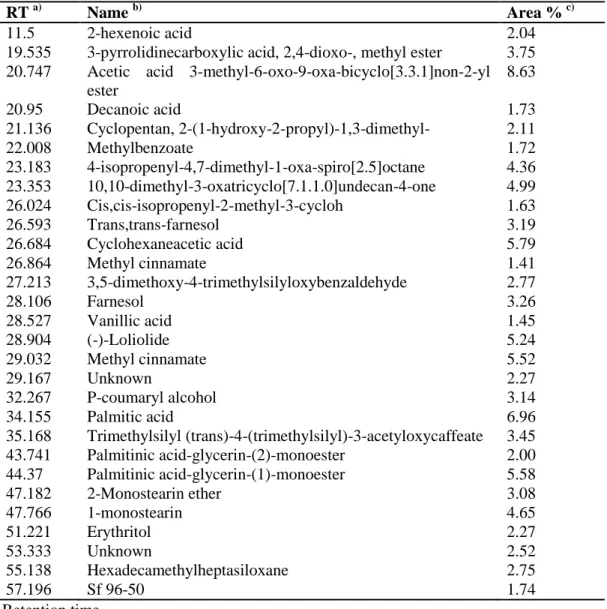
3.5. Compositional analysis by GC–MS. To investigate which compounds cause the apoptotic effect induced by the CFLL, compositional analysis by GC-MS was performed. Analyses of the CFLL identified 27 compounds, including acetic acid 3-methyl-6-oxo-9-oxa-bicyclo[3.3.1]non-2-yl ester (8.63%), palmitic acid (6.9%), cyclohexaneacetic acid (5.79%) (Table 1). Several of these 27 compounds are reported to have anticancer effects. Trans,trans-farnesol was reported to induce apoptosis in human lung carcinoma cells H460 (Joo et al., 2015), farnesol to inhibit colon carcinogenesis in rats (Rao et al., 2002), palmitic acid to show anticancer effect on human leukemic cells (Harada et al., 2002). Moreover, (-)-loliolide was reported to have antioxidant activity and a protective effect on a monkey kidney fibroblast cell line (Yang et al., 2011) and vanillic acid was reported to have antioxidant (Chou et al., 2010) and anti-inflammatory effects (Kim et al., 2011) effects. These data puts spotlight on the synergistic and additive effect of natural compounds in cancer therapy.
23
Table 1. Compounds identified from the chloroform fraction of lemon leaves (CFLL) by GC-MS
RT a) Name b) Area % c)
11.5 2-hexenoic acid 2.04
19.535 3-pyrrolidinecarboxylic acid, 2,4-dioxo-, methyl ester 3.75
20.747 Acetic acid 3-methyl-6-oxo-9-oxa-bicyclo[3.3.1]non-2-yl ester 8.63 20.95 Decanoic acid 1.73 21.136 Cyclopentan, 2-(1-hydroxy-2-propyl)-1,3-dimethyl- 2.11 22.008 Methylbenzoate 1.72 23.183 4-isopropenyl-4,7-dimethyl-1-oxa-spiro[2.5]octane 4.36 23.353 10,10-dimethyl-3-oxatricyclo[7.1.1.0]undecan-4-one 4.99 26.024 Cis,cis-isopropenyl-2-methyl-3-cycloh 1.63 26.593 Trans,trans-farnesol 3.19 26.684 Cyclohexaneacetic acid 5.79 26.864 Methyl cinnamate 1.41 27.213 3,5-dimethoxy-4-trimethylsilyloxybenzaldehyde 2.77 28.106 Farnesol 3.26 28.527 Vanillic acid 1.45 28.904 (-)-Loliolide 5.24 29.032 Methyl cinnamate 5.52 29.167 Unknown 2.27 32.267 P-coumaryl alcohol 3.14 34.155 Palmitic acid 6.96 35.168 Trimethylsilyl (trans)-4-(trimethylsilyl)-3-acetyloxycaffeate 3.45 43.741 Palmitinic acid-glycerin-(2)-monoester 2.00 44.37 Palmitinic acid-glycerin-(1)-monoester 5.58 47.182 2-Monostearin ether 3.08 47.766 1-monostearin 4.65 51.221 Erythritol 2.27 53.333 Unknown 2.52 55.138 Hexadecamethylheptasiloxane 2.75 57.196 Sf 96-50 1.74 a) Retention time b)
Compounds tentatively identified based on parent molecular ions, retention times, retention
indices and elution order, and fragmented spectra compared with the literature
c)
Peak area percentage (peak area relative to the total peak area %)
24
In conclusion, the CFLL suppresses the proliferation of the AGS human gastric cancer cell line via induction of apoptosis by altering Bax/Bcl-2 ratio in favor of apoptosis, cleavage of Bax, PARP, caspase 3 and caspase 9. To gain a better understanding of the anticancer activity of CFLL, compositional analysis using GC-MS was performed. The reported anticancer active components of the CFLL were trans,trans-farnesol (3.19%), farnesol (3.26%), vanillic acid (1.45%), (-)-loliolide (5.24%) and palmitic acid (6.96%). The modes of action of these bioactive compounds combined are still unclear and understanding the detailed mechanisms of their action will be helpful in providing useful information for their possible application in both cancer prevention and in cancer therapy.
25 References
1. Chou TH, Ding HY, Hung WJ, and Liang CH (2010) Antioxidative characteristics and inhibition of α-melanocyte-stimulating hormone-stimulated melanogenesis of vanillin and vanillic acid from Origanum vulgare. Exp Dermatol 19, 742-50.
2. Cragg GM, Newman DJ, and Snader KM (1997) Natural products in drug discovery and development. J Nat Prod 60, 52-60.
3. da Rocha AB, Lopes RM, and Schwartsmann G (2001) Natural products in anticancer therapy. Curr Opinion Pharmacol 1, 364-9.
4. Farnsworth NR, Akerele O, Bingel AS, Soejarto DD, and Guo Z (1985) Medicinal plants in therapy. Bull World Health Organ 63, 965-81.
5. Gamet-Payrastre L, Li P, Lumeau S, Cassar G, Dupont MA, Chevolleau S et al. (2000) Sulforaphane, a naturally occurring isothiocyanate, induce cell cycle arrest and apoptosis in HT29 human colon cancer cells. Cancer Res 60, 1426-33.
6. González-Molina E, Domínguez-Perles R, Moreno DA, and García-Viguera C (2010) Natural bioactive compounds of Citrus limon for food and health. J Pharm Biomed Anal 51, 327-45.
7. Green DR (2000) Apoptotic pathways: Paper wraps stone blunts scissors, Cell 102, 1-4.
26
8. Harada H, Yamashita U, Kurihara H, Fukushi E, Kawabata J, and Kamei Y (2002) Antitumor activity of palmitic acid found as a selective cytotoxic substance in a marine red alga, Anticancer Res 22, 2587-90.
9. Hosni K, Hassen I, M'Rabet Y, Sebei H, and Casabianca H (2013) Genetic relationship between some Tunisian Citrus species based on their leaf volatile oil constituents. Biochem Syst Ecol 50, 65-71.
10. Hwang SL, Shih PH, and Yen GC (2012) Neuroprotective effects of Citrus flavonoids. J Agric Food Chem 60, 877-85.
11. Joo JH, Liao G, Collins JB, Grissom SF, and Jetten AM (2007) Farnesol-Induced Apoptosis in Human Lung Carcinoma Cells Is Coupled to the Endoplasmic Reticulum Stress Response. Cancer Res 67, 7929-36.
12. Kelloff GJ, Crowell JA, Steele VE, Lubet RA, Malone WA, Boone CW et al. (2000) Progress in cancer chemoprevention: Development of diet-derived chemopreventive agents. J Nutr 130, (Suppl. 2S) 467S-71S.
13. Kerr JF, Wyllie AH, and Currie AR (1972) Apoptosis: a basic biological phenomenon with wide-ranging implications in tissue kinetics. Br J Cancer 26, 239-57.
14. Kerr JF, Winterford CM, and Harmon BV (1994) Apoptosis its significance in cancer and cancer therapy. Cancer 73, 2013–2026.
15. Kim MC, Kim SJ, Kim DS, Jeon YD, Park SJ, Lee HS et al. (2011) Vanillic acid inhibits inflammatory mediators by suppressing NF-κB in
27
lipopolysaccharide-stimulated mouse peritoneal macrophages. Immunopharmacol Immunotoxicol 33, 525-32.
16. Kim MC, Lee HJ, Lim B, Ha KT, Kim SY, So I et al. (2014) Quercetin induces apoptosis by inhibiting MAPKs and TRPM7 channels in AGS cells. Int J Mol Med 33, 1657-63.
17. Kitamura Y, Inden M, Miyamura A, Kakimura J, Taniguchi T, and Shimohama S (2002) Possible involvement of both mitochondria- and endoplasmic reticulum-dependent caspase pathways in rotenone-induced apoptosis in human neuroblastoma SH-SY5Y cells. Neurosci Lett 333, 25-8.
18. Lowe SW and Lin AW (2000) Apoptosis in cancer. Carcinogenesis 21, 485-95. 19. Martínez-Arribas F, Núñez-Villar MJ, Lucas AR, Sánchez J, Tejerina A, and
Schneider J (2003) Immunofluorometric study of Bcl-2 and Bax expression in clinical fresh tumor samples from breast cancer patients. Apoptosis 12, 1543-68.
20. Miyake Y, Yamamoto K, Morimitsu Y, and Osawa T (1997) Characteristics of Antioxidative Flavonoid Glycosides in Lemon Fruit. Food Sci Technol Int Tokyo 4, 48-53.
21. Newman DJ and Cragg GM (2007) Natural products as sources of new drugs over the last 25 years. J Nat Prod 70, 461-77.
22. Ogata S, Miyake Y, Yamamoto K, Okumura K, and Taguchi H (2000) Apoptosis Induced by the flavonoid from Lemon Fruit (Citrus limon BURM.f.)
28
and its metabolites in HL-60 Cells. Biosci Biotechnol 64, 1075-8.
23. Rao CV, Newmark HL, and Reddy BS (2002) Chemopreventive effect of farnesol and lanosterol on colon carcinogenesis. Cancer Detect Prevent 26, 419-25.
24. Sawamura M (2000) Volatile components of essential oils of the Citrus genus. Recent Res Dev Agric Food Chem 4, 131-64.
25. Sporn MB and Suh N (2000) Chemoprevention of cancer. Carcinogenesis 21, 525-30.
26. Stewart BW and Kleihues P (2003) World Cancer Report. Lyon: IARC Press. 27. Torres IF, Bastida F, Hernández T, and García C (2015) The effects of fresh and stabilized pruning wastes on the biomass, structure and activity of the soil microbial community in a semiarid climate. Appl Soil Ecol 89, 1-9.
28. Vekiari SA, Protopapadakis EE, Papadopoulou P, Papanicolaou D, Panou C, and Vamvakias M (2002) Composition and seasonal variation of the essential oil from leaves and peel of a Cretan lemon variety. J Agric Food Chem 50, 147-53.
29. Wilson RM and Danishefsky SJ (2006) Small molecule natural products in the discovery of therapeutic agents: the synthesis connection. J Org Chem 71, 8329-51.
30. Wilson RM and Danishefsky SJ (2007) Applications of total synthesis toward the discovery of clinically useful anticancer agents. Chem Soc Rev 36,
1207-29 26.
31. Wogan GN, Hecht SS, Felton JS, Conney AH, and Loeb LA (2004) Environmental and chemical carcinogenesis. Semin Cancer Biol 14, 473-86. 32. Wood DE and Newcomb EW (2000) Cleavage of Bax Enhances Its Cell
Death Function. Exp Cell Res 256, 375-82.
33. Wyllie AH, Kerr JF, and Currie AR (1980) Cell death: the significance of apoptosis. Int Rev Cytol 68, 251-306.
34. Yang X, Kang MC, Lee KW, Kang SM, Lee WW, and Jeon YJ (2011) Antioxidant activity and cell protective effect of loliolide isolated from Sargassum ringgoldianum subsp. Coreanum. Algae 26, 201-8.
30 Acknowledgement
First I want to thank for Professor, Somi Kim Cho for teaching me during my master’s program and for giving me the chance to do this work under her supervision. I want also to thank Professor, Jeon You-jin for introducing me to Professor Kim and for his help during my stay in Korea. Special thanks to Dr. Jeong Yong Moon for all the effort he made and all experiences he transferred to me and finally thank you all FGB lab members.