서 론
대황은 한국, 중국 등에서 예로부터 약재로 사용되어온
약용식물로 항염(Moon et al. 2006), 항알러지(Matsuda et al. 2004), 당뇨예방(Bae et al. 2015), 항암작용(Dorsey and Kao 2007; Huang et al. 2007; Yu et al. 2008) 등의 다양한 효
능이 보고되고 있다. 대황의 이러한 효능은 주로 약리성분들
의 항산화작용(Matsuda et al. 2001; Iizuka et al. 2004; Wang et al. 2014)과 항염과 같은 세포반응(Moon et al. 2006) 등이
복합적으로 작용하여 나타나는 결과로 해석되고 있다. 지금
까지 알려진 대황의 약리성분은 약 200여 가지에 이르는데 이들 대부분은 anthrones, anthraquinones, stilbenes, tannins,
acylglucosides, pyranones 등의 기본 구조에 다양한 작용기 가 결합된 유도체의 형태로 존재하며 대황의 약리작용을 나 타내는 성분은 주로 anthrones과 anthraquinones 계열의 유 도체로 알려져 있다(Zheng et al. 2013). 세노사이드(sennoside)는 대황의 anthrone 화합물 중 가 장 많은 부분을 차지하고 있는 배당체이다. 대황의 주요 약
리작용을 나타내는 emodin과 rhein 계통의 anthraquinones 물질들은 대부분 세노사이드의 분해, 산화 그리고 작용기 의 치환에 의하여 생성되는 물질이며, 대황 자체에서의 함 유량은 세노사이드에 비하여 상대적으로 낮다(Iizuka et al. 2004). 세노사이드는 경구섭취 후 장내에서 미생물, 소화효 소 등에 의하여 다양한 anthraquinones 계열의 물질로 변환 되면서 약리효과를 나타내며(Leng-Peschlow 1986, 1989; Yagi et al. 1997) 열처리 등의 가공공정에 의해서도 변환된 다(MaDougall et al. 2010). 최근의 대황의 약리효과에 대한
세노사이드
A
방사선분해 산물의
HepG2
및
PC-3
세포에 대한 영향
김 동 호1,* · 조 민 호1 1한국원자력연구원 첨단방사선연구소 생명공학연구부Effects of the Radiolysis Products of Sennoside A on HepG2
and PC-3 Cells
Dong Ho Kim
1,* and Min Ho Jo
11Research Division for Biotechnology, Korea Atomic Energy Research Institute, Jeongeup 56212, Korea
Abstract - Radiolysis of sennoside A was carried out by gamma irradiation and the anti-cancer activities of the radiolysis product were evaluated. An aqueous solution of sennoside A was exposed to 0.5~3kGy of gamma irradiation and the radiolysis products were analyzed by HPLC. A fraction of radiolysis product(RLF) of sennoside A was isolated and the RLF was presumed as a rhein-8-β-D-glucoside. The anticancer effect of the RLF was compared with the sennoside and rhein using a in vitro assay system of human prostate cancer cells(PC-3) and human hepatoma HepG2 cells. The cell viability of PC-3 and HepG2 cell was significantly decreased to 12.4±1.2% and 32.4±2.1%, respectively, by the treatment of 0.6μM of RLF. The sennoside A(range from 0 to 25μM) had no cytotoxic effect on PC-3 and HepG2 cells, while the rhein had the effect on HepG2 cells with a LD50 at 80μM.
Key words : Sennoside, Radiolysis, Rhein-8-β-D-glucoside, Anticancer effect
─ 83 ─
* Corresponding author: Dong Ho Kim, Tel. +82-63-570-3200, Fax. +82-63-570-3149, E-mail. [email protected]
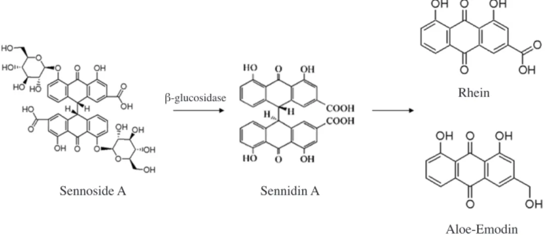
연구는 단일성분의 분리(Zhang and Liu 2004; Koyama et al. 2007; Ye et al. 2007; Gao et al. 2009), 단일성분의 효능검 증(Kon et al. 2014) 및 각 약리성분의 생체내 대사에 따른 약리효과 발현(Song et al. 2010) 분야로 집중되는 추세이다. 한편, 배당체의 분해 및 생체내 대사반응을 이용하면 특 정물질의 전구체가 되는 배당체로부터 특이성과 약리활성 이 높은 천연물신약을 생산할 수 있는데 항암효과가 있는 것으로 보고된 대황의 anthraquinones 계열 화합물인 rhein 의 경우도 효소와 미생물을 이용하여 세노사이드로부터 생 산이 가능하다. Fig. 1에 나타낸 바와 같이 세노사이드 A 배 당체에 β-glucosidase를 처리하여 2개의 포도당을 제거하면 sennidine A가 생성되고, 이어 NAD(P)H-dependent flavin enzyme 등에 의해 생성된 환원형 flavin이 sennidine A의 C-C결합을 비효소적으로 개열시키면 rheinanthrone이 생 성된다. Rheinanthrone은 산화 또는 작용기 치환을 통하여 rhein 또는 emodine으로 변환이 가능하다(Leng-Peschlow 1986; Leng-Peschlow 1989; Yagi et al. 1997). 이러한 배당 체의 체내 대사는 인삼의 saponin 배당체를 비롯한 여러 배 당체에서도 잘 알려져 있다(Hur and Kim 2011). 그러나 이 러한 생물화학반응 공정은 시간과 비용이 많이 소요되고 품 질관리가 어려운 문제점이 있어 이를 대체, 보완할 수 있는
물리적 공정, 천연물의 화학합성 등의 기술개발도 필요하다
(Kuhnert and Molod 2005; MaDougall et al. 2010).
이온화방사선은 강력한 물리적 에너지의 전달이므로 반 응시간이 짧고 물질과 반응하여 다양한 화학반응을 유도할 수 있어 천연물의 구조변환과 개질 분야에서 유망한 대체공 정이 될 수 있을 것으로 전망되고 있다. 이온화 방사선의 반 응은 주로 방사선에 의한 물의 방사선분해 유도물질에 의하 여 진행된다. 물의 방사선분해 반응산물 중 hydroxyl radical 은 매우 강력한 산화제로 작용하여 식품의 영양성분뿐만 아 니라 향기성분이나 기능성 약리성분 화합물과 반응하여 다 양한 구조변환을 유도할 수 있으며 aqueous electron 또한 높은 반응력을 나타낸다. 천연물과 관련된 이 분야의 초기 연구목표는 주로 식품의 방사선 살균과 연계하여 식품 성 분의 방사선분해(Jilan et al. 1995), 과실류의 페놀화합물과 같은 특정 성분의 변화(Breit fellner et al. 2003), 향기성분 의 변화(Ananthakumar et al. 2006) 등과 같이 방사선조사식 품의 품질변화 지표를 분석하는 데 집중되었으나, 최근에는 기능성 소재 생산을 위한 천연물의 구조변환 연구로 연구분 야가 확대되고 있다(Hai et al. 2003). 본 연구에서는 배당체로부터 약리성분을 나타내는 유도 체를 생산할 수 있는 생물화학적 공정의 대체 및 신규 공정 으로 이온화방사선이라는 물리적 공정의 적용가능성을 검 증하기 위하여, 대황의 주성분을 이루고 있는 세노사이드 A 표준품에 감마선을 조사한 후의 구조 및 약리효능 변화를 HPLC 분석, HepG2 및 PC-3 세포에 대한 세포독성 평가를 통하여 제시하고자 하였다.
재료 및 방법
1. 시료 및 감마선 조사본 실험에 사용된 세노사이드 A와 rhein은 Sigma Aldrich 사(Sigma, St. Louis, MO, USA)에서 구입하였다. 방사선 조 사를 위한 세노사이드 A 수용액은 세노사이드 A 1mg을 50% 메탄올(MtOH) 10ml에 녹인 후 syringe filter(pore size 0.2μm, Whatman, UK)에 여과하여 제조하였으며, 방사선 조사 직전 세노사이드 A 50% 메탄올 수용액에 최종농도가 0.05%가 되도록 과산화수소를 첨가하였다. 시료의 감마선 조사는 한국원자력연구원 첨단방사선연구소의 60Co 감마 선 조사시설(AECL; IR-79, MDS Nordion International Co. Ltd., Ottawa, ON, Canada)을 이용하여 1kGyh-1의 선량율
로 0, 0.5, 1, 2, 3kGy의 총 흡수선량을 얻도록 하였으며, 이 때 흡수선량의 확인은 ceric/cerous 선량계를 이용하여 측정
Fig. 1. A schematic diagram of the biochemical conversion of sennoside to rhein and/or emodin derivatives.
Sennoside A Sennidin A
Aloe-Emodin
하였다(±5%). 2. HPLC 분석
세노사이드 A 및 방사선을 조사한 세노사이드 A 수용액
의 분석은 Koyama 등(Koyama et al. 2007)의 방법에 따라 Agillent HPLC 시스템(Series 1200, Agilent technology, Palo Alto, CA, USA)에서 실시하였다. HPLC 분석 시 컬럼은 C18 column, 검출기는 Agillent UV detector(203nm), eluent solvent는 acetonitrile-water gradient(65min.)를 사용하였으 며 oven 온도는 35℃, 이동상 유속은 1.0mlmin-1, 시료 주 입량은 10μl로 설정하였다.
3. 세포배양
사람의 전립선 암세포(PC-3)와 간암세포(HepG2)는 Ame-rican Type Culture Collection(ATCC, Rockville, MD)에서 구입하였으며, 10% FBS(Fetal bovine serum; Gibco, USA) 를 첨가한 RPMI 1640 배지(Gibco, USA)에 세포를 접종하 여 공기 95%-CO2 5%, 배양온도 37℃ 조건으로 배양하였다.
4. 세포 생존율 측정
세포생존율은 MTT
[3-(4,5-dimethylthiazol-2yl)-2,5-di-phenyltetrazolium bromide] 분석방법으로 측정하였다. 사람 의 전립선암세포인 PC-3 세포와 간암세포인 HepG2세포를 각 well 당 1×10⁴ cells로 24 well 배양판에 배양하고 rhein
표준물질, 세노사이드 A 그리고 방사선을 조사한 세노사이 드 A를 농도별로 처리하여 48시간 동안 배양하였다. 48시 간 후에 기존의 배지를 버리고, MTT가 100μgml-1 함유되 어있는 RPMI 배지를 채워 3시간 배양시킨 다음 다시 배지 를 제거한 후 iso-propanol를 각 well 당 500μl씩 분주하고 570nm에서 흡광도를 측정하였다. 모든 실험에 표시된 결과 는 3번 수행하였으며, 통계분석(STASTICA)은 mean±SD 로 표시하였고, ANOVA에 의해 분석하였다. 통계적 유의성 은 p≤0.05로 판정하였다.
결 과
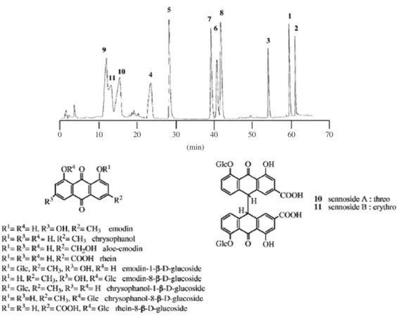
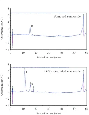
1. 세노사이드 A의 방사선 분해 0.01% 세노사이드 A 수용액(dissolved in 50% MeOH-0.05% H2O2)에 0.5, 1, 2, 3kGy의 총 흡수선량을 얻도록 감 마선을 처리한 후 HPLC chromatogram을 실시한 결과, Fig. 2에 나타낸 바와 같이 세노사이드 A 이외의 새로운 피크가 검출되어 방사선 조사에 의하여 세노사이드 A의 구조적 변Fig. 2. HPLC chromatograms and molecular structure of the standard compounds of some anthrones and anthraquinones in rhubarb. Adapted
and reprinted by permission of authors of Koyama et al. 2007. Copyright(2007) Elsevier B.V.
0 10 20 30 40 50 60 70
화가 이루어졌음을 확인할 수 있었다. 아울러, 방사선 조사 선량을 달리하였을 때에도 신규로 생성된 피크의 검출시간
은 동일하였으며, 피크 면적의 비교 결과 세노사이드 A로
부터 신규 물질로의 전환률은 1kGy에서 약 80% 정도로 가장 높은 결과를 나타내어(data not shown) 세포실험에서 는 1kGy 조사 분획을 시료로 사용하였다. 한편, 본 연구의 HPLC 분석은 Koyama 등(Koyama et al. 2007)의 방법에 따라 수행된 바, Koyama 등이 보고한 11종의 anthrone 및 anthraquinone 표준물질의 분리시간(Fig. 2)과 본 연구에서 측정된 세노사이드 A의 방사선분해산물 피크의 분리시간 (Fig. 3)을 비교한 결과 세노사이드 A의 방사선분해산물은 rhein-8-β-D-glucoside일 가능성이 높은 것으로 사료되었다. 2. PC-3 및 HepG2 세포독성 평가
세노사이드 A와 rhein 표준물질 그리고 rhein-8-β-D-glu-coside로 추정되는 세노사이드 A 방사선분해산물 분획에 대하여 사람의 전립선 암세포(PC-3)와 간암세포(HepG2) 에 대한 in vitro 세포독성 평가를 실시하였다. PC-3 세포 에 대한 세포독성 시험결과, 세노사이드 A는 24μM까지 세 포독성이 거의 없었으나 세노사이드 A 방사선분해산물 분 획(RLF)은 0.6μM의 처리구에서 90% 내외(cell viability= 12.4±1.2%)의 세포독성 효과를 나타내었다(Fig. 4). HepG2 세포에 대한 세포독성 시험에서도 세노사이드 A는 24μM까 지 세포독성이 거의 없었으나 세노사이드 A 방사선분해산물 분획(RLF)은 0.6μM의 처리구에서 70% 내외(cell viability 32.4±2.1%), 3μM의 처리구에서 85% 수준의 세포독성 효 과를 나타내었다(Fig. 5). 한편, 대황의 약리성분 중 항암효 과를 갖는 것으로 가장 잘 알려진 rhein의 PC-3 및 HepG2 세포에 대한 세포독성을 측정한 결과 rhein은 PC-3 세포에 대해서는 세포독성이 나타나지 않았으나 HepG2 세포에 대 해서는 80μM의 농도에서 LD50 값을 보였다(Fig. 6). 따라 서 본 실험에서 얻어진 세노사이드 A의 방사선분해산물은
Fig. 3. HPLC chromatograms of standard sennoside and 1kGy
ir-radiated sennoside. Component number as indicated Figure 2.
Standard sennoside
1 kGy irradiated sennoside
Absorbance
(mAU)
Retention time(min)
Retention time(min)
0 10 20 30 40 50 60 0 10 20 30 40 50 60 Absorbance (mAU) 8 6 4 2 0 -2 -4 8 6 4 2 0 -2 -4
Fig. 4. The cytotoxicity effects of sennoside A and a fraction of
ra-diolysis product of sennoside(RLF) on PC-3 cell. The cell viability was significantly decreased to 12.4±1.2% by the treatment of 0.6μM of RLF. Cell viability (%) 140 120 100 80 60 40 20 0
Concentration of sennoside A and RLF(μM)
0 0.12 0.6 3 6 12 24
PC-3
Sennoside A RLF
Fig. 5. The cytotoxicity effects of sennoside A and a fraction of
radiolysis product of sennoside(RLF) on HepG2 cell. The cell viability was significantly decreased to 32.4±2.1% by the treatment of 0.6μM of RLF. Cell viability (%) 140 120 100 80 60 40 20 0
Concentration of sennoside A and RLF(μM)
0 0.12 0.6 3 6 12 24
HepG2
Sennoside A RLF
rhein에 비하여 전립선 암세포(PC-3)와 간암세포(HepG2) 에 대하여 100배 이상의 세포독성 효과를 갖는 것으로 확인 되었다. 또한 현재까지 알려진 천연물 유래 항암 소재 가운 데 가장 우수한 효과를 보이는 것의 하나인 taxol의 간암세 포에 대한 LD50 값이 0.1~1μM 수준으로 보고된 것(He et al. 2012)과 비교하였을 때 본 연구에서 생산된 세노사이드 A 방사선분해산물의 암세포에 대한 독성효과가 매우 높은 것으로 평가된다.
고 찰
본 연구에서 분리된 세노사이드 A의 방사선분해산물은 rhein-8-β-D-glucoside일 가능성이 높은 것으로 사료되었다. 이는 두 개의 glycoside가 탄소결합으로 연결된 bianthrone glycoside 구조인 세노사이드 A가 방사선에 의하여 생성된 라디칼의 산화작용에 의하여 탄소결합이 끊어져 anthrone glycoside로 분해된다고 해석될 수 있다. 방사선에 의한 탄 소결합구조의 분해는 탄소사슬의 길이를 달리한 alkane chain의 방사선 분해에 관한 연구(Allayarov et al. 2007) 등 에서 보고된 바 있다. 또한 본 연구에서 rhein-8-β-D-gluco-side로 추정되는 피크 이외의 aglycon anthraquinone 물질 들은 검출되지 않은 것으로 보아 세노사이드 A에 결합된 glucose는 본 실험 조건(1kGy)의 방사선에 대해서는 영향 을 받지 않으며 세노사이드 A의 탄소결합에만 특이적으로 작용하는 것으로 해석된다. 그러나 향후 세노사이드 A 방사 선분해산물을 기능성식품 또는 의약품으로 사용하거나 방 사선 분해를 생산공정으로 응용하기 위해서는 보다 상세한 구조분석과 동정, 그리고 세노사이드의 방사선 분해에 대한 물리화학적 반응기작 규명이 요구되며 유사한 결합구조를 가진 배당체에 대한 비교연구 또한 필요하다. 방사선 조사식품의 품질변화 연구에서 비롯된 방사선의 방사선분해 연구는 천연물의약 소재 개발 분야로 연구가 확 대되고 있다(Hai et al. 2003; Hur and Kim 2011). Hur 등(Hur and Kim 2011)은 홍삼의 에탄올 추출물에 감마선을 조사하 였을 때 홍삼 에탄올 추출물은 30μgml-1에서도 세포독성 을 보이지 않았으나, 감마선 조사를 처리한 홍삼 추출물은 3 μgml-1부터 PC-3 세포에서 세포생존율을 감소시켰다고 보 고하여 본 연구와 유사한 경향을 보였다. 본 연구에서는 대 황의 주성분인 세노사이드 A에 방사선을 조사하였을 때 세 노사이드 A가 rhein-8-β-D-glucoside로 추정되는 구조로 전 환되고, 이는 기존에 대황의 항암성분으로 보고된 rhein에 비하여 항암효과가 더 높은 것을 확인할 수 있었다. 나아가 방사선 조사를 통하여 유사 배당체로부터 새로운 약리효과 를 나타내는 물질의 발굴 또는 생산이 가능할 것으로 사료 되었으나 이를 검증하고 산업적 활용이 가능한 수준으로 발 전시키기 위해서는 방사선분해산물의 구조분석 및 항암효 과 기전규명 등의 보다 세부적인 연구가 필요하다.결 론
0.01% 세노사이드 A 수용액에 1kGy의 감마선을 조사한 후 HPLC chromatogram을 실시한 결과 방사선분해산물로 rhein-8-β-D-glucoside로 추정되는 물질이 생성됨을 확인하 였다. 세노사이드 A와 rhein 표준물질 그리고 rhein-8-β-D-glucoside로 추정되는 세노사이드 A 방사선분해산물 분획에 대하여 사람의 전립선 암세포(PC-3)와 간암세포(HepG2)에 대한 in vitro 세포독성 평가 결과 세노사이드 A는 0~24 μM의 농도에서 세포독성을 나타내지 않았으나 세노사이드 A 방사선분해산물 분획은 0.6μM의 농도에서도 PC-3(세포 생존율 12.4±1.2%) 및 HepG2(세포생존율 32.4±2.1%) 세 포에 대하여 높은 세포독성을 나타내었다. Rhein은 0~80 μM의 농도에서 PC-3 세포에 대해서는 세포독성이 관찰되 지 않았으며 HepG2 세포에 대해서는 80μM의 농도에서 LD50 수준의 독성을 나타내었다. 본 연구에서는 세노사이드A에 방사선을 조사하였을 때 세노사이드 A가 rhein-8-β-D-glucoside로 추정되는 구조로 전환되고 이는 기존에 보고된 rhein에 비하여 항암효과가 더 높은 것을 확인할 수 있었다.
사 사
본 연구는 한국원자력연구원 기본연구사업 연구비 지원
에 의하여 수행되었으며 이에 감사드립니다.
Fig. 6. The cytotoxicity effects of standard rhein on HepG2 and
PC-3 cell. The rhein had the effect on HepG2 cell with a LD50 at 80μM. Cell viability (%) 140 120 100 80 60 40 20 0 Concentration of rhein(μM) 0 2 10 20 40 60 80 HepG2 PC-3
참 고 문 헌
Allayarov SR, Konovalikhin SV, Olkhov YA, Jackson VE, Kispert LD, Dixon DA, Ila D and Lappan U. 2007. Degra-dation of γ-irradiated linear perfluoroalkanes at high dos-age. J. Fluorine Chem. 128:575-586.
Ananthakumar A, Variyar PS and Sharma A. 2006. Estimation of aroma glycosides of nutmeg and their changes during radiation processing. J. Chromatogr. A. 1108:252-257. Bae UJ, Song MY, Jang HY, Lim JM, Lee SY, Ryu JH and Park
BH. 2015. Emodin isolated from Rheum palmatum prevents cytokine-induced β-cell damage and the development of type 1 diabetes. J. Funct. Foods. 16:9-19.
Breitfellner F, Solar S and Sontag G. 2003. Radiation induced chemical changes of phenolic compounds in strawberries. Radiat. Phys. Chem. 67:497-499.
Dorsey JF and Kao GD. 2007. Aloe(-emodin) for cancer? More than just comforting salve. Cancer Biol. Ther. 6:89-90. Gao XY, Jiang Y, Lu JQ and Tu PF. 2009. One single standard
substance for the determination of multiple anthraquinone derivatives in rhubarb using high-performance liquid chro-matography-diode array detection. J. Chromatogr. A. 1216: 2118-2123.
Hai L, Diep TB, Nagasawa N, Yoshii F and Kume T. 2003. Ra-diation depolymerization of chitosan to prepare oligomers. Nucl. Instr. and Meth. in Phys. Res. B. 208:466-470. He L, Ling Y, Fu L, Yin D, Wang X and Zhang Y. 2012.
Syn-thesis and biological evaluation of novel derivatives of gam-bogic acid as anti-hepatocellular carcinoma agents. Bioorg. Med. Chem. Lett. 22:289-292.
Huang Q, Lu G, Shen HM, Chung MCM and Ong CN. 2007. Anti-cancer properties of anthraquinones from rhubarb. Med. Res. Rev. 27:609-630.
Hur JM and Kim D. 2011. The Ethanol Extract of Red Ginseng Enhances Anti-Tumor Effects Using Co-60 Gamma Irradi-ation. J. Appl. Biol. Chem. 54:15-20.
Iizuka A, Iijima OT, Kondo K, Itakura H, Yoshie F, Miyamoto H, Kubo M, Higuchi M, Takeda H and Matsumiya T. 2004. Evaluation of Rhubarb using antioxidative activity as an index of pharmacological usefulness. J. Ethnopharmacol. 91:89-94.
Jilan W, Xujia Z, Rongyao Y and Yongke H. 1995. Radiolysis of herb. Radiat. Phys. Chem. 46:275-279.
Kon R, Ikarashi N, Nagoya C, Takayama T, Kusunoki Y, Ishii M, Ueda H, Ochiai W, Machida Y, Sugita K and Sugiyama K. 2014. Rheinanthrone, a metabolite of sennoside A, trig-gers macrophage activation to decrease aquaporin-3 ex-pression in the colon, causing the laxative effect of rhubarb extract. J. Ethnopharmacol. 152:190-200.
Koyama J, Morita I and Kobayashi N. 2007. Simultaneous determination of anthraquinones in rhubarb by
high-perfor-mance liquid chromatography and capillary electrophore-sis. J. Chromatogr. A. 1145:183-189.
Kuhnert N and Molod HY. 2005. An efficient total synthesis of chrysophanol and the sennoside C aglycon. Tetrahedron Lett. 46:7571-7573.
Leng-Peschlow E. 1986. Acceleration of large intestine transit time in rats by sennosides and related compounds. J. Pharm. Pharmacol. 38:369-373.
Leng-Peschlow E. 1989. Effects of sennosides A+B and bisac-odyl on rat large intestine. Pharmacol. 38:310-318. MaDougall GJ, Dobson P and Jordan-Mahy N. 2010. Effect of
different cooking regimes on rhubarb polyphenols. Food Chem. 119:758-764.
Matsuda H, Morikawa T, Toguchida I, Park JY, Harima S and Yoshikawa M. 2001. Antioxidant constituents from rhu-barb: Structural requirements of stilbenes for the activity and structures of two new antraquinone glucosides. Bioorg. Med. Chem. 9:41-50.
Matsuda H, Tewtrakul S, Morikawa T and Yoshikawa M. 2004. Anti-allergic activity of stilbenes from Korean rhubarb (Rheum undulatum L.): structure requirements for inhibi-tion of antigen-induced degranulainhibi-tion and their effects on the release of TNF-α and IL-4 in RBL-2H3 cells. Bioorg. Med. Chem. 12:4871-4876.
Moon MK, Kang DG, Lee JK, Kim JS and Lee HS. 2006. Vaso-dilatory and anti-inflammatory effects of the aqueous extract of rhubarb via a NO-cGMP pathway. Life Sci. 78:1550-1557.
Song R, Xu L, Xu F, Li Z, Dong H, Tian Y and Zhang Z. 2010. In vivo metabolism study of rhubarb decoction in rat using high-performance liquid chromatography with UV photo-diode-array and mass-spectrometric detection: A strategy for systematic analysis of metabolites from traditional Chinese medicines in biological samples. J. Chromatogr. A. 1217: 7144-7152.
Wang JL, Liu MF, Li XX, Wang Q and Li JF. 2014. Anti-ox-idative activities of ethanol extracts from both wild plant and suspension cell cultures of Rheum franzenbachii. Chi-nese Herbal Med. 6(2):115-119.
Yagi T, Yamauchi K and Kuwano S. 1997. The synergistic pur-gative action of aloe-emodin anthrone and rheinanthrone in mice: synergism in large intestinal propulsion and water secretion. J. Pharm. Pharmacol. 49:22-25.
Ye M, Han J, Chen H, Zheng J and Guo D. 2007. Analysis of phenolic compounds in rhubarbs using liquid choromatog-raphy coupled with electrospray ionization mass spectrom-etry. J. Am. Soc. Mass Spectrom. 18:82-91.
Yu HM, Liu YF, Cheng YF, Hu LK and Hou M. 2008. Effects of rhubarb extract on radiation induced lung toxicity via decreasing transforming growth factor-beta-1 and inter-leukin-6 in lung cancer patients treated with radiotherapy.
Lung Cancer 59:219-226.
Zhang HX and Liu MC. 2004. Separation procedures for the pharmacologically active components of rhubarb. J. Chro-matogr. B. 812:175-181.
Zheng QX, Wu HF, Guo J, Nan HJ, Chen SL, Yang JS and Xu XD. 2013. Review of rhubarbs: Chemistry and
pharmacol-ogy. Chinese Herbal Med. 5(1):9-32.
Received: 6 May 2016 Revised: 7 June 2016 Revision accepted: 12 June 2016