http://www.j-circ.or.jp
as with other risk factors for sudden cardiac death.3,7–9 Recent studies have further demonstrated that myocardial fibrosis as measured on CMRI is an independent predictor of all-cause and cardiac mortality even in low or asymptomatic HCM pa-tients.7,10 Although the relationship between the presence and the extent of LGE and ventricular arrhythmias and sudden cardiac death has been well documented, few previous studies have evaluated the relationship between LGE and left ventricu-lar (LV) functional reserve in HCM patients. In patients with HCM, already impaired myocardial relaxation does not aug-yocardial fibrosis, along with maug-yocardial disarray,
is a characteristic histological finding in hypertro-phic cardiomyopathy (HCM).1 It can lead to car-diac chamber stiffness, and its extent has been suggested to be an important determinant of systolic and diastolic dysfunction in HCM.2–4 The presence and the extent of myocardial fibrosis can now be non-invasively assessed with cardiovascular mag-netic resonance imaging (CMRI) using late gadolinium en-hancement (LGE).5,6 Several studies have shown that LGE is associated with non-sustained ventricular tachycardia as well
M
Received November 7, 2012; revised manuscript received January 23, 2013; accepted February 19, 2013; released online April 2, 2013 Time for primary review: 28 days
Cardiology Division, Gachon University of Medicine and Science, Incheon (J.M.); Department of Radiology (Y.J.H., Y.-J.K.) and Cardiol-ogy Division (C.Y.S., Y.J., N.C., S.-Y.C., J.-W.H.), Yonsei University College of Medicine, Seoul, Korea
Mailing address: Jong-Won Ha, MD, PhD, Cardiology Division, Severance Cardiovascular Hospital, Severance Biomedical Science Insti-tute, Yonsei University College of Medicine, 134 Shinchon-dong, Seodaemun-gu, 120-752 Seoul, Republic of Korea. E-mail: jwha@ yuhs.ac
ISSN-1346-9843 doi: 10.1253/circj.CJ-12-1378
All rights are reserved to the Japanese Circulation Society. For permissions, please e-mail: [email protected]
Extent of Late Gadolinium Enhancement on
Cardiovascular Magnetic Resonance Imaging and
Its Relation to Left Ventricular Longitudinal Functional
Reserve During Exercise in Patients With
Hypertrophic Cardiomyopathy
Jeonggeun Moon, MD; Yoo Jin Hong, MD; Young-Jin Kim, MD, PhD;
Chi Young Shim, MD, PhD; Yangsoo Jang, MD, PhD; Namsik Chung, MD, PhD;
Seung-Yun Cho, MD, PhD; Jong-Won Ha, MD, PhD
Background: The aim of this study was to investigate whether the extent of late gadolinium enhancement (LGE) on cardiovascular magnetic resonance imaging reflecting myocardial fibrosis correlates with left ventricular (LV) longitudinal function during exercise in hypertrophic cardiomyopathy (HCM).
Methods and Results: Mitral annular velocities (E’ and S’) were measured on echocardiography at rest and during graded bicycle exercise (25 W, 3-min increments) in 46 HCM patients (mean age, 53 years; 32 men). LV longitudinal diastolic and systolic functional reserve indices were calculated as ΔE’×E’base and ΔS’×S’base, where ΔE’ and ΔS’ are
the changes in E’ and S’ from baseline to 50 W of exercise, respectively. The patients were divided into 2 groups according to the extent of LGE (as “percentage of LV mass containing LGE”: %LV with LGE; range, 0–37%; median, 6%): group 1 (n=23), %LV with LGE <6%, and group 2, %LV with LGE ≥6%. Baseline echocardiographic parameters were similar between the 2 groups, but changes in E’ and S’ during exercise were smaller in group 2 (ΔE’: 2.8±1.8 cm/s vs.1.5±1.0 cm/s, P=0.007; ΔS’: 2.2±1.2 cm/s vs. 0.9±0.8 cm/s, P<0.0001). LV functional reserve indices were also significantly lower in group 2 (ΔE’×E’base: 12.8±7.7 vs. 5.5±3.4, P=0.001; ΔS’×S’base: 12.6±7.4 vs. 4.7±4.5,
P<0.0001).
Conclusions: LV longitudinal function during exercise is influenced by the extent of LGE in HCM. Myocardial fibro-sis may represent a pathologic substrate that determines LV functional reserve in patients with HCM. (Circ J 2013; 77: 1742 – 1749)
Key Words: Echocardiography; Exercise; Hypertrophic cardiomyopathy; Left ventricular function; Magnetic reso-nance imaging
ducer during rest and exercise. Standard 2-D measurements (LV end-diastolic and end-systolic dimensions, ventricular sep-tum and posterior wall thickness, left atrial [LA] volume, and LVOT diameter) and Doppler parameters were obtained with the patient in the left lateral decubitus position. LV ejection fraction (EF) was calculated using the modified method of Quinones et al.14 LA volume was determined using the prolate ellipsoid formula and was indexed to body surface area.15 From the apical window, a 1–2-mm pulsed Doppler sample volume was placed at the mitral valve tip, and mitral flow velocities from 5 to 10 cardiac cycles were recorded. The mitral inflow velocities were traced, and the following variables were ob-tained: peak velocity of early (E) and late (A) filling, and de-celeration time of the E wave velocity. The peak instantaneous LV outflow gradient was estimated with continuous wave Doppler under basal conditions, and LVOT obstruction was defined as peak instantaneous outflow gradient ≥30 mmHg.16,17 Tricuspid regurgitant jet velocity was also obtained to estimate pulmonary artery systolic pressure using continuous wave Doppler, if measurable. After obtaining the rest images from the standard parasternal and apical views, a multistage supine bicycle exercise test was performed with a variable load bi-cycle ergometer (Medical Positioning, Kansas City, MO, USA). Patients pedaled at a constant rate beginning at a workload of 25 W with an increment of 25 W every 3 min. Mitral annular velocity was measured on Doppler tissue imaging (DTI) using the pulsed wave Doppler mode. The filter was set to exclude high-frequency signal, and the Nyquist limit was adjusted to a range of 15–20 cm/s. Gain and sample volume were minimized to allow a clear tissue signal with minimal background noise. Early diastolic (E’) and systolic (S’) velocities of the mitral annulus were measured on apical 4-chamber view with a 2–5-mm sample volume placed at the septal corner of the mitral annulus. These measurements were performed at baseline and at each stage of exercise in the same sequence.
ment LV longitudinal function as much as seen in normal in-dividuals during exercise.11 The anatomic substrate of this lack of diastolic reserve during exercise, however, has not been explored before. In the present study, we hypothesized that the extent of myocardial fibrosis would correlate with the magni-tude of augmentation in LV longitudinal function during ex-ercise in patients with HCM.
Methods
SubjectsAll 46 patients with HCM (mean age, 53 years; 32 men; 10 with LV outflow tract [LVOT] obstruction) were consecu-tively enrolled. The diagnosis of HCM was made according to the World Health Organization/International Society and Federation of Cardiology criteria.12 Patients with apical HCM or atrial fibrillation were excluded. Patient who could not per-form bicycle exercise for any reason or who had specific con-traindications for CMRI such as implanted cardioverter- defibrillators or pacemakers, metallic fragments, known claus-trophobia or renal insufficiency were also excluded. None of the patients had a history of alcohol septal ablation or surgical septal myectomy. CMRI and comprehensive echocardiography, including exercise Doppler evaluation, were performed in all patients. Prescribed medications, which were grossly similar between the 2 groups, were discontinued before exercise echo-cardiography. This study was approved by the institutional ethics committee. The study complies with the Declaration of Helsinki, and informed consent was obtained from all subjects. 2-D and Exercise Doppler Echocardiography (Diastolic Stress Echocardiography)
Diastolic stress echocardiography13 was performed as follows. Echocardiography was carried out using an ultrasound system (System 7, GE Vingmed, Horten, Norway) with 2.5-MHz
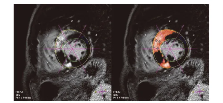
trans-Figure 1. Quantification of late gadolinium enhancement (LGE) using the automatic thresholding software (ViewForum version 4.1; Philips Medical Systems, The Netherlands). Left ventricular (LV) myocardial mass was determined (g) and the extent of LGE was expressed as LGE mass (g) and percentage of LV mass containing LGE (%LV with LGE).
Intera; Philips Medical Systems, Best, The Netherlands) 10–15 min after 0.2 mmol/kg of a gadolinium-based contrast media. Short-axis slices of the heart were obtained without any gaps, encompassing the entire LV. LGE was identified on a segmented inversion recovery T1-weighted turbo field-echo sequence with the following parameters (Look-Locker): slice thickness, 10 mm; typical repetition time, 5.3 ms; typical echo time, 1.6 ms; flip angle, 15°; field of view, 36 cm; number of signal averages, 2; acquisition matrix, 320×256; and recon-struction matrix, 512×512. Quantification of LGE was done using the automatic thresholding software (ViewForum, ver-sion 4.1; Philips Medical Systems) by 2 radiologists in con-sensus who were unaware of the patients’ clinical and echo-cardiographic data (Figure 1). At first, raw image data were analyzed by each radiologist separately. After that, normal myo-cardium was designated on consensus and the dedicated soft-LV longitudinal diastolic and systolic functional reserve
in-dices were calculated using the following formulas:18,19 diastolic functional reserve index = ΔE’ × E’base,
where ΔE’ is the change in E’ from baseline to 25 W or 50 W of exercise, and
systolic functional reserve index = ΔS’ × S’base,
where ΔS’ is the change in S’ from baseline to 25 W or 50 W of exercise.
All data were digitally stored and analyzed by 2 experi-enced echocardiographers who were blinded to the clinical and CMRI data.
CMRI and LGE Quantification
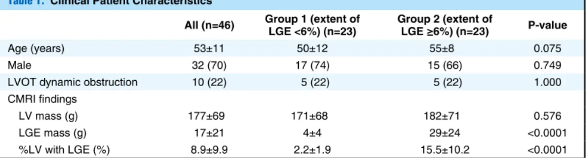
LGE images were acquired with a 1.5-T CMRI unit (Gyroscan Table 1. Clinical Patient Characteristics
All (n=46) Group 1 (extent of LGE <6%) (n=23) Group 2 (extent of LGE ≥6%) (n=23) P-value
Age (years) 53±11 50±12 55±8 0.075
Male 32 (70) 17 (74) 15 (66) 0.749
LVOT dynamic obstruction 10 (22) 5 (22) 5 (22) 1.000 CMRI findings
LV mass (g) 177±69 171±68 182±71 0.576 LGE mass (g) 17±21 4±4 29±24 <0.0001 %LV with LGE (%) 8.9±9.9 2.2±1.9 15.5±10.2 <0.0001 Data given as n (%) or mean ± SD. CMRI, cardiovascular magnetic resonance imaging; LGE, late gadolinium enhancement; LV, left ventricle/ventricular; %LV with LGE, percentage of LV volume containing LGE (ie, extent of LGE); LVOT, left ventricular outflow tract.
Table 2. Echocardiographic Findings
Group 1 (extent of
LGE <6%) (n=23) Group 2 (extent of LGE ≥6%) (n=23) P-value
LVEDD (mm) 48±4 45±6 0.052
LVESD (mm) 30±4 29±5 0.324
LVEF (%) 69±6 68±6 0.576
IVS (mm) 14±4 16±4 0.176
PW (mm) 11±2 11±2 0.889
Maximum wall thickness (mm) 19±4 20±4 0.667
LAVI (ml/m2) 30±9 38±16 0.099 E (m/s) 0.62±0.16 0.57±0.23 0.343 A (m/s) 0.59±0.12 0.58±0.23 0.929 E/A 1.1±0.5 1.1±0.5 0.880 DT (ms) 189±36 230±75 0.023 TR (m/s) 2.3±0.3 2.3±0.3 0.762 E’ (cm/s) 4.6±1.3 3.8±1.4 0.054 E/E’ 14±5 17±8 0.150 S’ (cm/s) 5.8±1.2 5.0±1.0 0.022 Longitudinal DFRI at 25W 7.0±5.5 4.0±4.7 0.060 Longitudinal DFRI at 50W 12.8±7.7 5.5±3.4 0.001 Longitudinal SFRI at 25W 3.7±6.2 2.6±3.5 0.475 Longitudinal SFRI at 50W 12.6±7.4 4.7±4.5 <0.0001 Data given as mean ± SD. A, late diastolic mitral inflow; DFRI, diastolic functional reserve index; DT, deceleration time of E wave; E, early diastolic mitral inflow; E’, early diastolic mitral annulus velocity; IVS, interventricular septum; LAVI, left atrial volume index; LVEDD, LV diastolic dimension; LVEF, LV ejection fraction; LVESD, LV end-systolic dimension; PW, posterior wall; S’, end-systolic mitral annulus velocity; SFRI, end-systolic functional reserve index; TR, tricuspid regurgitation. Other abbreviations as in Table 1.
results from the first observer. P<0.05 was considered to be significant.
Results
LGE was observed in 38 of 46 patients (83%), but its degree was variable (%LV with LGE: range, 0–37%; median, 6%). Patients were divided into 2 groups according to the extent of LGE: group 1, %LV with LGE <6%; and group 2, %LV with LGE ≥6%. Table 1 lists the clinical characteristics and CMRI findings of the patients. Demographic findings and LV mass assessed on CMRI were comparable between the 2 groups except the extent of LGE (Table 1). Echocardiographic find-ings are listed in Table 2. The baseline echocardiographic parameters were also similar between the 2 groups, except deceleration time of E wave and S’. LV longitudinal diastolic and systolic functional reserve indices at 25 W were similar between the groups, but LV functional reserve indices at 50 W were significantly lower in group 2 (Table 2). Table 3 lists the hemodynamic responses during supine bicycle exercise. In both groups, heart rate, systolic and diastolic blood pressures increased during exercise compared with those at rest, but no significant differences were found between the 2 groups. Exercise duration was also similar between the 2 groups (Table 3). We analyzed the relationship between exercise du-ware determined the mean (μ) and standard deviation (σ) of
pixel value of the area (ie, region of interest). After that, we defined the enhanced area (LGE) as signal intensity above the threshold T, where T = μ + 3σ. LV mass was determined (g),
and the extent of LGE was expressed as LGE mass (g) and percentage of LV mass containing LGE (%LV with LGE). Statistical Analysis
Continuous data are expressed as mean ± SD, and normality tests were performed for each variable. Categorical variables were summarized as a percentage of the group total. The base-line characteristics of the 2 groups were compared using the 2-sample t-test for continuous variables, and chi-square test and Fisher’s exact test for categorical variables. Differences in Doppler indexes between rest and exercise were compared using repeated measures analysis of variance. Pearson’s bi-variate correlation analysis was used to determine the correla-tion between variables. Multiple linear regression analysis was performed to test the association of diastolic/systolic func-tional reserve index with other parameters that were clinically meaningful or statistically significant including %LV with LGE. Intra- and interobserver variability for DTI measure-ment were calculated by analysis of 10 random images by 1 investigator blinded to the first analysis and additional analy-sis of images by a second observer who was unaware of the
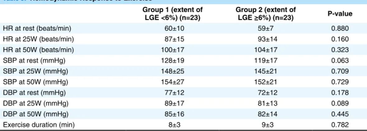
Table 3. Hemodynamic Response to Exercise
Group 1 (extent of
LGE <6%) (n=23) Group 2 (extent of LGE ≥6%) (n=23) P-value
HR at rest (beats/min) 60±10 59±7 0.880 HR at 25W (beats/min) 87±15 93±14 0.160 HR at 50W (beats/min) 100±17 104±17 0.323 SBP at rest (mmHg) 128±19 119±17 0.063 SBP at 25W (mmHg) 148±25 145±21 0.709 SBP at 50W (mmHg) 154±27 152±21 0.729 DBP at rest (mmHg) 77±12 72±12 0.178 DBP at 25W (mmHg) 89±17 81±13 0.089 DBP at 50W (mmHg) 85±16 82±14 0.445 Exercise duration (min) 8±3 9±3 0.782 Data given as mean ± SD. DBP, diastolic blood pressure; HR, heart rate; SBP, systolic blood pressure. Other abbre-viations as in Table 1.
Table 4. Independent Predictors of DFRI and SFRI at 50W Workload
β t P-value
ΔE’×E’base at 50W (Adjusted R square=0.096)
%LV with LGE* –0.360* –2.396* 0.021*
Age 0.018 0.115 0.909
Male 0.175 1.147 0.258
LVOT dynamic obstruction –0.140 –0.957 0.344
DT –0.129 –0.757 0.453
ΔS’×S’base at 50W (Adjusted R square=0.061)
%LV with LGE* –0.318* –2.045* 0.048*
Age –0.204 –1.259 0.216
Male 0.135 0.854 0.398
LVOT dynamic obstruction –0.124 –0.824 0.415
DT –0.009 –0.052 0.958
*Iindependent predictor. DFRI, diastolic functional reserve index (ΔE’×E’base); SFRI, systolic functional reserve index
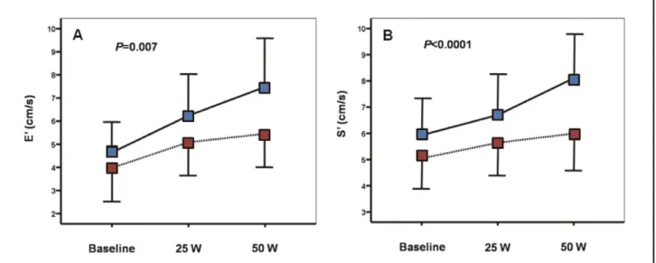
rest, E’ was smaller in group 2 (P=0.054), suggesting more impaired LV relaxation in those patients. In both groups, sig-nificant augmentation in E’ was observed during exercise, but the increase in E’ during exercise was less pronounced in group 2, and ΔE’ at 50 W of exercise was significantly smaller in group 2 (Figure 2A). Similarly, the baseline S’ was sig-nificantly smaller in group 2 (P=0.022), suggesting reduced longitudinal contraction in these subjects. During exercise, S’ was significantly increased in both groups, but the magnitude of increase in S’ during exercise and ΔS’ at 50 W of exercise were smaller in group 2 when compared with group 1 (Figure 2B). In simple correlation analysis, the extent of LGE ration and LGE or LV longitudinal functional reserve and
found that there was no significant correlation between these parameters (exercise duration and %LV with LGE, R=0.069, P=0.650; exercise duration with diastolic functional reserve index at 50 W, R=0.149, P=0.322; exercise duration with sys-tolic functional reserve index at 50 W, R=0.184, P=0.227). Multiple linear regression analysis found that %LV with LGE was the only independent predictor of diastolic/systolic func-tional reserve index at 50 W workload even after age, gender, presence of LVOT obstruction and mitral DT were controlled for (Table 4).
The changes in LV performance are shown in Figure 2. At
Figure 2. Changes in E’ and S’ from baseline to exercise (– –, group 1; - - - -, group 2). Increases in E’ and S’ with exercise were less pronounced in group 2.
Figure 3. Relationship between extent of late gadolinium enhancement (LGE; percentage of left ventricular [LV] volume contain-ing LGE: %LV with LGE) and the diastolic functional reserve index (ΔE’×E’base) at 50 W workload. %LV with LGE had a weak but
significant negative linear correlation with ΔE’×E’base at 50 W in the overall cohort. The same was true in patients with and without
LVEF because it is load independent and reflects subendocar-dial fiber function. The contraction and relaxation of LV are abnormal not only at rest23 but also during exercise11 in HCM. We have shown that LV longitudinal functional reserve was reduced in HCM patients when compared with age- and sex-matched normal control subjects11 by assessing mitral annular velocity during exercise using DTI. The underlying histo-pathological substrate responsible for the abnormal LV func-tional reserve during exercise in HCM, however, is still un-clear. The present results suggest that the extent of myocardial fibrosis may represent a pathological substrate which deter-mines LV performance during exercise in patients with HCM. Myocardial Fibrosis and LV Longitudinal Functional Reserve in HCM
Myocardial scarring has been considered in part to reflect the long-term consequence of myocyte death and replacement collagen accumulation as a repair process,24 and thus the pres-ence and the extent of LGE on CMRI have been reported to be associated with systolic and diastolic dysfunction in HCM patients.2–4,25 But even though LGE is regarded as a determi-nant of LV dysfunction in HCM, the relationship between the extent of LGE and the degree of LV dysfunction assessed at rest is relatively weak.3,8,26 In contrast, several current CMRI studies have nicely shown the correlation between LGE and impaired LV longitudinal function in patients with different kinds of cardiomyopathy.27–29 In the present study, we have further shown that the extent of myocardial fibrosis affects myocardial function during exercise in HCM patients. Although LV longitudinal movement was more depressed in patients with a larger volume of LGE even at rest, the augmentation in LV longitudinal function was more blunted in patients with a larger extent of LGE. The difference between group 1 and group 2 became greater as the intensity of exercise increased. The present results suggest that the presence and the extent of myocardial fibrosis may be determinants of exertional LV functional reserve in patients with HCM.
showed a significant inverse correlation with LV diastolic (R=–0.369, P=0.012; Figure 3) and systolic functional reserve indices (R=–0.317, P=0.034; Figure 4). Intra- and interob-server variability for DTI measurement were 5±3% and 7±4%, respectively.
We additionally analyzed the changes in LV long-axis per-formance according to the location of LGE (ie, septal involve-ment of myocardial fibrosis), because we determined E’ and S’ only at the septal corner of mitral annulus. The augmen-tation of E’ and S’ during exercise (from baseline to 50 W workload) was significantly blunted in patients who had LGE at the interventricular septum (n=32) than in patients without septal myocardial fibrosis (n=14; P=0.001 for E’ and P=0.030 for S’).
Discussion
We investigated the relationship between the extent of LGE on CMRI and LV functional reserve during exercise in pa-tients with HCM and found that the augmentation of LV lon-gitudinal function during exercise was more blunted in pa-tients who had a larger extent of LGE. In addition, the magnitude of augmentation in LV long-axis movement was inversely correlated with the extent of LGE. The present study is the first to demonstrate the relationship between the burden of myocardial scar, non-invasively assessed on CMRI, and LV longitudinal functional reserve during exercise in patients with HCM.
Impaired Diastolic and Systolic Function in HCM
The cardinal pathophysiologic feature of HCM is diastolic dysfunction, which is seen in the majority of HCM patients.20 Not only the diastolic function but also myocardial contractile property are known to be reduced in HCM, despite normal LVEF. Interestingly, even in the subclinical stage, mutation carriers have been found to have impaired myocardial relax-ation and contractile property in the absence of myocardial hypertrophy.21,22 The assessment of LV long-axis movement is superior to classic echocardiographic parameters such as
Figure 4. Relationship between extent of late gadolinium enhancement (LGE; percentage of left ventricular [LV] volume contain-ing LGE: %LV with LGE) and systolic functional reserve index (ΔS’×S’base) at 50 W workload. %LV with LGE had a weak but
sig-nificant negative linear correlation with ΔS’×S’base at 50 W in the overall cohort. The linear correlation was more prominent in patients
opathy: Myocardial fibre disarray and myocardial fibrosis. Br Heart J 1980; 44: 433 – 443.
2. Olivotto I, Maron BJ, Appelbaum E, Harrigan CJ, Salton C, Gibson CM, et al. Spectrum and clinical significance of systolic function and myocardial fibrosis assessed by cardiovascular magnetic resonance in hypertrophic cardiomyopathy. Am J Cardiol 2010; 106: 261 – 267. 3. Maron MS, Appelbaum E, Harrigan CJ, Buros J, Gibson CM, Hanna C, et al. Clinical profile and significance of delayed enhancement in hypertrophic cardiomyopathy. Circ Heart Fail 2008; 1: 184 – 191. 4. Lombardi R, Betocchi S, Losi MA, Tocchetti CG, Aversa M, Miranda
M, et al. Myocardial collagen turnover in hypertrophic cardiomy-opathy. Circulation 2003; 108: 1455 – 1460.
5. Moon JC, McKenna WJ, McCrohon JA, Elliott PM, Smith GC, Pennell DJ. Toward clinical risk assessment in hypertrophic cardio-myopathy with gadolinium cardiovascular magnetic resonance. J Am Coll Cardiol 2003; 41: 1561 – 1567.
6. Moon JC, Reed E, Sheppard MN, Elkington AG, Ho SY, Burke M, et al. The histologic basis of late gadolinium enhancement cardiovas-cular magnetic resonance in hypertrophic cardiomyopathy. J Am Coll Cardiol 2004; 43: 2260 – 2264.
7. Bruder O, Wagner A, Jensen CJ, Schneider S, Ong P, Kispert EM, et al. Myocardial scar visualized by cardiovascular magnetic reso-nance imaging predicts major adverse events in patients with hyper-trophic cardiomyopathy. J Am Coll Cardiol 2010; 56: 875 – 887. 8. Choudhury L, Mahrholdt H, Wagner A, Choi KM, Elliott MD, Klocke
FJ, et al. Myocardial scarring in asymptomatic or mildly symptom-atic patients with hypertrophic cardiomyopathy. J Am Coll Cardiol 2002; 40: 2156 – 2164.
9. Adabag AS, Maron BJ, Appelbaum E, Harrigan CJ, Buros JL, Gibson CM, et al. Occurrence and frequency of arrhythmias in hypertrophic cardiomyopathy in relation to delayed enhancement on cardiovascu-lar magnetic resonance. J Am Coll Cardiol 2008; 51: 1369 – 1374. 10. O’Hanlon R, Grasso A, Roughton M, Moon JC, Clark S, Wage R, et
al. Prognostic significance of myocardial fibrosis in hypertrophic cardiomyopathy. J Am Coll Cardiol 2010; 56: 867 – 874.
11. Ha JW, Ahn JA, Kim JM, Choi EY, Kang SM, Rim SJ, et al. Abnor-mal longitudinal myocardial functional reserve assessed by exercise tissue Doppler echocardiography in patients with hypertrophic car-diomyopathy. J Am Soc Echocardiogr 2006; 19: 1314 – 1319. 12. Richardson P, McKenna W, Bristow M, Maisch B, Mautner B,
O’Connell J, et al. Report of the 1995 World Health Organization/ International Society and Federation of Cardiology Task Force on the Definition and Classification of Cardiomyopathies. Circulation 1996; 93: 841 – 842.
13. Ha JW, Oh JK, Pellikka PA, Ommen SR, Stussy VL, Bailey KR, et al. Diastolic stress echocardiography: A novel noninvasive diagnostic test for diastolic dysfunction using supine bicycle exercise Doppler echocardiography. J Am Soc Echocardiogr 2005; 18: 63 – 68. 14. Quinones MA, Waggoner AD, Reduto LA, Nelson JG, Young JB,
Winters WL Jr, et al. A new, simplified and accurate method for determining ejection fraction with two-dimensional echocardiogra-phy. Circulation 1981; 64: 744 – 753.
15. Lang RM, Bierig M, Devereux RB, Flachskampf FA, Foster E, Pellikka PA, et al. Recommendations for chamber quantification: A report from the American Society of Echocardiography’s Guidelines and Standards Committee and the Chamber Quantification Writing Group, developed in conjunction with the European Association of Echocardiography, a branch of the European Society of Cardiology. J Am Soc Echocardiogr 2005; 18: 1440 – 1463.
16. Rigopoulos AG, Seggewiss H. A decade of percutaneous septal abla-tion in hypertrophic cardiomyopathy. Circ J 2011; 75: 28 – 37. 17. Maron MS, Olivotto I, Betocchi S, Casey SA, Lesser JR, Losi MA,
et al. Effect of left ventricular outflow tract obstruction on clinical outcome in hypertrophic cardiomyopathy. N Engl J Med 2003; 348: 295 – 303.
18. Choi EY, Shim CY, Kim SA, Rhee SJ, Choi D, Rim SJ, et al. Passive leg-raise is helpful to identify impaired diastolic functional reserve during exercise in patients with abnormal myocardial relaxation. J Am Soc Echocardiogr 2010; 23: 523 – 530.
19. Choi EY, Ha JW, Rim SJ, Kim SA, Yoon SJ, Shim CY, et al. Incre-mental value of left ventricular diastolic function reserve index for predicting exercise capacity in patients with hypertrophic cardiomy-opathy. J Am Soc Echocardiogr 2008; 21: 487 – 492.
20. Maron BJ, McKenna WJ, Danielson GK, Kappenberger LJ, Kuhn HJ, Seidman CE, et al. American College of Cardiology/European Soci-ety of Cardiology clinical expert consensus document on hypertro-phic cardiomyopathy: A report of the American College of Cardiol-ogy Foundation Task Force on Clinical Expert Consensus Documents and the European Society of Cardiology Committee for Practice Guidelines. J Am Coll Cardiol 2003; 42: 1687 – 1713.
LGE on CMRI in Patients With HCM: Clinical Perspectives In HCM, characteristic histological changes such as myocar-dial fibrosis, small vessel disease, and myocarmyocar-dial disarray are seen, which ultimately result in non-compliant LV and sig-nificant diastolic dysfunction. Moon et al have shown that regions of myocardial LGE on CMRI represent regions of increased myocardial collagen, which is a major component of myocardial fibrosis, but not disarray on histology of an explanted heart from an HCM patient.6 Therefore, CMRI with LGE is considered to be a robust imaging modality to quanti-tatively measure myocardial scar burden in various cardiac diseases including HCM. Previous investigations in ungeno-typed HCM patients have shown a good correlation between the extent of LGE and both the clinical risk of sudden cardiac death and the presence of heart failure,5,30,31 and thus it was subsequently proposed as a prognostic factor.5,9,26 Based on the present results, we suggest that the extent of LGE on CMRI is a determinant of LV performance during exercise in HCM patients and that changes in LV long-axis movement may serve as a clinically useful complimentary tool to assess the early pathophysiologic changes in LV systolic or diastolic function, which are influenced by the burden of myocardial fibrosis.
Study Limitations
First, the current investigation had a small sample size, and the present subjects may not represent all HCM patients because Yonsei University Hospital is a tertiary referral center. Sec-ond, it would have been better to evaluate regional myocar-dial performance and changes during exercise with strain anal-ysis, although it has technical limitations, particularly during faster heart rate with exercise. Third, some of the present pa-tients had LVOT dynamic obstruction, which may have af-fected the loading condition of LV and the changes in LV longitudinal performances during exercise. The proportion of patients with LVOT dynamic obstruction, however, was not different between the 2 groups (22% vs. 22%, respectively), and, thus, its influence may not have been significant. Finally, although it would be better to evaluate the potential associa-tion between the severity of LVOT obstrucassocia-tion, instead of presence of LVOT obstruction, and burden of myocardial scar as in a recent study by Biagini et al,32 we did not have suffi-cient data for quantitative analysis of LVOT flow velocity during exercise for all patients.
Conclusion
Augmentation of LV longitudinal function during exercise is more blunted in HCM patients with greater extent of LGE on CMRI. The extent of myocardial fibrosis may represent a pathologic substrate that determines LV longitudinal func-tional reserve during exercise in patients with HCM.
Acknowledgments
We would like to thank the Research Affairs Department of Yonsei Uni-versity College of Medicine for excellent assistance in statistical analysis. This work was supported by a grant from the Korean Science and Engi-neering Foundation (M10642120001-06N4212-00110).
Disclosures None.
References
1. St John Sutton MG, Lie JT, Anderson KR, O’Brien PC, Frye RL. Histopathological specificity of hypertrophic obstructive
cardiomy-ral function in hypertrophic cardiomyopathy: Mechanistic insights from MRI late gadolinium enhancement and high-resolution displace-ment encoding with stimulated echoes strain maps. Circ Cardiovasc Imaging 2011; 4: 425 – 434.
28. Holmstrom M, Kivisto S, Helio T, Jurkko R, Kaartinen M, Antila M, et al. Late gadolinium enhanced cardiovascular magnetic resonance of lamin A/C gene mutation related dilated cardiomyopathy. J Car-diovasc Magn Reson 2011; 13: 30.
29. Rudolph A, Abdel-Aty H, Bohl S, Boye P, Zagrosek A, Dietz R, et al. Noninvasive detection of fibrosis applying contrast-enhanced cardiac magnetic resonance in different forms of left ventricular hypertrophy relation to remodeling. J Am Coll Cardiol 2009; 53: 284 – 291.
30. Tanaka M, Fujiwara H, Onodera T, Wu DJ, Hamashima Y, Kawai C. Quantitative analysis of myocardial fibrosis in normals, hyperten-sive hearts, and hypertrophic cardiomyopathy. Br Heart J 1986; 55: 575 – 581.
31. Teraoka K, Hirano M, Ookubo H, Sasaki K, Katsuyama H, Amino M, et al. Delayed contrast enhancement of MRI in hypertrophic cardio-myopathy. Magn Reson Imaging 2004; 22: 155 – 161.
32. Biagini E, Lorenzini M, Olivotto I, Rocchi G, Lovato L, Lai F, et al. Effects of myocardial fibrosis assessed by MRI on dynamic left ventricular outflow tract obstruction in patients with hypertrophic cardiomyopathy: A retrospective database analysis. BMJ Open 2012; 2: e001267.
21. Nagueh SF, Bachinski LL, Meyer D, Hill R, Zoghbi WA, Tam JW, et al. Tissue Doppler imaging consistently detects myocardial abnor-malities in patients with hypertrophic cardiomyopathy and provides a novel means for an early diagnosis before and independently of hypertrophy. Circulation 2001; 104: 128 – 130.
22. Nagueh SF, McFalls J, Meyer D, Hill R, Zoghbi WA, Tam JW, et al. Tissue Doppler imaging predicts the development of hypertrophic cardiomyopathy in subjects with subclinical disease. Circulation 2003; 108: 395 – 398.
23. Di Bella G, Minutoli F, Pingitore A, Zito C, Mazzeo A, Aquaro GD, et al. Endocardial and epicardial deformations in cardiac amyloidosis and hypertrophic cardiomyopathy. Circ J 2011; 75: 1200 – 1208. 24. Girolami F, Ho CY, Semsarian C, Baldi M, Will ML, Baldini K, et
al. Clinical features and outcome of hypertrophic cardiomyopathy associated with triple sarcomere protein gene mutations. J Am Coll Cardiol 2010; 55: 1444 – 1453.
25. Choi DS, Ha JW, Choi B, Yang WI, Choi EY, Rim SJ, et al. Extent of late gadolinium enhancement in cardiovascular magnetic reso-nance and its relation with left ventricular diastolic function in pa-tients with hypertrophic cardiomyopathy. Circ J 2008; 72: 1449 – 1453. 26. Rubinshtein R, Glockner JF, Ommen SR, Araoz PA, Ackerman MJ,
Sorajja P, et al. Characteristics and clinical significance of late gado-linium enhancement by contrast-enhanced magnetic resonance imag-ing in patients with hypertrophic cardiomyopathy. Circ Heart Fail 2010; 3: 51 – 58.
![Figure 4. Relationship between extent of late gadolinium enhancement (LGE; percentage of left ventricular [LV] volume contain- contain-ing LGE: %LV with LGE) and systolic functional reserve index (ΔS’×S’ base ) at 50 W workload](https://thumb-ap.123doks.com/thumbv2/123dokinfo/5074025.72404/6.879.81.805.81.343/relationship-gadolinium-enhancement-percentage-ventricular-systolic-functional-workload.webp)
