Noninvasive Prenatal Diagnosis of Duchenne
Muscular Dystrophy:
Comprehensive Genetic Diagnosis
in Carrier, Proband, and Fetus
Seong-Keun Yoo,1,2†Byung Chan Lim,3,4†Jiyoung Byeun,3Hee Hwang,3Ki Joong Kim,3
Yong Seung Hwang,3JoonHo Lee,5Joong Shin Park,5Yong-Sun Lee,6Junghyun Namkung,6Jungsun Park,6
Seungbok Lee,1Jong-Yeon Shin,1,7Jeong-Sun Seo,1,2,7,8,9Jong-Il Kim,1,8,9*and Jong Hee Chae3,4*
BACKGROUND: Noninvasive prenatal diagnosis of
mono-genic disorders using maternal plasma and targeted mas-sively parallel sequencing is being investigated actively. We previously demonstrated that comprehensive genetic diagnosis of a Duchenne muscular dystrophy (DMD) patient is feasible using a single targeted sequencing plat-form. Here we demonstrate the applicability of this ap-proach to carrier detection and noninvasive prenatal diagnosis.
METHODS: Custom solution-based target enrichment
was designed to cover the entire dystrophin (DMD) gene region. Targeted massively parallel sequencing was per-formed using genomic DNA from 4 mother and proband pairs to test whether carrier status could be detected reli-ably. Maternal plasma DNA at varying gestational weeks was collected from the same families and sequenced using the same targeted platform to predict the inheritance of the DMD mutation by their fetus. Overrepresentation of an inherited allele was determined by comparing the al-lele fraction of 2 phased haplotypes after examining and correcting for the recombination event.
RESULTS: The carrier status of deletion/duplication and point mutations was detected reliably through using a single targeted massively parallel sequencing platform. Whether the fetus had inherited the DMD mutation was predicted correctly in all 4 families as early as 6 weeks and 5 days of gestation. In one of these, detection of the
recombination event and reconstruction of the phased haplotype produced a correct diagnosis.
CONCLUSIONS: Noninvasive prenatal diagnosis of DMD
is feasible using a single targeted massively parallel se-quencing platform with tiling design.
© 2015 American Association for Clinical Chemistry
Discovery of the presence of cell-free fetal DNA (cffDNA)10in maternal plasma offers a powerful tool for the development of noninvasive prenatal genetic diagno-sis (1 ). The application to prenatal diagnodiagno-sis has been accelerated by the introduction of massively parallel se-quencing technology (2, 3 ). Prenatal tests capable of de-tecting aneuploidies using cffDNA have been commer-cialized and are highly sensitive and accurate (4 ). Several studies have confirmed the accuracy of whole-genome sequencing and sequencing after target enrichment of cffDNA by demonstrating the relatively even distribu-tion of fetal and maternal DNA across the entire genome (5–7 ). This result provides the basis for extending the applications to monogenic disorders, which comprise a larger proportion of genetic diseases than chromosomal aneuploidies.
However, unlike the rapid incorporation of aneu-ploidy detection into clinical practice, the application to monogenic disorders is far more complex and has many obstacles to overcome. Technically, the low and variable
1Genomic Medicine Institute (GMI), Medical Research Center, Seoul National Univer-sity, Seoul, Korea;2
Interdisciplinary Program in Bioinformatics, Seoul National Univer-sity, Seoul, Korea;3
Department of Pediatrics, Seoul National University College of Med-icine, Seoul National University Children’s Hospital, Seoul, Korea;4Institute of Repro-ductive Medicine and Population, Medical Research Center, Seoul National University, Seoul, Korea;5
Department of Obstetrics and Gynecology, Seoul National University College of Medicine, Seoul National University Children’s Hospital, Seoul, Korea; 6Bioinformatics Technology Lab, Healthcare Group, Future Technology R&D Division, SK Telecom, Sungnam, Korea;7Macrogen, Seoul, Korea;8Department of Biomedical Sci-ences, Seoul National University Graduate School, Seoul, Korea;9
Department of Biochemis-try and Molecular Biology, Seoul National University College of Medicine, Seoul, Korea. * Address correspondence to: J-I.K. at Genomic Medicine Institute, Medical Research
Cen-ter, Seoul National University College of Medicine, 101 Daehak-ro, Jongno-gu, Seoul
110-799, Korea. Fax +82-2-741-5423; e-mail jongil@snu.ac.kr. J.H.C. at Department of Pediatrics, Seoul National University College of Medicine, Seoul National University Hos-pital, 101 Daehak-ro, Jongno-gu, Seoul 110-744, Korea. Fax +82-2-743-3455; e-mail chaeped1@snu.ac.kr.
†Seong-Keun Yoo and Byung Chan Lim contributed equally to the work, and both should be considered as first authors.
Received November 18, 2014; accepted March 9, 2015. Previously published online at DOI: 10.1373/clinchem.2014.236380 © 2015 American Association for Clinical Chemistry
10
Nonstandard abbreviations: cffDNA, cell-free fetal DNA; DMD, Duchenne muscular dys-trophy; IRB, institutional review board; indel, small insertion and deletion; GATK, Gene Analysis Toolkit; SNV, single-nucleotide variant; HapA, haplotype A; bcp, Bayesian change point; DMD-02-9-wk, DMD-02 at 9 weeks; cM, centimorgan.
Clinical Chemistry 61:6
fraction of cffDNA in maternal plasma limits the reliable detection of fetal variants at the single-nucleotide level. Moreover, complex ethical and socioeconomic issues limit the implementation of noninvasive genome-wide screening in the prenatal diagnosis of monogenic disor-ders in pregnant women without a known increased risk. Therefore, for clinical applications, the ideal platform needs to be a targeted design that can ensure deep cover-age and be equally applicable to the proband and carrier, and for prenatal diagnosis.
We have developed a method that allows the com-prehensive genetic diagnosis of a Duchenne muscular dystrophy (DMD) patient. We have shown that this method is feasible when used with a targeted massively parallel sequencing platform (8 ). Targeting the entire exonic and intronic regions produced nearly continuous uniform coverage across the dystrophin (DMD)11gene,
enabling identification of both large deletions/duplica-tions and point mutadeletions/duplica-tions. Because this method is sensi-tive enough to detect a dosage imbalance, the mother’s carrier status could be easily identified with the same approach. In addition, because approximately 1000 het-erozygote sites can be used to analyze maternal X alleles at 2 phases, this method may be also applicable to prenatal diagnosis using cffDNA by detecting haplotype imbal-ances between 2 phased haplotypes in the DMD gene. This haplotype-based imbalance analysis via either whole-genome sequencing or targeted sequencing of ma-ternal plasma DNA has been substantiated in models of several diseases, including-thalassemia, congenital ad-renal hyperplasia, and congenital deafness (5, 9 –11 ). Specifically, New et al. adopted a similar approach using targeted sequencing and a tiling design for the noninva-sive prenatal diagnosis of congenital adrenal hyperplasia inherited with an autosomal recessive pattern (11 ).
In the present study, we attempted to demonstrate the feasibility of using the targeted massively parallel se-quencing platform for carrier detection and noninvasive prenatal diagnosis of DMD.
Materials and Methods
PATIENTS
The 4 DMD families receiving a prenatal diagnosis were prospectively recruited. Each family cohort consisted of a proband and the carrier mother. DMD mutations in the families included both large deletion/duplication and point mutations (Table 1). The experiment was designed and performed in 2 parts. First, genomic DNA from 4 mother and DMD proband pairs was sequenced to test whether an inherited DMD mutation from the carrier mother could be detected confidently. Second, maternal plasma DNA from the 4 carrier mothers at varying weeks of gestation was sequenced to determine whether inher-itance of a DMD mutation from a carrier mother could be predicted in her fetus. Fetal genomic DNA obtained from either chorionic villi sampling or amniocentesis was used to validate the results of the maternal plasma DNA sequencing. All procedures were performed as routine prenatal diagnosis. Additional informed consent was ob-tained for the study that used maternal plasma DNA and fetal DNA. The institutional review board (IRB) ap-proved the study protocol (IRB no. 1302-055-464).
TARGET ENRICHMENT AND MASSIVELY PARALLEL SEQUENCING
Maternal plasma (8 –10 mL) was obtained as described previously (12 ). To construct the DNA library, we used the SureSelectXT reagent kit (Agilent Technologies) and 0.5–1 g of plasma DNA for each case. Because the library-preparation section in the SureSelect protocol was designed primarily for genomic DNA, we modified it by diluting all reagents in the kit to prepare the plasma DNA library. This protocol was better suited for small amounts 11Human genes: DMD, dystrophin; ZFX, zinc finger protein, X-linked; ZFY, zinc finger
protein, Y-linked; F8, coagulation factor VIII, procoagulant component. Study
number
Maternal plasma DNA sequencing (gestational age)
Fetal DNA sequencing Probandb Mother Proband Mother
DMD-01 Exon 49–52 deletion Carrier + + + (6 weeks 5 days, 17 weeks 1 day) +
DMD-02 Exon 2 duplication Carrier + + + (9 weeks 3 days, 12 weeks 1 day) +
DMD-03 Exon 3–7 deletion Carrier + + + (8 weeks 5 days, 11 weeks 3 days) +
DMD-04 c.649 + 2T>C Carrier + + + (7 weeks 1 day) +
aMLPA, multiple ligation-dependent probe amplification. b
of input DNA. The adapter-ligated DNA was purified directly with the spin columns provided in the QIAquick PCR purification kit (Qiagen) without further size selec-tion. Four-cycle PCR and SureSelect primers were then used to amplify the adapter-ligated DNA.
We quantified the DNA libraries using a Qubit 2.0 fluorometer (Invitrogen), and we used the DNA 1000 kit with a 2100 bioanalyzer (Agilent) to check the size dis-tribution of the libraries. We generated 0.3– 0.5g of an amplified plasma DNA library for each sample, with an approximate mean size of 270 base pairs. Targeted se-quence enrichment was performed using the SureSelect custom kit (Agilent). The custom capture probes targeted entire transcribed DMD, zinc finger protein, X-linked (ZFX), and zinc finger protein, Y-linked (ZFY) regions according to 4 gene databases (RefSeq, Ensembl, CCDS, and GENCODE) and were designed using Agilent SureDesign (https://earray.chem.agilent.com/suredesign). The following parameters were used for the capture de-sign sequences: density, 2; masking, least stringent; and boosting, balanced. We incubated 300 ng of the ampli-fied plasma DNA library with the capture probes for 24 h at 65 °C, in accordance with the manufacturer’s instructions. After hybridization, we selected the cap-tured targets by pulling down the biotinylated probe– target hybrids with streptavidin-coated magnetic beads (Dynabeads M-280 Streptavidin; Invitrogen) and puri-fied the targets using a MinElute PCR purification kit (Qiagen). Finally, we enriched the targeted DNA librar-ies using 12-cycle PCR amplification with SureSelect PCR primers (Agilent). The PCR products were purified using the QIAquick PCR Purification Kit. The library was paired-end sequenced on the Illumina HiSeq 2000 sequencing system. The sequenced paired-end reads were submitted to the EBI European Nucleotide Archive (ENA) database with accession number PRJEB7629 (direct access: http://www.ebi.ac.uk/ena/data/view/ PRJEB7629).
VARIANT CALLING
Paired-end sequencing reads were aligned to the human genome (Genome Reference Consortium Human Refer-ence 37) with Bowtie2 aligner (v.2.2.3) (13 ). Picard Tools (http://picard.sourceforge.net) was used to remove PCR-duplicated reads, and duplicate-free BAM files were indexed by using SAMtools (v.0.1.19) (14 ). Local realignment around small insertions and deletions (in-dels) and base quality score recalibration were achieved using the Genome Analysis Toolkit (GATK, v.3.2–2). Variant calling was performed using GATK Haplotype-Caller. We filtered out low-quality variant calls using GATK VariantFiltration with parameters described by GATK Best Practice (http://www.broadinstitute.org/ gatk/guide/best-practices) (15 ). Using our in-house script, we also filtered out variants with a genotype
qual-ityⱕ30 and read depth ⱕ200. Lastly, we used ANNO-VAR to annotate the unfiltered variants against the Ref-Seq gene set (16 ).
STRUCTURAL VARIATION DETECTION
Pindel (0.2.4.w) was used to detect structural variations (17 ). Only structural variations with a supportive read countⱖ50 and minimum length ⱖ1000 on the DMD gene were selected as pathogenic candidates. Compared with coverage plots visualized by the UCSC genome browser, large deletions/duplications were confirmed (18 ).
HAPLOTYPE CONSTRUCTION
Because of hemizygosity in males, we directly phased the maternal haplotypes of the DMD region. Using heterozy-gous single-nucleotide variants (SNVs) in the genomic DNA sequencing from the carrier mothers and probands, we classified the inherited haplotype that contained a deleterious mutation as haplotype A (HapA) and the other haplotype without a mutation as haplotype B (HapB).
MEASUREMENT OF FRACTIONAL FETAL DNA CONCENTRATION
In addition to the DMD gene, capturing the ZFX and ZFY genes provided a measurement of the fractional fetal DNA concentration. Using mean read depth of 2 zinc finger genes (ZFX and ZFY ) with a minimum mapping quality score of 20 and base quality score of 20, we cal-culated the fractional fetal DNA concentration as:
Fractional fetal DNA concentration
⫽ 2⫻ ZFY
ZFX⫹ ZFY⫻ 100%.
FETAL GENOTYPE PREDICTION
The DMD gene is known to have a high recombination rate, and tests to detect the recombination event and recombination point were performed before fetal geno-type prediction. To prevent the occurrence of a predic-tion error for the recombinapredic-tion point because of outlier values originating from duplicated or repetitive regions, we used the R package ‘qcc’ for outlier removal (19 ). After outlier detection, we predicted the change point in the read fraction values using the R package ‘bcp’ (Bayes-ian change point) (20 ). After detection of the recombi-nation event, we reconstructed haplotypes with and without deleterious DMD mutations and designated these as HapA* and HapB*, respectively. Subsequently, we predicted the fetal genotype by identifying the allele fraction imbalance between 2 haplotypes obtained from maternal plasma sequencing. Because the inherited allele would be overrepresented in relation to the fetal DNA fraction in the maternal plasma, the fetal genotype was Noninvasive Prenatal Diagnosis of Duchenne Muscular Dystrophy
determined by estimating which haplotype was overrep-resented. If the overrepresented haplotype was the one harboring the DMD mutation, the fetus could be pre-dicted to have inherited the DMD mutation (Fig. 1).
The statistical significance of the allele fraction im-balance was estimated using a 1-tailed Student’s paired t-test or Wilcoxon signed-rank test depending on the assumption of normality. All statistical tests were per-formed with outlier-removed datasets.
Results
CARRIER DETECTION FROM GENOMIC DNA SEQUENCING OF MOTHER AND PROBAND PAIRS
Targeted deep sequencing of 4 mother and proband pairs revealed uniform coverage across the DMD gene. A sum-mary of the basic sequencing of the 4 pairs is provided (see Table 1 in the Data Supplement that accompa-nies the online version of this report at http://www.
Genotype of mother
Targeted massively parallel sequencing
Read fraction calculation Genotype of proband Normal Normal fetus Affected fetus Carrier Haplotype construction
Targeted massively parallel sequencing
Recombination event estimation
Presence of recombination event Absence of recombination event
Haplotype reconstruction HapA overrepresentation HapB overrepresentation Normal fetus Affected fetus HapA* overrepresentation HapB* overrepresentation
clinchem.org/content/vol61/issue6). Large deletions/ duplications were identified on visual inspection from a coverage plot across the DMD gene in both probands and carrier mothers (see online Supplemental Fig. 1). The breakpoints were estimated successfully using the struc-tural variation detection software Pindel (see online Sup-plemental Table 2). With the use of visual inspection and breakpoint estimation, the predicted deleted or dupli-cated exons in all pairs were identical to the previous results detected using the multiple ligation-dependent probe amplification method. In agreement with the pre-vious result, probands with a deletion mutation had nearly zero read depth at the deletion site, whereas the carriers with a deletion mutation had about half the read depth compared with the baseline read depth outside the deleted region (DMD-01 and DMD-03). The read depth height of the carrier with a duplication mutation was positioned between that of the baseline and proband with a duplication mutation (DMD-02). An inherited splice site mutation was also identified in the DMD-04-proband and the carrier mother (see online Supplemental Fig. 2). The number of heterozygous or hemizygous SNVs in carriers and probands ranged from 700 to 1200 (see online Supplemental Table 3). We successfully con-structed 2 maternal haplotypes in the DMD gene using heterozygous SNVs and their proband haplotype. The mean read depth ratio of ZFY to ZFX ranged from 0.95 to 0.98 in male probands and was 0 in female carriers, indicating that these zinc finger genes could be used as a reliable indicator of the fractional cffDNA concentration in the subsequent study using maternal plasma (see on-line Supplemental Table 4).
FETAL GENOTYPE PREDICTION BY MATERNAL PLASMA DNA SEQUENCING
Seven plasma DNA samples obtained from 4 pregnant carriers at different gestational weeks were sequenced. The sequencing results showed uniform and high cover-age across the DMD gene (see online Supplemental Fig. 3 and online Supplemental Table 1). The number of SNVs and indels were also compatible with the genomic DNA sequencing data (see online Supplemental Table 3). Frac-tional cffDNA concentration estimated by calculating the mean read depth of ZFX and ZFY ranged from 5.8% to 9.7% (see online Supplemental Table 4). Higher du-plicated read rates (18%–28%) were noted in 7 plasma samples used for DNA sequencing (see online Supple-mental Table 5). This difference may have originated from additional PCR cycles used during library prepara-tion and target enrichment because of the low concentra-tion of the input plasma DNA.
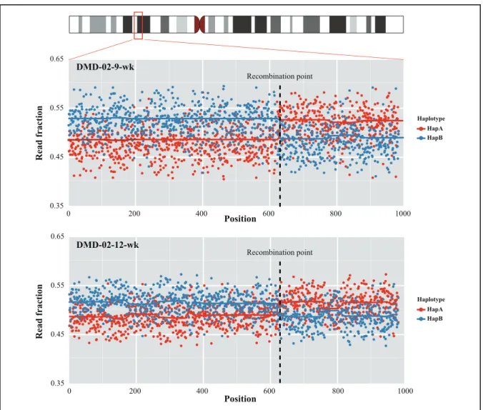
Before examining the haplotype imbalance between the 2 phased maternal haplotypes, we investigated the recombination event within the DMD region using the R package, as described in Materials and Methods. The bcp
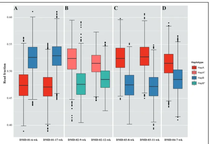
algorithm estimated a significant change point in the read fraction in the sequencing data from DMD-02 at 9 weeks (DMD-02-9-wk) and DMD-02-12-wk, which was also evident from a scatterplot of the read fraction distribu-tion of the phased haplotype (Fig. 2). Subsequent analysis suggested the recombination point between chromo-somal X positions 32 321 115 and 32 346 373 based on the bcp algorithm. The haplotypes of DMD-02-9-wk and DMD-02-12-wk sequencing data were recon-structed using the recombination point information. Be-cause all the other plasma sequencing data revealed 1 large segment of 2 haplotypes, all remaining families, with the exception of DMD-02, used the same haplotype that was phased from the proband sequencing data (see online Supplemental Fig. 4, A–C). We next attempted to predict the fetal genotype by comparing the allele frac-tion between the 2 haplotypes in the maternal plasma. In both DMD-01-6-wk and DMD-01-17-wk, the allele fraction of HapB was significantly higher, indicating in-heritance of the nonmutated haplotype by the fetus (Fig. 3A). All remaining samples supported the inheritance of a mutated haplotype by the fetuses, including DMD-02-9-wk and DMD-02-12-wk, the haplotypes of which were reconstructed based on a recombination event pre-diction (Fig. 3, B–D). All the allele fraction differences and statistical test results are provided (see online Supple-mental Table 6). The fetal genotypes predicted from the 4 DMD families matched exactly the fetal genomic DNA sequencing data (see online Supplemental Fig. 5 and on-line Supplemental Table 2).
Discussion
In the present study, we demonstrated that targeted deep sequencing makes feasible not only genetic diagnosis of a DMD patient but also carrier detection and noninvasive prenatal diagnosis. Considering that the prenatal diagno-sis of monogenic disorders is still performed in a family-based setting in which a genetically confirmed proband or carrier has been identified, this method has a practical advantage that proband diagnosis, carrier detection, and noninvasive prenatal diagnosis can be accomplished effi-ciently with a single platform.
For clinical implementation of noninvasive prenatal diagnosis of X-linked recessive disorders including DMD, refining the detection method of dosage imbal-ance caused by presence of a small fraction of cffDNA is a critical step in identifying the maternally derived geno-type. Tsui et al. used digital PCR to detect the slight overrepresentation of a coagulation factor VIII, proco-agulant component (F8) mutation in pregnant women who are carriers of hemophilia mutations (12 ). The rel-ative mutation dosage analysis based on a sequential probability ratio test was used. Although it is simple and does not require haplotype information, the detecting Noninvasive Prenatal Diagnosis of Duchenne Muscular Dystrophy
probe must be individualized according to the specific mutation type. Further, multiple tests of each sample should be conducted to obtain sufficient statistical power, especially when the fetal DNA fraction is low, as in the early gestational weeks. Relative haplotype dosage analysis may be an alternative option for identifying the slight overrepresentation of an inherited maternal mutation or allele because the genome-wide or targeted massively parallel sequencing approach can produce many informative SNVs for haplotyping. Lam et al. used this approach to identify the maternal inheritance of a mutation in a -thalassemia model (9). Although -thalassemia is a disease with autosomal recessive inheritance, a method that identifies the maternal inher-itance pattern would be equally applicable to X-linked recessive diseases. Instead of using the separate
haplotyp-ing method employed by Lam et al., New et al. sequenced a parent and patient trio, and then used the resulting haplotype information for maternal DNA analysis in the targeted platform used in the trio analysis (11 ). This diagnostic flow could be incorporated more easily into the current genetic diagnosis and counseling process.
Several gene-specific factors inherent to DMD should be considered for clinical implementation of non-invasive prenatal diagnosis. First, large deletion/duplica-tion mutadeletion/duplica-tions constitute about two-thirds of the DMD mutation spectrum. Because a dosage imbalance already exists in DMD carriers with a large deletion/duplication mutation, it would be difficult to perform relative muta-tion dosage analysis using digital PCR. Thus, measuring the haplotype imbalance of the DMD region outside the mutation would be plausible. Second, the recombination
0.35 0.45 0.55 0 200 400 600 80 000 0.35 0.45 0.55 0.65 0 200 400 600 80 0 1 0 1000 DMD-02-12-wk Haplotype HapA HapB Haplotype HapA HapB Recombination point Read fraction Read fraction Position Position
Fig. 2. Detection of a recombination event and haplotype reconstruction in the DMD-02 family.
Read fraction distribution of 2 haplotypes. Each haplotype was divided into 2 large segments. The black dotted lines indicate the recombina-tion point predicted by the bcp algorithm. The karyogram representing chromosome X was generated by ggbio [Yin et al. (24 )].
rate of DMD is about 4 times higher than the recombi-nation rate with chromosome X and with the whole ge-nome: 4.73 centimorgans (cM)/Mb, 1.21 cM/Mb, and 1.26 cM/Mb, respectively (21, 22 ). If a recombination event occurred within the DMD region, it would greatly affect the dosage imbalance analysis and could result in an incorrect prediction. Fetal genotype prediction with-out considering the inheritance of recombinant haplo-types would cause nonsignificant haplotype imbalance or contradictory result; it is possible to misdiagnose a non-DMD fetus as harboring a non-DMD mutation and vice versa (see online Supplemental Fig. 6 and online Supplemental Table 7). Thus, targeting the whole DMD region with a tiling design is preferable to ensure the reliable detection of a recombination event within the DMD region. Third, because the DMD region is hemizygous for male pro-bands, all phased heterozygous SNVs are informative for dosage imbalance analysis provided that the recombina-tion event is checked and corrected for before analysis.
Considering the abovementioned gene-specific fac-tors, we postulate that our approach targeting the whole
DMD region with tiling design provide the best approach for noninvasive prenatal diagnosis of DMD. Instead of using multiple haplotype blocks for repeated relative hap-lotype dosage analysis (5, 11 ), we hypothesized that the whole DMD gene could be analyzed as 1 large haplotype block, and we compared directly the allele fractions of 2 phased maternal heterozygous alleles. Although the study reported by New et al., in which multiple haplotype blocks were used, demonstrated the clinical applicability of that design to an array of autosomal recessive disorders (11 ), we believe that the current approach using 1 large haplotype block is a simpler and more straightforward method, at least for DMD. Although the fetal genotypes from all 4 DMD families were accurately predicted, the application of the current approach to routine molecular testing may have several limitations, in particular regard-ing noninvasive prenatal diagnosis. First, the current proband-based phasing approach may lead to misdiagno-sis if separate recombination occurs both in a proband and in a new fetus. In addition, it was basically impossible to discern whether the recombination event observed in 0.40 0.45 0.50 0.55 0.60 DMD-01-6-wk DMD-01-17-wk DMD-02-9-wk DMD-02-12-wk DMD-03-8-wk DMD-03-11-wk DMD-04-7-wk Read fraction Haplotype HapA HapB HapB* HapA*
A
B
C
D
Fig. 3. Fetal genotype prediction.
(A), HapB is overrepresented in DMD-01 maternal plasma samples (allele fraction differences: 5.0% and 5.6%). HapA or HapA* are overrep-resented in the remaining maternal plasma samples. (B), DMD-02 (allele fraction differences: 4.4% and 2.9%). (C), DMD-03 (allele fraction differences: 5.6% and 4.9%). (D), DMD-04 (allele fraction difference: 2.9%).
still be correctly predicted. This disadvantage could be partly overcome by adding the grandfather to maternal haplotype phasing, as the maternal X haplotype inherited from the grandfather would theoretically be free of re-combination. This approach of using the grandparents for phasing was introduced in a recent article by Meng et al. (10 ). Second, as female patients or carriers of DMD mutations with no known proband have been increas-ingly identified, the current proband-based phasing ap-proach also has a practical limitation in that setting. An alternative method that uses other family members for maternal haplotype phasing could be introduced to over-come this limitation, although it may still not be appli-cable to all at-risk couples without a proband. A grand-father or normal male child may be used for phasing via the same approach. Moreover, a carrier female or normal female child may also be used for phasing when the pa-ternal genotype is available. Third, the optimal timing of testing and the least fetal DNA fraction required should be determined and validated in extended DMD families with various DMD mutations. In the current study, the earliest gestational time and the lowest fetal fraction that allowed successful noninvasive prenatal diagnosis were 6 weeks and 5 days and 5.8%, respectively. New et al. re-ported a successful case of prenatal diagnosis at 5 weeks and 6 days of gestation with a fetal fraction of 1.4% (11 ). We believe that our results are compatible with those of the study performed by New et al. in terms of resolution, considering that 5– 6 weeks of gestation might be the earliest period at which prenatal tests may be offered. However, data collection in extended families is needed, as the current study used only 4 families compared with the 14 families reported by New et al. Fourth, because the informative SNVs that are required for dosage imbalance analysis of maternally inherited alleles might be limited in number and located at greater distance, the current ap-proach using 1 large haplotype block may be biased by false-recombination prediction. Thus, the extension of the applicability of the current approach to autosomal recessive disorders should be demonstrated separately.
clinic. Currently, no curative therapy is available for DMD, although some therapeutic molecules are under clinical trial (23 ). However, considering the current method is best fitted to clinical circumstances in which the presence of an affected proband is the reason for prenatal testing, noninvasive determination of fetal geno-type in the early gestational weeks could provide an autonomy-based reproductive option to the parents.
Despite the need to overcome these various hurdles, our approach for the comprehensive genetic diagnosis of the proband and noninvasive prenatal diagnosis using a single massively parallel targeted sequencing platform may provide a practical model for implementation of next-generation sequencing technology to clinical genetics.
Author Contributions: All authors confirmed they have contributed to
the intellectual content of this paper and have met the following 3 require-ments: (a) significant contributions to the conception and design, acquisi-tion of data, or analysis and interpretaacquisi-tion of data; (b) drafting or revising the article for intellectual content; and (c) final approval of the published article.
Authors’ Disclosures or Potential Conflicts of Interest: Upon
man-uscript submission, all authors completed the author disclosure form. Dis-closures and/or potential conflicts of interest:
Employment or Leadership: None declared. Consultant or Advisory Role: None declared. Stock Ownership: None declared.
Honoraria: None declared.
Research Funding: J.H. Chae, grant from the Korea Health
Technol-ogy R&D Project through the Korea Health Industry Development Institute, funded by the Ministry of Health and Welfare, Republic of Korea (grant number: HI13C-1468-050013); and Seoul National University Hospital and SK Telecom grant (grant number: 800-20120447).
Expert Testimony: None declared. Patents: None declared.
Role of Sponsor: The funding organizations played no role in the
design of study, choice of enrolled patients, review and interpretation of data, or preparation or approval of manuscript.
References 1. Lo YM, Corbetta N, Chamberlain PF, Rai V, Sargent IL,
Redman CW, Wainscoat JS. Presence of fetal DNA in maternal plasma and serum. Lancet 1997;350:485–7.
2. Chiu RW, Lo YM. Clinical applications of maternal plasma
fetal DNA analysis: translating the fruits of 15 years of re-search. Clin Chem Lab Med 2013;51:197–204.
3. Lo YM. Non-invasive prenatal testing using massively
parallel sequencing of maternal plasma DNA: from mo-lecular karyotyping to fetal whole-genome sequencing. Reprod Biomed Online 2013;27:593– 8.
4. American College of Obstetricians and Gynecologists
Committee on Genetics. Committee Opinion No. 545:
noninvasive prenatal testing for fetal aneuploidy. Ob-stet Gynecol 2012;120:1532– 4.
5. Lo YM, Chan KC, Sun H, Chen EZ, Jiang P, Lun FM, et al.
Maternal plasma DNA sequencing reveals the genome-wide genetic and mutational profile of the fetus. Sci Transl Med 2010;2:61ra91.
6. Liao GJ, Lun FM, Zheng YW, Chan KC, Leung TY, Lau TK,
et al. Targeted massively parallel sequencing of mater-nal plasma DNA permits efficient and unbiased detec-tion of fetal alleles. Clin Chem 2011;57:92–101.
7. Kitzman JO, Snyder MW, Ventura M, Lewis AP, Qiu R,
Simmons LE, et al. Noninvasive whole-genome
se-quencing of a human fetus. Sci Transl Med 2012;4: 137ra76.
8. Lim BC, Lee S, Shin JY, Kim JI, Hwang H, Kim KJ,
et al. Genetic diagnosis of Duchenne and Becker mus-cular dystrophy using next-generation sequencing technology: comprehensive mutational search in a sin-gle platform. J Med Genet 2011;48:731– 6.
9. Lam KW, Jiang P, Liao GJ, Chan KC, Leung TY, Chiu RW,
Lo YM. Noninvasive prenatal diagnosis of monogenic diseases by targeted massively parallel sequencing of maternal plasma: application to-thalassemia. Clin Chem 2012;58:1467–75.
10. Meng M, Li X, Ge H, Chen F, Han M, Zhang Y, et al.
Noninvasive prenatal testing for autosomal recessive conditions by maternal plasma sequencing in a case of congenital deafness. Genet Med 2014;99:E1022–30.
11. New MI, Tong YK, Yuen T, Jiang P, Pina C, Chan KC, et al.
Noninvasive prenatal diagnosis of congenital adrenal hyperplasia using cell-free fetal DNA in maternal plasma. J Clin Endocrinol Metab 2014;99:E1022–30.
12. Tsui NB, Kadir RA, Chan KC, Chi C, Mellars G,
Tudden-ham EG, et al. Noninvasive prenatal diagnosis of hemo-philia by microfluidics digital PCR analysis of maternal plasma DNA. Blood 2011;117:3684 –91.
13. Langmead B, Salzberg SL. Fast gapped-read alignment
with bowtie 2. Nat Methods 2012;9:357–9.
14. Li H, Handsaker B, Wysoker A, Fennell T, Ruan J, Homer
N, et al. The Sequence Alignment/Map format and SAMtools. Bioinformatics 2009;25:2078 –9.
15. DePristo MA, Banks E, Poplin R, Garimella KV, Maguire
JR, Hartl C, et al. A framework for variation discovery and genotyping using next-generation DNA sequenc-ing data. Nat Genet 2011;43:491– 8.
16. Wang K, Li M, Hakonarson H. Annovar: functional
an-notation of genetic variants from high-throughput se-quencing data. Nucleic Acids Res 2010;38:e164.
17. Ye K, Schulz MH, Long Q, Apweiler R, Ning Z. Pindel:
a pattern growth approach to detect break points of large deletions and medium sized insertions from paired-end short reads. Bioinformatics 2009;25: 2865–71.
18. Kent WJ, Sugnet CW, Furey TS, Roskin KM, Pringle TH,
Zahler AM, Haussler D. The human genome browser at UCSC. Genome Res 2002;12:996 –1006.
19. Scrucca, L. qcc: an R package for quality control
chart-ing and statistical process control. R News 2004;4/
1:11–7.
20. Erdman C, Emerson JW. A fast Bayesian change point
analysis for the segmentation of microarray data. Bioin-formatics 2008;24:2143– 8.
21. Jensen-Seaman MI, Furey TS, Payseur BA, Lu Y, Roskin
KM, Chen CF, et al. Comparative recombination rates in the rat, mouse, and human genomes. Genome Res 2004;14:528 –38.
22. International HapMap Consortium. The International
HapMap Project. Nature 2003;426:789 –96.
23. Jarmin S, Kymalainen H, Popplewell L, Dickson G. New
developments in the use of gene therapy to treat Duch-enne muscular dystrophy. Expert Opin Biol Ther 2014; 14:209 –30.
24. Yin T, Cook D, Lawrence M. ggbio: an R package for
extending the grammar of graphics for genomic data. Genome Biol 2012;13:R77.